Downloaded 263 times


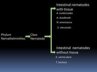


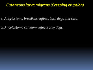


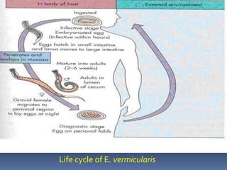


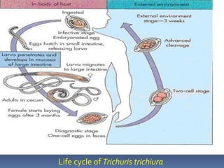


This document discusses several intestinal nematodes (roundworms) that can infect humans. It provides details on the morphology, life cycles, modes of transmission, symptoms, and diagnosis of Ascaris lumbricoides (the large roundworm), the two hookworm species (Ancylostoma duodenale and Necator americanus), Strongyloides stercoralis, Enterobius vermicularis (the pinworm), and Trichuris trichiura (the whipworm). Each worm has a unique life cycle involving eggs, larvae, and adult stages, and they are transmitted through ingestion of contaminated food, water, or soil. Symptoms vary but often involve abdominal pain, diarrhea, and


![Trypanosoma [1]](https://cdn.slidesharecdn.com/ss_thumbnails/trypanosomaseminar1-170312074241-thumbnail.jpg?width=640&height=640&fit=bounds)


