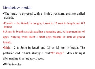
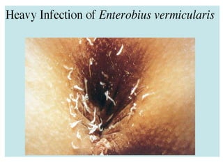
Enterobius vermicularis
By: Osman H. Ali
Introduction
•Enterobius vermicularis, the human pinworm, threadworm or seat worm,
formerly called Oxyuris vermicularis has been known from ancient times.
•The name Enterobius vermicularis means a tiny worm living in the intestine
(Greek enteron—intestine, bios-life and vermiculus—small worm).
•The term Oxyuris means ‘sharp tail’, a feature of the female worm, from
which the name ‘pinworm’ is also derived.
•It is worldwide in distribution. Unlike the usual situation where helminthic
infections are more prevalent in the poor people of the tropics, E.vermicularis
is one worm infestation which is far more common in the affluent nations in
the cold and temperate regions.
Introduction
•E. vermicularis is parasitic only to humans.
•Adults inhabit the ileocecus, that is, cecum and adjacent
ascending colon and distal ileum.
•The infection of E. vermicularis may cause Enterobiasis.
•World-wide distribution, it is commonly found in
kindergarten and primary school students.
•It has a round body with cylindrical ends and a complete
digestive system including mouth and anus
•It has separate sexes, the female is usually larger than a male.
Introduction
•Right after mating, the male dies. Therefore, the male worms are
rarely seen.
•The female worms migrate out the anus depositing eggs on the
perianal skin. Humans get this infection by mouth and by
autoinfection.
Morphology -- Adult
•The adults are short, white, fusiform
worms with pointed ends, looking like
bits of white thread.
•The mouth is surrounded by three
wing- like cuticular expansions
(cervical alae) which are transversely
striated.
Morphology -- Adult
•The body is covered with a highly resistant coating called
cuticle.
•Female – the female is longer, 8 mm to 12 mm in length and 0.3
mm to
0.5 mm in breath straight and has a tapering end. A large number of
eggs varying from 4600 -17000 eggs present in uteri of gravid
female.
•Male - 2 to 5mm in length and 0.1 to 0.2 mm in breath. The
posterior end in blunt, sharply curved “6” shape” . Males die right
after mating, thus are rarely seen.
•White in color
Morphology -- Egg
•Eggs are colorless, transparent and non-bile stained. They are
measure 50 to 60m by 25 µm. The are planoconvex (one side
flattened and one
side convex).
•The egg shell is very thin, hyaline and transparent
and contain typically coiled larva.
Distribution:
•Cosmopolitan.
•More common in temperate and cold climate than warm
climate.
Habitat:
LARGE INTESTINE
- Caecum
- Appendix
- Ascending colon
molt molt 3 times
Adults Newly laid
eggs
Infective
eggs
Larvae Adults
6h
Life cycle
Pathogenesis and Pathology
A.Pathogenesis of adult worm.
•Adult worm produce little pathological changes in the intestine.
Less frequently it may cause mild catarrhal inflammation
(inflammation of the mucous membranes) of intestinal mucosa
and their sites of attachment. Occasionally, minute ulcer may
developed in caecum and appendix.
•In sensitized person, absorption of metabolites secreted by
worm gives rise to allergic manifestation.
B. Pathogenecity of eggs.
•Significant pathological lesions are caused by the eggs. The eggs adhere
well on surface of skin and by their mucoid secretions cause irritation and
characteristic nocturnal perianal or perineal itching.
Clinical manifestation.
•Most patients are asymptomatic.
•In asymptomatic cases, the most common complaint is perianal and
perineal pruritus, usually nocturnal or in the early morning.
•Other symptoms includes abdominal pain, irritability and restlessness.
• All these symptoms are caused by female worms laying eggs on anal area.
• In children in severe infection, behavioral changes such as sleep
disturbances, neurosis, nail bite, grinding teeth at night etc.
• Adult worms also cause appendicitis.
Complications:
In heavy infection following complication are seen:
• Impetigo (pustules) or excoriation due to perineal scratching.
• Vulvo-vaginitis, salpingitis (inflammation of the fallopian tubes) and
prostatitis due to ectopic migration of gravid female.
• Urethritis and Endometritis.
Prognosis:
•E. Vermicularis infection is rarely fatal. Cure rate with proper
treatment is above 90%. However, reinfestation is common.
Diagnosis:
The history of pruritus and demonstration of small white thread
like worms in the undergarments are suggestive of E.
vermicularis infection in children.
Laboratory diagnosis:
Parasitic diagnosis.
Microscopic demonstration of characteristic eggs in the perianal
or perineal scraping is the methods of choice for the diagnosis of
enterobiasis. Anal or perianal specimens can be collected by either
National Institute of health (NIH) swab, cellophane swab or
Scotch tape swab method.
National Institute of health (NIH) swab
•Transparent adhesive tape pressed firmly against perianal skin and
then spread on to a microscope slide.
Treatments:
•Pyrantel pamoate is the drug of choice. It paralyses the worms.
•Effective oral dose is 5mg/kg.
•Mebendazole single dose 100mg .
•Albendazole, piperazine are also frequently used.
•In heavy infection, these drugs are repeated after interval of 4
weeks.
•All the members of the family may need treatments.
Preventions:
•Treatment of infected children and other members of the
family.
•Improved personal hygiene and cleanliness such as cuttings
nails, hand washing and washing the bed linens night dress etc.
•Teaching the children not to put the fingers or other object in
their mouth.
enterobiusvermicularis.pptx

enterobiusvermicularis.pptx

  • 1.
  • 2.
    Introduction •Enterobius vermicularis, thehuman pinworm, threadworm or seat worm, formerly called Oxyuris vermicularis has been known from ancient times. •The name Enterobius vermicularis means a tiny worm living in the intestine (Greek enteron—intestine, bios-life and vermiculus—small worm). •The term Oxyuris means ‘sharp tail’, a feature of the female worm, from which the name ‘pinworm’ is also derived. •It is worldwide in distribution. Unlike the usual situation where helminthic infections are more prevalent in the poor people of the tropics, E.vermicularis is one worm infestation which is far more common in the affluent nations in the cold and temperate regions.
  • 3.
    Introduction •E. vermicularis isparasitic only to humans. •Adults inhabit the ileocecus, that is, cecum and adjacent ascending colon and distal ileum. •The infection of E. vermicularis may cause Enterobiasis. •World-wide distribution, it is commonly found in kindergarten and primary school students. •It has a round body with cylindrical ends and a complete digestive system including mouth and anus •It has separate sexes, the female is usually larger than a male.
  • 4.
    Introduction •Right after mating,the male dies. Therefore, the male worms are rarely seen. •The female worms migrate out the anus depositing eggs on the perianal skin. Humans get this infection by mouth and by autoinfection.
  • 5.
    Morphology -- Adult •Theadults are short, white, fusiform worms with pointed ends, looking like bits of white thread. •The mouth is surrounded by three wing- like cuticular expansions (cervical alae) which are transversely striated.
  • 6.
    Morphology -- Adult •Thebody is covered with a highly resistant coating called cuticle. •Female – the female is longer, 8 mm to 12 mm in length and 0.3 mm to 0.5 mm in breath straight and has a tapering end. A large number of eggs varying from 4600 -17000 eggs present in uteri of gravid female. •Male - 2 to 5mm in length and 0.1 to 0.2 mm in breath. The posterior end in blunt, sharply curved “6” shape” . Males die right after mating, thus are rarely seen. •White in color
  • 7.
    Morphology -- Egg •Eggsare colorless, transparent and non-bile stained. They are measure 50 to 60m by 25 µm. The are planoconvex (one side flattened and one side convex). •The egg shell is very thin, hyaline and transparent and contain typically coiled larva.
  • 8.
    Distribution: •Cosmopolitan. •More common intemperate and cold climate than warm climate. Habitat: LARGE INTESTINE - Caecum - Appendix - Ascending colon
  • 9.
    molt molt 3times Adults Newly laid eggs Infective eggs Larvae Adults 6h Life cycle
  • 10.
    Pathogenesis and Pathology A.Pathogenesisof adult worm. •Adult worm produce little pathological changes in the intestine. Less frequently it may cause mild catarrhal inflammation (inflammation of the mucous membranes) of intestinal mucosa and their sites of attachment. Occasionally, minute ulcer may developed in caecum and appendix. •In sensitized person, absorption of metabolites secreted by worm gives rise to allergic manifestation.
  • 12.
    B. Pathogenecity ofeggs. •Significant pathological lesions are caused by the eggs. The eggs adhere well on surface of skin and by their mucoid secretions cause irritation and characteristic nocturnal perianal or perineal itching. Clinical manifestation. •Most patients are asymptomatic. •In asymptomatic cases, the most common complaint is perianal and perineal pruritus, usually nocturnal or in the early morning. •Other symptoms includes abdominal pain, irritability and restlessness.
  • 13.
    • All thesesymptoms are caused by female worms laying eggs on anal area. • In children in severe infection, behavioral changes such as sleep disturbances, neurosis, nail bite, grinding teeth at night etc. • Adult worms also cause appendicitis. Complications: In heavy infection following complication are seen: • Impetigo (pustules) or excoriation due to perineal scratching. • Vulvo-vaginitis, salpingitis (inflammation of the fallopian tubes) and prostatitis due to ectopic migration of gravid female. • Urethritis and Endometritis.
  • 14.
    Prognosis: •E. Vermicularis infectionis rarely fatal. Cure rate with proper treatment is above 90%. However, reinfestation is common.
  • 15.
    Diagnosis: The history ofpruritus and demonstration of small white thread like worms in the undergarments are suggestive of E. vermicularis infection in children. Laboratory diagnosis: Parasitic diagnosis. Microscopic demonstration of characteristic eggs in the perianal or perineal scraping is the methods of choice for the diagnosis of enterobiasis. Anal or perianal specimens can be collected by either National Institute of health (NIH) swab, cellophane swab or Scotch tape swab method.
  • 16.
    National Institute ofhealth (NIH) swab •Transparent adhesive tape pressed firmly against perianal skin and then spread on to a microscope slide.
  • 17.
    Treatments: •Pyrantel pamoate isthe drug of choice. It paralyses the worms. •Effective oral dose is 5mg/kg. •Mebendazole single dose 100mg . •Albendazole, piperazine are also frequently used. •In heavy infection, these drugs are repeated after interval of 4 weeks. •All the members of the family may need treatments.
  • 18.
    Preventions: •Treatment of infectedchildren and other members of the family. •Improved personal hygiene and cleanliness such as cuttings nails, hand washing and washing the bed linens night dress etc. •Teaching the children not to put the fingers or other object in their mouth.