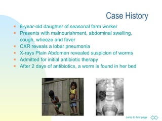
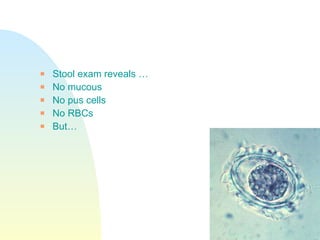
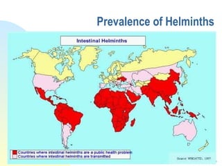
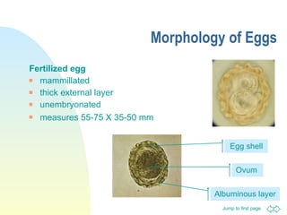
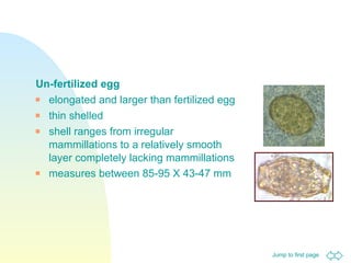
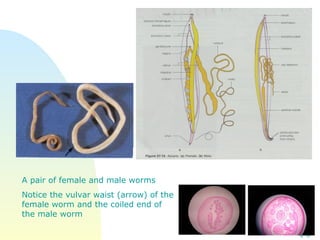
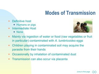
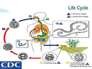
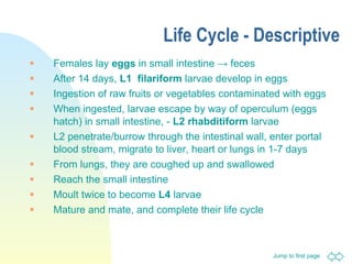
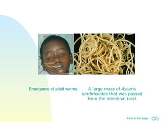
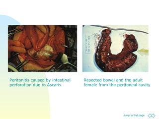
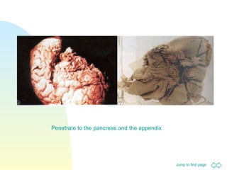
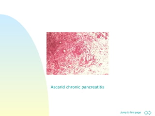
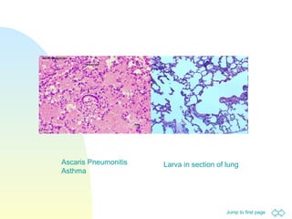
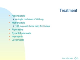
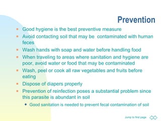
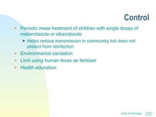
Case History 6-year-old daughter of seasonal farm worker Presents with malnourishment, abdominal swelling, cough, wheeze and fever CXR reveals a lobar pneumonia X-rays Plain Abdomen revealed suspicion of worms Admitted for initial antibiotic therapy After 2 days of antibiotics, a worm is found in her bed
Stool exam reveals … No mucous No pus cells No RBCs But…
Ascaris lumbricoides Dr Kamran Afzal Classified Microbiologist
Nematode The intestinal nematodes Ascaris Hook worms Pin worm Whip worm The blood- and tissue dwelling nematodes Filaria Trichinella
Prevalence of Helminths
Family Ascaridae Two species, one in humans and the other in pigs Ascaris lumbricoides  - humans Because of size and cosmopolitan distribution, these are well known parasites Rural > urban Children > adults
Prevalence Most common helminthic human infection - Worldwide High prevalence in underdeveloped countries that have poor sanitation Occurs during rainy months, tropical and subtropical countries Largest nematode to infect the human intestine A common cream colored roundworm that is parasitic in the intestines of humans An estimated 1 billion people are infected   1 out of 4 people in the world
Epidemiology Indiscriminate defecation particularly near areas of habitation seeds the soil with eggs Children become infected by ingesting soil or putting soiled items in the mouth The eggs may contaminate unwashed vegetables and water supplies Eggs can be carried by cockroaches, flies, wind, and even on money
Morphology of Eggs Fertilized egg mammillated  thick external layer  unembryonated  measures 55-75 X 35-50 mm   Albuminous layer Egg shell Ovum
Un-fertilized egg elongated and larger than fertilized egg  thin shelled  shell ranges from irregular mammillations to a relatively smooth layer completely lacking mammillations  measures between 85-95 X 43-47 mm
Characteristics of Eggs Eggs can survive for prolonged periods as long as warm, shade, moist conditions are available  Eggs are resistant to low temperatures, desiccation, and strong chemicals Can remain viable for up to 10 years Eggs are resistant to usual methods of chemical water purification, removed by filtration and killed by boiling Eggs can embryonate in solutions of formalin, H 2 SO 4  and HCl Eggs embryonate but do not hatch until ingested by man Developing larvae are destroyed by sunlight, high temperatures and desiccation
Morphology of Worms Adult worm Tapered ends; length 15 to 35 cm  Characterized by three large rounded lips Female is larger in size and has a genital girdle  Ovaries are extensive, contain up to 27 million eggs at a given time Female lays eggs into host intestine  200,000 per day passed out in host feces Feed on semi-digested contents in the gut Evidence shows that they can bite the intestinal mucus membrane and feed on blood and tissue fluids
A pair of female and male worms  Notice the vulvar waist (arrow) of the female worm and the coiled end of the male worm
Modes of Transmission Definitive host  Humans or pigs Intermediate Host None Mainly via ingestion of water or food (raw vegetables or fruit in particular) contaminated with  A. lumbricoides  eggs Children playing in contaminated soil may acquire the parasite from their hands Occasionally by inhalation of contaminated dust Transmission can also occur via placenta
Life Cycle
Life Cycle - Descriptive Females lay  eggs  in small intestine  ->  feces After 14 days,  L1   filariform  larvae develop in eggs Ingestion of raw fruits or vegetables contaminated with eggs When ingested, larvae escape by way of operculum (eggs hatch) in small intestine, -  L2   rhabditiform  larvae L2 penetrate/burrow through the intestinal wall, enter portal blood stream, migrate to liver, heart or lungs in 1-7 days From lungs, they are coughed up and swallowed  Reach the small intestine Moult twice to become  L4  larvae Mature and mate, and complete their life cycle
Pathology Migration of larvae Little damage is caused by the penetration  Some larvae migrate to ectopic sites and dependent upon number and location, cause various inflammatory responses, leading to very severe allergic reactions  Spleen, liver, lymph nodes and brain Transplacental migration can also occur
In the lungs Worms destroy capillaries in the lungs, causing hemorrhage Heavy infections can lead to pools of blood which block air sacs Migration of white blood cells lead to more congestion; a condition known as Ascaris pneumonitis  Loeffler's pneumonia Lung tissue destroyed and bacterial infections occur, may be fatal
How the worms cause Pathology Normal worm activities - Rob the host of nutrients  Overcrowding leads to wandering  Females wander in search of males If worms migrate to stomach, acid irritates them leading to nausea, abdominal pain, restlessness and allergic reactions Penetration of the intestine or appendix can lead to peritonitis which is often fatal Aspiration of a vomited worm can result in death If worms migrate to lungs, they can cause extensive damage and possible death
Symptoms Symptoms associated with larval migration Migration of larvae in lungs may cause hemorrhagic/ eosinophilic pneumonia, cough (Loeffler's Syndrome) Breathing difficulties and fever Complications caused by parasite proteins that are highly allergenic - asthmatic attacks, pulmonary infiltration and urticaria (hives )
Symptoms associated with adult parasite in the intestine Usually asymptomatic (85%) Vague abdominal discomfort, nausea in mild cases Malnutrition in host especially in children in severe cases Heavy worm loads can retard physical and mental development Sometimes fatality may occur when mass of worms cause intestinal obstruction
Symptoms associated with worm migration Worms retain motility, do not attach Migration of adult worms may cause signs and symptoms of perforation, peritonitis, appendicitis or extrahepatic biliary obstruction Severe inflammatory reactions mark the migratory route Dermatological and allergic reactions can occur
Host immune response Innate Immune Response Macrophages, neutrophils and most importantly eosinophils The worms would be coated with IgG or IgE which would increase the release of eosinophil granules  Adaptive Immune Response High IL-4 production, high levels of IgE, eosinophilia and mastocytosis  Prior infection does not confer protective immunity
Complications Intestinal obstruction, volvulus, intussusception  Obstruction of intrahepatic and extrahepatic bile ducts Peritonitis caused by intestinal perforation Chronic pancreatitis Acute or chronic appendicitis Pneumonitis, bronchitis and asthma
Emergence of adult worms A large mass of  Ascaris lumbricoides  that was passed from the intestinal tract
Intestinal blockage in heavy infections - intestinal obstruction  Worms may knot up causing blockage which may be fatal
CS of a liver specimen contains many adult worms of  A.lumbricoides  obstructing the intrahepatic and extrahepatic bile ducts A.lumbricoides  in common bile duct
An autopsy specimen shows intestinal obstruction by many adult worms of  A.lumbricoides Markedly distended intestinal loop and worms protruding from the perforated wound
Peritonitis caused by intestinal perforation due to Ascaris Resected bowel and the adult female from the peritoneal cavity
Penetrate to the pancreas and the appendix
Ascarid chronic pancreatitis
Larva in section of lung Ascaris Pneumonitis Asthma
 
Laboratory Diagnosis Macroscopic identification Of adults passed in stool or through the mouth or nose Larval worms  Detection in sputum   Stool Microscopy   Eggs may be identified on direct stool examination Eosinophilia Eosinophilia can be found, particularly during larval migration through the lungs
Imaging In heavily infested individuals, particularly children, large collections of worms may be detectable on plain film of the abdomen Ultrasound Ultrasound exams can help to diagnose hepatobiliary or pancreatic ascariasis Single worms, bundles of worms, or pseudotumor-like appearance Individual body segments of worms may be seen
Endoscopic Retrograde Cholangiopancreatography (ERCP) A duodenoscope with a snare to extract the worm out of the patient
Treatment Albendazole A single oral dose of 400 mg Mebenazole 100 mg orally twice daily for 3 days Piperazine  Pyrantel pamoate Ivermectin Levamisole
Prevention Good hygiene is the best preventive measure Avoid contacting soil that may be  contaminated with human feces Wash hands with soap and water before handling food When traveling to areas where sanitation and hygiene are poor, avoid water or food that may be contaminated Wash, peel or cook all raw vegetables and fruits before eating Dispose of diapers properly Prevention of reinfection poses a substantial problem since this parasite is abundant in soil  Good sanitation is needed to prevent fecal contamination of soil
Control Periodic mass treatment of children with single doses of mebendazole or albendazole Helps reduce transmission in community but does not protect from reinfection Environmental sanitation Limit using human feces as fertilizer Health education
How many people in the world are estimated to be infected with  A. lumbricoides  ?
Who are the definitive host/s of this parasite?
Name 2 modes of transmission?
What morphological difference can be seen in fertile and infertile eggs?
Name the symptoms caused by larvae migration in the lungs
What is the drug of choice for this parasite?
What are some of the methods of prevention?

Ascaris.ppt

  • 1.
    Case History 6-year-olddaughter of seasonal farm worker Presents with malnourishment, abdominal swelling, cough, wheeze and fever CXR reveals a lobar pneumonia X-rays Plain Abdomen revealed suspicion of worms Admitted for initial antibiotic therapy After 2 days of antibiotics, a worm is found in her bed
  • 2.
    Stool exam reveals… No mucous No pus cells No RBCs But…
  • 3.
    Ascaris lumbricoides DrKamran Afzal Classified Microbiologist
  • 4.
    Nematode The intestinalnematodes Ascaris Hook worms Pin worm Whip worm The blood- and tissue dwelling nematodes Filaria Trichinella
  • 5.
  • 6.
    Family Ascaridae Twospecies, one in humans and the other in pigs Ascaris lumbricoides - humans Because of size and cosmopolitan distribution, these are well known parasites Rural > urban Children > adults
  • 7.
    Prevalence Most commonhelminthic human infection - Worldwide High prevalence in underdeveloped countries that have poor sanitation Occurs during rainy months, tropical and subtropical countries Largest nematode to infect the human intestine A common cream colored roundworm that is parasitic in the intestines of humans An estimated 1 billion people are infected 1 out of 4 people in the world
  • 8.
    Epidemiology Indiscriminate defecationparticularly near areas of habitation seeds the soil with eggs Children become infected by ingesting soil or putting soiled items in the mouth The eggs may contaminate unwashed vegetables and water supplies Eggs can be carried by cockroaches, flies, wind, and even on money
  • 9.
    Morphology of EggsFertilized egg mammillated thick external layer unembryonated measures 55-75 X 35-50 mm Albuminous layer Egg shell Ovum
  • 10.
    Un-fertilized egg elongatedand larger than fertilized egg thin shelled shell ranges from irregular mammillations to a relatively smooth layer completely lacking mammillations measures between 85-95 X 43-47 mm
  • 11.
    Characteristics of EggsEggs can survive for prolonged periods as long as warm, shade, moist conditions are available Eggs are resistant to low temperatures, desiccation, and strong chemicals Can remain viable for up to 10 years Eggs are resistant to usual methods of chemical water purification, removed by filtration and killed by boiling Eggs can embryonate in solutions of formalin, H 2 SO 4 and HCl Eggs embryonate but do not hatch until ingested by man Developing larvae are destroyed by sunlight, high temperatures and desiccation
  • 12.
    Morphology of WormsAdult worm Tapered ends; length 15 to 35 cm Characterized by three large rounded lips Female is larger in size and has a genital girdle Ovaries are extensive, contain up to 27 million eggs at a given time Female lays eggs into host intestine 200,000 per day passed out in host feces Feed on semi-digested contents in the gut Evidence shows that they can bite the intestinal mucus membrane and feed on blood and tissue fluids
  • 13.
    A pair offemale and male worms Notice the vulvar waist (arrow) of the female worm and the coiled end of the male worm
  • 14.
    Modes of TransmissionDefinitive host Humans or pigs Intermediate Host None Mainly via ingestion of water or food (raw vegetables or fruit in particular) contaminated with A. lumbricoides eggs Children playing in contaminated soil may acquire the parasite from their hands Occasionally by inhalation of contaminated dust Transmission can also occur via placenta
  • 15.
  • 16.
    Life Cycle -Descriptive Females lay eggs in small intestine -> feces After 14 days, L1 filariform larvae develop in eggs Ingestion of raw fruits or vegetables contaminated with eggs When ingested, larvae escape by way of operculum (eggs hatch) in small intestine, - L2 rhabditiform larvae L2 penetrate/burrow through the intestinal wall, enter portal blood stream, migrate to liver, heart or lungs in 1-7 days From lungs, they are coughed up and swallowed Reach the small intestine Moult twice to become L4 larvae Mature and mate, and complete their life cycle
  • 17.
    Pathology Migration oflarvae Little damage is caused by the penetration Some larvae migrate to ectopic sites and dependent upon number and location, cause various inflammatory responses, leading to very severe allergic reactions Spleen, liver, lymph nodes and brain Transplacental migration can also occur
  • 18.
    In the lungsWorms destroy capillaries in the lungs, causing hemorrhage Heavy infections can lead to pools of blood which block air sacs Migration of white blood cells lead to more congestion; a condition known as Ascaris pneumonitis Loeffler's pneumonia Lung tissue destroyed and bacterial infections occur, may be fatal
  • 19.
    How the wormscause Pathology Normal worm activities - Rob the host of nutrients Overcrowding leads to wandering Females wander in search of males If worms migrate to stomach, acid irritates them leading to nausea, abdominal pain, restlessness and allergic reactions Penetration of the intestine or appendix can lead to peritonitis which is often fatal Aspiration of a vomited worm can result in death If worms migrate to lungs, they can cause extensive damage and possible death
  • 20.
    Symptoms Symptoms associatedwith larval migration Migration of larvae in lungs may cause hemorrhagic/ eosinophilic pneumonia, cough (Loeffler's Syndrome) Breathing difficulties and fever Complications caused by parasite proteins that are highly allergenic - asthmatic attacks, pulmonary infiltration and urticaria (hives )
  • 21.
    Symptoms associated withadult parasite in the intestine Usually asymptomatic (85%) Vague abdominal discomfort, nausea in mild cases Malnutrition in host especially in children in severe cases Heavy worm loads can retard physical and mental development Sometimes fatality may occur when mass of worms cause intestinal obstruction
  • 22.
    Symptoms associated withworm migration Worms retain motility, do not attach Migration of adult worms may cause signs and symptoms of perforation, peritonitis, appendicitis or extrahepatic biliary obstruction Severe inflammatory reactions mark the migratory route Dermatological and allergic reactions can occur
  • 23.
    Host immune responseInnate Immune Response Macrophages, neutrophils and most importantly eosinophils The worms would be coated with IgG or IgE which would increase the release of eosinophil granules Adaptive Immune Response High IL-4 production, high levels of IgE, eosinophilia and mastocytosis Prior infection does not confer protective immunity
  • 24.
    Complications Intestinal obstruction,volvulus, intussusception Obstruction of intrahepatic and extrahepatic bile ducts Peritonitis caused by intestinal perforation Chronic pancreatitis Acute or chronic appendicitis Pneumonitis, bronchitis and asthma
  • 25.
    Emergence of adultworms A large mass of Ascaris lumbricoides that was passed from the intestinal tract
  • 26.
    Intestinal blockage inheavy infections - intestinal obstruction Worms may knot up causing blockage which may be fatal
  • 27.
    CS of aliver specimen contains many adult worms of A.lumbricoides obstructing the intrahepatic and extrahepatic bile ducts A.lumbricoides in common bile duct
  • 28.
    An autopsy specimenshows intestinal obstruction by many adult worms of A.lumbricoides Markedly distended intestinal loop and worms protruding from the perforated wound
  • 29.
    Peritonitis caused byintestinal perforation due to Ascaris Resected bowel and the adult female from the peritoneal cavity
  • 30.
    Penetrate to thepancreas and the appendix
  • 31.
  • 32.
    Larva in sectionof lung Ascaris Pneumonitis Asthma
  • 33.
  • 34.
    Laboratory Diagnosis Macroscopicidentification Of adults passed in stool or through the mouth or nose Larval worms Detection in sputum Stool Microscopy Eggs may be identified on direct stool examination Eosinophilia Eosinophilia can be found, particularly during larval migration through the lungs
  • 35.
    Imaging In heavilyinfested individuals, particularly children, large collections of worms may be detectable on plain film of the abdomen Ultrasound Ultrasound exams can help to diagnose hepatobiliary or pancreatic ascariasis Single worms, bundles of worms, or pseudotumor-like appearance Individual body segments of worms may be seen
  • 36.
    Endoscopic Retrograde Cholangiopancreatography(ERCP) A duodenoscope with a snare to extract the worm out of the patient
  • 37.
    Treatment Albendazole Asingle oral dose of 400 mg Mebenazole 100 mg orally twice daily for 3 days Piperazine Pyrantel pamoate Ivermectin Levamisole
  • 38.
    Prevention Good hygieneis the best preventive measure Avoid contacting soil that may be contaminated with human feces Wash hands with soap and water before handling food When traveling to areas where sanitation and hygiene are poor, avoid water or food that may be contaminated Wash, peel or cook all raw vegetables and fruits before eating Dispose of diapers properly Prevention of reinfection poses a substantial problem since this parasite is abundant in soil Good sanitation is needed to prevent fecal contamination of soil
  • 39.
    Control Periodic masstreatment of children with single doses of mebendazole or albendazole Helps reduce transmission in community but does not protect from reinfection Environmental sanitation Limit using human feces as fertilizer Health education
  • 40.
    How many peoplein the world are estimated to be infected with A. lumbricoides ?
  • 41.
    Who are thedefinitive host/s of this parasite?
  • 42.
    Name 2 modesof transmission?
  • 43.
    What morphological differencecan be seen in fertile and infertile eggs?
  • 44.
    Name the symptomscaused by larvae migration in the lungs
  • 45.
    What is thedrug of choice for this parasite?
  • 46.
    What are someof the methods of prevention?