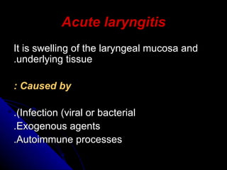
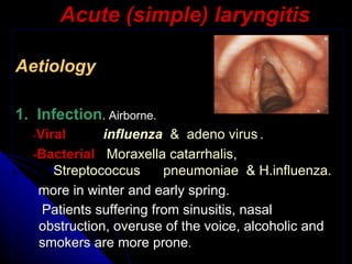
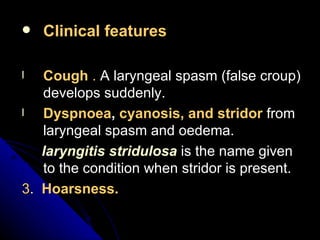
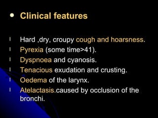
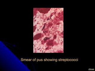
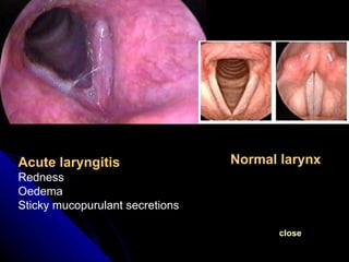
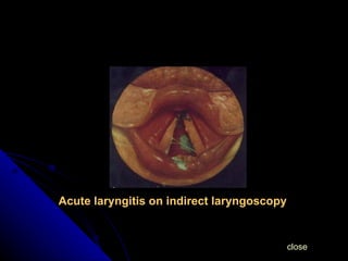
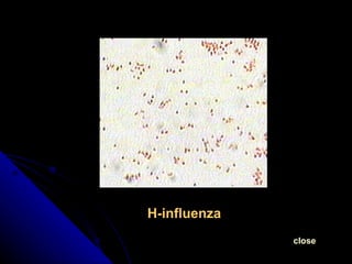
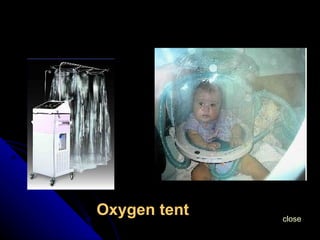
ACUTE LARYNGITIS Prepared by Dr.Hiwa As’ad
Acute laryngitis It is swelling of the laryngeal mucosa and underlying tissue. Caused by : Infection (viral or bacterial). Exogenous agents. Autoimmune processes.
Clinical entities Acute simple laryngitis. Acute laryngotracheobronchitis  (croup). Subglottic laryngitis  (pseudocroup). Acute epiglottitis.   Diphtheric laryngitis. Membranous laryngitis. Herps zoster of the larynx.
Acute (simple) laryngitis Aetiology 1.  Infection . Airborne. - Viral   influenza    &  adeno virus  . - Bacterial   Moraxella catarrhalis,  Streptococcus      pneumoniae   &  H.influenza. more in winter and early spring.  Patients suffering from sinusitis, nasal obstruction, overuse of the voice, alcoholic and smokers are more prone .
2.   Trauma  (vocal abuse &/or endoscopic manipulation). 3.   Irritation  from inhaled fumes or gas, including tobacco smoke.
Pathology   The laryngeal mucosa shows all signs of acute inflammation: Extravasation of fluid. Infiltration by polymorphnuclear leucocytes. Later plasma cells and lymphocytes predominates. The underlying muscles, the perichondrium, and the cricoarytenoid joints may be affected. The epithelium may be destroyed and exfoliated. Full recovery is usuall. Sometimes fibrosis will results leading to permanent damage to the laryngeal mucosa which can be the beginning of chronic laryngitis.
Pathological changes in the mucosa Redness   of the mucosa. Oedema  of the mucosa. Sticky mucopurulant exudate . Slight abrasions. Purulent exudation  in severe forms(septic laryngitis). Fibrinous laryngitis  in influenza, there are white plaques on the surface of the cords and the laryngeal inlet. Perichondritis  may follow the purulant form by H.streptococcus.
Clinical fearures Hoarsness  (high-pitched husky voice). Discomfort   in the throat. Pain  is slight or absent. Dysphagia  if epiglottis &/or arytenoid are markedly involved. Dyspnoea  in severe oedema. Dry and irritant  cough . Generalized symptoms  (malaise and fever , toxaemia is rare) more in bacterial infections. Symmetrical  redness &/or sticky secretions  on both vocal cords, at  indirect laryngoscopy . The clinical coarse in children can be rapidly progressive.
Progress Usually resolves in a few days. The hoarsness may persist for as long as 2 weeks after apparent resolution. A functional aphonia may follow specially in women. In severe cases the inflammation spreads to the lung in aged patients.
Treatment   Local  (supportive) Voice rest  (a quiet unforced whisper is allowed). Steam inhalations. M enthol  loosen viscid secretions. Aspirin. Warm application  to the neck. Codeine  to suppress dry cough.
General Rest  and sedatives. Avoidance  of alcohol and tobacco. Systemic antibiotics  in cases of bacterial infection - Penicillin 500 mg 4 times daily, - Doxycycline 200 mg daily or - Erythromycin 500 mg twice daily
Acute simple laryngitis in children More serious because of: Anatomical difference of infantile larynx. Rich lymphatic drainage. The neuromuscular mechanism is more easily upset and spasm more easily provoked. The child is less liable to expel secretions by cough.
Clinical features Cough  .  A laryngeal spasm (false croup) develops suddenly. Dyspnoea ,  cyanosis, and stridor  from laryngeal spasm and oedema. laryngitis stridulosa  is the name given to the condition when stridor is present. 3 .  Hoarsness.
Treatment  The same as in adult. In addition to oxygenation and securing the airway when there is stridor and cyanosis by intubation or tracheostomy. Home
Subglottic laryngitis (pseudocroup) Also called  spasmodic cough. Is common  <3 years  of age. The symptoms are  alarming. The cause is unkown but is  associated with influenza viruses  infections. There will be substantial  swelling of the subglottic space.
Clinical features Starts abruptly in a child with history of URTI. The child wakes up with dry cough and increasing stridor. No or mild fever. Restless, nervous and crying child.
Treatment   Sedatives are given to the parents and never to the child. Parenteral corticosteroids? Taking the child to a room with moist ear (e.g bathroom). In an emergency endotracheal intubation for 1-2 days. Home
Membranous laryngitis Rare, closely linked with croup , sometimes called  (pseudomembranous croup). Caused by : *Pseudomonus aeruginosa. *streptococci. *Vincent’s organisms. A confluent  membrane  covering the surface of the larynx and  when removed no bleeding  or ulceration occur. The main site is the  supraglottis.
Clinical features Simillar to other forms of laryngitis. The constitutional symptoms accompanied by anorexia and thirst. Moderate fever. Painfull swallowing. Cough. There may be stridor.
Diagnosis  is established by by bacteriology. Treatment  penicillins or sulphoneamides. Home
Acute epiglottitis Definition   special form of acute laryngitis, in which the inflammatory changes affect mainly the loosely attached mucosa of the epiglottis. Pathology  Localized oedema may obstruct the airway.    - H.influenza   is the usuall causative organism. - B-Haemolytic streptococci   rarely. Submucous abscesses may form.
Acute epiglottitis
Incidence 1:17.000  children. 1:100.000 adults. .  Vaccination is reducing its incidence.
Clinical features History is short and abrupt. Fever >40 C. Dyspnoea and stridor (progressive and alarming). Pain on swallowing (commoner than respiratory obstruction in adults). Drooling of saliva. The patient is preferring the sitting position
Radiologically diagnosed by thump print sign on lateral view of neck X-ray
Treatment Constant supervision in hospital when stridor is present. Inhalation of moist air. Throat swab and blood cultures. IV antibiotics in high doses (Amoxycillin). Endotrachial intubation , may be difficult. Tracheostomy. Home
Acute laryngotracheobronchitis (Croup)   Aetiology -Affects infants and small children up to the age of  7 . -Caused by  parainfluenza virus . -secondary bacterial infection  (haemolytic streptococcus)  superimpose by the  3 rd  day  which makes the condition worse.
Clinical features Hard ,dry, croupy  cough and hoarsness . Pyrexia  (some time>41). Dyspnoea  and cyanosis. Tenacious  exudation and crusting. Oedema  of the larynx. Atelactasis. caused by occlusion of the bronchi.
Narrowinng of the air column in the subglottic space (steeple sign) on x-ray of the neck AP view
The icreased muscular energy consumption required for breathing and coughing, with CO 2  retension leads to combination of metabolic and respiratory acidosis which paralyses the central regulation of respiration. During the initial phase the child is restless and cyanotic, in the later stages there may be an apparent improvement when the child becomes tired and calm.the retension of CO 2  causes a change of colour from cyanotic to pale and it is the sign of imminent disaster. A small child with a temp.>38.5 C and stridor should be admitted to hospital for observation
Treatment   Admission. Rest and reasurance. Systemic antibiotics(Penicillin) and anti-pyretics. Humidification. Oxygen preferably in a tent . Fluid by mouth or IV. IV steroids? Nasotracheal tube or tracheostomy. Removal of secretions by: Bronchoscopy with removal of secretions by suction  or forceps. Tracheostomy with intermittent suction. Home
Diphtheric laryngitis Usually an extension of faucial infection. Aetiology   corynbacterium diphtheriae . In children younger than 10 years of age and occasionally young adults. It has been less frequent since  universal immunization.
Histological changes in laryngeal diphtheria A-membrane B-submucosa infiltrated by leucocytes C-cartilage
Clinical features The onset is insidious. Cough of a hoarse, croupy nature. Stridor follows accompanied by cyanosis and recession of the chest wall. Pyrexia rarely above 37.8 C. Weak and rapid pulse. Greyish-white membrane  and bleeding when removed.
Diagnosis  By identifying the organism in swabs from the membrane. Treatment  Antitoxin injections IM or IV (20.000-100.000 units according to the age). Systemic penicillin. Oxygen. Tracheostomy. Home
Herpes zoster of the larynx Rare condition caused by a  neurotrophic virus . The  superior laryngeal branch of the vagus nerve and the pharyngeal plexus  may be involved specially in debilitated persons.
Clinical features Pain in the throat. Dysphagia. Fever and malaise. Vesicles on the epiglottis, arytenoid, and ventricular bands (unilateral). Palsies of the vocal cords from involvement of the motor branch to the cricothyroid muscle or of the recurrent laryngeal nerve.
Treatment  Oral acyclovir 800 mg 5 times daily for 1 week if a painful cord palsy is diagnosed within the first 3 days of onset. Home
Herpes zoster virus on electron microscopy close
Influenza virus on electron microscopy close
Smear of pus showing streptococci close
Blood agar culture showing beta haemolysis close
close Acute laryngitis Redness Oedema Sticky mucopurulant secretions Normal larynx
H.influenza close
close Diphtheric laryngitis (Greish whiye membrane)
Acute laryngitis on indirect laryngoscopy close
H-influenza close
Oxygen tent close
Thank you

E.N.T 5th year, 3rd lecture (Dr. Hiwa)

  • 1.
    ACUTE LARYNGITIS Preparedby Dr.Hiwa As’ad
  • 2.
    Acute laryngitis Itis swelling of the laryngeal mucosa and underlying tissue. Caused by : Infection (viral or bacterial). Exogenous agents. Autoimmune processes.
  • 3.
    Clinical entities Acutesimple laryngitis. Acute laryngotracheobronchitis (croup). Subglottic laryngitis (pseudocroup). Acute epiglottitis. Diphtheric laryngitis. Membranous laryngitis. Herps zoster of the larynx.
  • 4.
    Acute (simple) laryngitisAetiology 1. Infection . Airborne. - Viral influenza & adeno virus . - Bacterial Moraxella catarrhalis, Streptococcus pneumoniae & H.influenza. more in winter and early spring. Patients suffering from sinusitis, nasal obstruction, overuse of the voice, alcoholic and smokers are more prone .
  • 5.
    2. Trauma (vocal abuse &/or endoscopic manipulation). 3. Irritation from inhaled fumes or gas, including tobacco smoke.
  • 6.
    Pathology The laryngeal mucosa shows all signs of acute inflammation: Extravasation of fluid. Infiltration by polymorphnuclear leucocytes. Later plasma cells and lymphocytes predominates. The underlying muscles, the perichondrium, and the cricoarytenoid joints may be affected. The epithelium may be destroyed and exfoliated. Full recovery is usuall. Sometimes fibrosis will results leading to permanent damage to the laryngeal mucosa which can be the beginning of chronic laryngitis.
  • 7.
    Pathological changes inthe mucosa Redness of the mucosa. Oedema of the mucosa. Sticky mucopurulant exudate . Slight abrasions. Purulent exudation in severe forms(septic laryngitis). Fibrinous laryngitis in influenza, there are white plaques on the surface of the cords and the laryngeal inlet. Perichondritis may follow the purulant form by H.streptococcus.
  • 8.
    Clinical fearures Hoarsness (high-pitched husky voice). Discomfort in the throat. Pain is slight or absent. Dysphagia if epiglottis &/or arytenoid are markedly involved. Dyspnoea in severe oedema. Dry and irritant cough . Generalized symptoms (malaise and fever , toxaemia is rare) more in bacterial infections. Symmetrical redness &/or sticky secretions on both vocal cords, at indirect laryngoscopy . The clinical coarse in children can be rapidly progressive.
  • 9.
    Progress Usually resolvesin a few days. The hoarsness may persist for as long as 2 weeks after apparent resolution. A functional aphonia may follow specially in women. In severe cases the inflammation spreads to the lung in aged patients.
  • 10.
    Treatment Local (supportive) Voice rest (a quiet unforced whisper is allowed). Steam inhalations. M enthol loosen viscid secretions. Aspirin. Warm application to the neck. Codeine to suppress dry cough.
  • 11.
    General Rest and sedatives. Avoidance of alcohol and tobacco. Systemic antibiotics in cases of bacterial infection - Penicillin 500 mg 4 times daily, - Doxycycline 200 mg daily or - Erythromycin 500 mg twice daily
  • 12.
    Acute simple laryngitisin children More serious because of: Anatomical difference of infantile larynx. Rich lymphatic drainage. The neuromuscular mechanism is more easily upset and spasm more easily provoked. The child is less liable to expel secretions by cough.
  • 13.
    Clinical features Cough . A laryngeal spasm (false croup) develops suddenly. Dyspnoea , cyanosis, and stridor from laryngeal spasm and oedema. laryngitis stridulosa is the name given to the condition when stridor is present. 3 . Hoarsness.
  • 14.
    Treatment Thesame as in adult. In addition to oxygenation and securing the airway when there is stridor and cyanosis by intubation or tracheostomy. Home
  • 15.
    Subglottic laryngitis (pseudocroup)Also called spasmodic cough. Is common <3 years of age. The symptoms are alarming. The cause is unkown but is associated with influenza viruses infections. There will be substantial swelling of the subglottic space.
  • 16.
    Clinical features Startsabruptly in a child with history of URTI. The child wakes up with dry cough and increasing stridor. No or mild fever. Restless, nervous and crying child.
  • 17.
    Treatment Sedatives are given to the parents and never to the child. Parenteral corticosteroids? Taking the child to a room with moist ear (e.g bathroom). In an emergency endotracheal intubation for 1-2 days. Home
  • 18.
    Membranous laryngitis Rare,closely linked with croup , sometimes called (pseudomembranous croup). Caused by : *Pseudomonus aeruginosa. *streptococci. *Vincent’s organisms. A confluent membrane covering the surface of the larynx and when removed no bleeding or ulceration occur. The main site is the supraglottis.
  • 19.
    Clinical features Simillarto other forms of laryngitis. The constitutional symptoms accompanied by anorexia and thirst. Moderate fever. Painfull swallowing. Cough. There may be stridor.
  • 20.
    Diagnosis isestablished by by bacteriology. Treatment penicillins or sulphoneamides. Home
  • 21.
    Acute epiglottitis Definition special form of acute laryngitis, in which the inflammatory changes affect mainly the loosely attached mucosa of the epiglottis. Pathology Localized oedema may obstruct the airway. - H.influenza is the usuall causative organism. - B-Haemolytic streptococci rarely. Submucous abscesses may form.
  • 22.
  • 23.
    Incidence 1:17.000 children. 1:100.000 adults. . Vaccination is reducing its incidence.
  • 24.
    Clinical features Historyis short and abrupt. Fever >40 C. Dyspnoea and stridor (progressive and alarming). Pain on swallowing (commoner than respiratory obstruction in adults). Drooling of saliva. The patient is preferring the sitting position
  • 25.
    Radiologically diagnosed bythump print sign on lateral view of neck X-ray
  • 26.
    Treatment Constant supervisionin hospital when stridor is present. Inhalation of moist air. Throat swab and blood cultures. IV antibiotics in high doses (Amoxycillin). Endotrachial intubation , may be difficult. Tracheostomy. Home
  • 27.
    Acute laryngotracheobronchitis (Croup) Aetiology -Affects infants and small children up to the age of 7 . -Caused by parainfluenza virus . -secondary bacterial infection (haemolytic streptococcus) superimpose by the 3 rd day which makes the condition worse.
  • 28.
    Clinical features Hard,dry, croupy cough and hoarsness . Pyrexia (some time>41). Dyspnoea and cyanosis. Tenacious exudation and crusting. Oedema of the larynx. Atelactasis. caused by occlusion of the bronchi.
  • 29.
    Narrowinng of theair column in the subglottic space (steeple sign) on x-ray of the neck AP view
  • 30.
    The icreased muscularenergy consumption required for breathing and coughing, with CO 2 retension leads to combination of metabolic and respiratory acidosis which paralyses the central regulation of respiration. During the initial phase the child is restless and cyanotic, in the later stages there may be an apparent improvement when the child becomes tired and calm.the retension of CO 2 causes a change of colour from cyanotic to pale and it is the sign of imminent disaster. A small child with a temp.>38.5 C and stridor should be admitted to hospital for observation
  • 31.
    Treatment Admission. Rest and reasurance. Systemic antibiotics(Penicillin) and anti-pyretics. Humidification. Oxygen preferably in a tent . Fluid by mouth or IV. IV steroids? Nasotracheal tube or tracheostomy. Removal of secretions by: Bronchoscopy with removal of secretions by suction or forceps. Tracheostomy with intermittent suction. Home
  • 32.
    Diphtheric laryngitis Usuallyan extension of faucial infection. Aetiology corynbacterium diphtheriae . In children younger than 10 years of age and occasionally young adults. It has been less frequent since universal immunization.
  • 33.
    Histological changes inlaryngeal diphtheria A-membrane B-submucosa infiltrated by leucocytes C-cartilage
  • 34.
    Clinical features Theonset is insidious. Cough of a hoarse, croupy nature. Stridor follows accompanied by cyanosis and recession of the chest wall. Pyrexia rarely above 37.8 C. Weak and rapid pulse. Greyish-white membrane and bleeding when removed.
  • 35.
    Diagnosis Byidentifying the organism in swabs from the membrane. Treatment Antitoxin injections IM or IV (20.000-100.000 units according to the age). Systemic penicillin. Oxygen. Tracheostomy. Home
  • 36.
    Herpes zoster ofthe larynx Rare condition caused by a neurotrophic virus . The superior laryngeal branch of the vagus nerve and the pharyngeal plexus may be involved specially in debilitated persons.
  • 37.
    Clinical features Painin the throat. Dysphagia. Fever and malaise. Vesicles on the epiglottis, arytenoid, and ventricular bands (unilateral). Palsies of the vocal cords from involvement of the motor branch to the cricothyroid muscle or of the recurrent laryngeal nerve.
  • 38.
    Treatment Oralacyclovir 800 mg 5 times daily for 1 week if a painful cord palsy is diagnosed within the first 3 days of onset. Home
  • 39.
    Herpes zoster viruson electron microscopy close
  • 40.
    Influenza virus onelectron microscopy close
  • 41.
    Smear of pusshowing streptococci close
  • 42.
    Blood agar cultureshowing beta haemolysis close
  • 43.
    close Acute laryngitisRedness Oedema Sticky mucopurulant secretions Normal larynx
  • 44.
  • 45.
    close Diphtheric laryngitis(Greish whiye membrane)
  • 46.
    Acute laryngitis onindirect laryngoscopy close
  • 47.
  • 48.
  • 49.