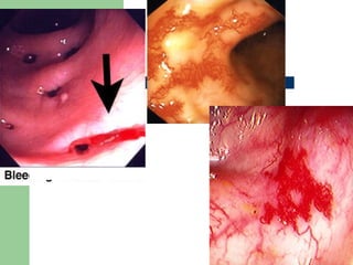
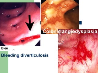
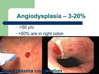
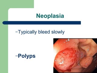
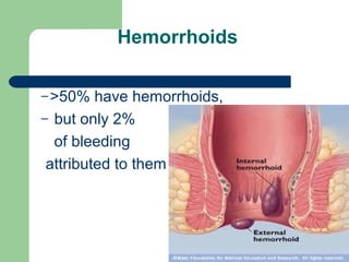
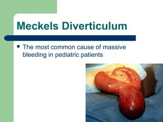
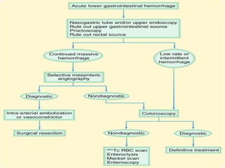
Lower GI Hemorrhage
ABDWAHID M SALIS,
M.D
LGI hemorrhage
Colon – 95-97%
Small bowel – 3-5%
Only 15% of massive GI bleeding
Finding the site
Intermittent bleeding common

Up to 42% have multiple sites
Hematochezia:
Bright red stool, called, is the sign of a fast
moving active GI bleed
Maroon color:
short time taken from the site of the bleed
and the exiting at the anus
Causes
Coagulopathy - specifically a bleeding
diathesis
Bleeding diverticulosis
Colonic angiodysplasia
Diverticulosis – 40-55%
90% stop spontaneously
10% rebleed in 1st
year
and 25% at 4 years
Angiodysplasia – 3-20%
– >50 y/o
–>50% are in right colon
argon plasma coagulation
Neoplasia
–Typically bleed slowly
–Polyps
Inflammatory conditions
15% of UC patients,
1% of chron’s patients
Ischaemic
Radiation
Infectious
AIDS rarely
Hemorrhoids
–>50% have hemorrhoids,
– but only 2%
of bleeding
attributed to them
Meckels Diverticulum
 The most common cause of massive
bleeding in pediatric patients
Evaluation
Same for UGI bleed
If unstable with hematochezia
need EGD 1st
Concealed Bleeding
Occasionally, a person with a LGIB will not
present with any signs of internal
bleeding.
 A Diagnostic or pre-assessment:
hypotension, tachycardia, angina, syncope,
weakness, confusion, stroke,
myocardial infarction/heart attack, and shock.
Laboratory test
 Hemoglobin, hematocrit, and platelets
 Partial thromboplastin time (PTT) and INR
Diagnostics
Colonoscopy
Video capsule endoscopy
Intraoperative endoscopy
Selective viseral angiography
Need >0.5 ml/min bleeding
40-75% sensitive if bleeding at time
of exam
Tagged RBC scan
Can detect bleeding at 0.1 ml/min
Meckel’s scan
Initial test for patients <30 years old
Enteroclysis
Ulcerations
Inflammation
CT scan
Tumors
 Inflammation
 Diverticuli
GI hemorrhage from unknown source
Only 2-5% are not
upper or lower
Treatment
Endoscopy:
Theraputic
Angiodysplasia
polypectomy sites
Angiographic
– Selective embolization for poor
surgical candidates
– Can lead to ischemic sites
requiring later resection
Surgery
Ongoing hemorrhage, >6 units
ongoing transfusion requirement
Site selection
Intraoperative endoscopy
Segmental resection
‫ل‬ ‫الحمد‬

Surgery 6th year, Tutorial (Dr. AbdulWahid)

Editor's Notes

  • #8 Others = meckel’s, juvinial polyps, solitary rectal ulcer, Dieulafoy’s lesion of the colon, portal colopathy, NSAIDs, intussusception, or bleeding following colonoscopic biopsy or polypectomy.
  • #9 Others = meckel’s, juvinial polyps, solitary rectal ulcer, Dieulafoy’s lesion of the colon, portal colopathy, NSAIDs, intussusception, or bleeding following colonoscopic biopsy or polypectomy.
  • #10 Others = meckel’s, juvinial polyps, solitary rectal ulcer, Dieulafoy’s lesion of the colon, portal colopathy, NSAIDs, intussusception, or bleeding following colonoscopic biopsy or polypectomy.
  • #11 Others = meckel’s, juvinial polyps, solitary rectal ulcer, Dieulafoy’s lesion of the colon, portal colopathy, NSAIDs, intussusception, or bleeding following colonoscopic biopsy or polypectomy.
  • #12 Others = meckel’s, juvinial polyps, solitary rectal ulcer, Dieulafoy’s lesion of the colon, portal colopathy, NSAIDs, intussusception, or bleeding following colonoscopic biopsy or polypectomy.