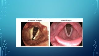
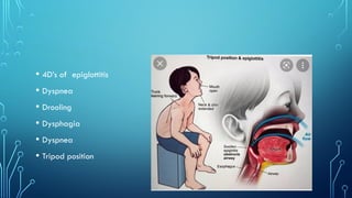
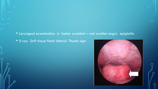
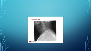
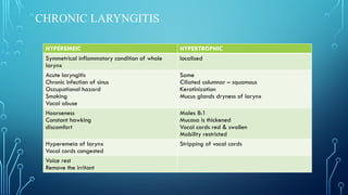
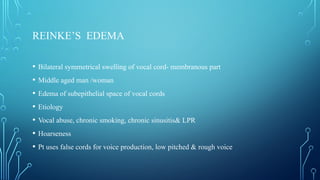
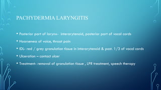
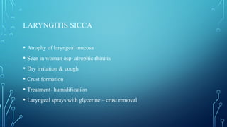
ACUTE INFLAMMATION OF
LARYNX
ACUTE LARYNGITIS
• Infections- Streptococcus pneumonia, H.influenza, Hemolytic streptococcus,
S.aureus
• Noninfectious- Vocal abuse, allergy, trauma, burns ( chemical /thermal)
• Clinical features- abrupt onset, hoarseness of voice, Pain and vocal fatigue,
fever
• Laryngeal edema, erythema , interarytenoid secretion
• Submucosal hemorrhage in vocal cord
MANAGEMENT
• Video laryngoscopy
• Flexible fiberoptic laryngoscopy
• Voice rest
• Smoking and alcohol cessation
• Vocal hygiene
• Steam inhalation
• Anti tussives
• Antibiotics and analgesics
• Steroids
ACUTE EPIGLOTTITIS
• Inflammation of the supraglottis
• Life threatening
• Children, H.influenza
• Adults less common
• Sudden in onset
• Fever- High grade , toxic look
• Dyspnea and stridor
• 4D’s of epiglottitis
• Dyspnea
• Drooling
• Dysphagia
• Dyspnea
• Tripod position
• Laryngeal examination is better avoided – red swollen angry epiglottis
• X-ray Soft tissue Neck lateral- Thumb sign
TREATMENT
• Immediate hospitalization
• Antibiotics – 3rd
generation cephalosporin
• Steroids
• Humidification with oxygen
• Intubation/ Tracheostomy
ACUTE LARYNGOTRACHEO BRONCHITIS
• Etiology- Parainfluenza type I and III
• Age- 6 months to 3 years
• Male
• Secondary bacterial infection with gram positive cocci
• Edema of loose areolar tissue of subglottic area, tenacious secretion occlude
the airway
• Onset is slow, non toxic
• Hoarseness and croupy cough- Barking seal like
• Fever low grade
• Respiratory difficulties is slow in onset with intercostal and suprasternal
recession
• Xray neck- AP view – Steeple sign
TREATMENT
• Immediate hospitalization
• Antibiotics – 3rd
generation cephalosporin
• Steroids
• Nebulisation with adrenaline
• Humidification with oxygen
• Intubation/ Tracheostomy
LARYNGEAL DIPHTHERIA
• Secondary to faucial diphtheria
• Less than 10yrs
• Infective material- nasopharyngeal secretion
• IP-2 to 6 days
• Portal of entry- respiratory or non respiratory
• Droplet and direct
• Period of infectivity-14 to 28 days from onset
Pseudomembrane occlude the airway
• Fever>101
• Toxemia and tachycardia
• Hoarse voice, croupy cough
• Inspiratory stridor and dyspnoea leading to significant upper airway obstruction.
• Membrane. Greyish white membrane is seen on the tonsil, pharynx and soft palate. It is
adherent and its removal leaves a bleeding surface. Similar membrane is seen over the larynx
and trachea.
• Cervical lymphadenopathy.- BULL’S NECK APPEARANCE
MANAGEMENT
• Isolation
• Diphtheria antitoxin -20,000 – 1,00,000 Units in I.v infusion
• DOC- Benzylpenicillin and erythromycin
• Intubation
• Tracheostomy
• Complete bed rest
• Three negative cultires at 24 he intervals
COMPLICATIONS
• Asphyxia ,, death
• Laryngeal, palatal paralysis
• Toxic myocarditis
PERTUSSIS
• Bordetella pertussis
• Exotoxin and endotoxin – stop cilia function and epithelial necrosis
• Spread by sneezing and coughing (Droplets)
• Notifiable disease
• IP- 7 to 14 days
• Period of communicable – 1 week after Exposure to 3 weeks after onset of cough
• Cough – paroxysmal attacks with gasping and whoop
• Throat swab and PCR
TREATMENT
• Erythromycin 7 to 14 days
• Steroids
• Beta 2 adrenergic
• Pertussis specific immunoglobulin
INFECTIOUS MONONUCLEOSIS
• EBV
• Affinity to B lymphocytes , Infect epithelium of oropharynx
• Transmitted through bodily secretions
• Common in adolescent
• IP 4 to 7 weeks
• Sore throat, tonsillar enlargement, periorbital edema and edema of the lower lips-
HOGALAND SIGN
• Palatal petechiae, hepatomegaly, splenomegaly
MANAGEMENT
• CBC, LFT, RFT
• Leucocytosis (50%), atypical leukocytes (10%, >20%)
• Monospot test, Paul bennell test
• Positivity to monospot test first 6weeks , and repeated after 7 days
• Repeated negative test-CMV, HIV, Rubella and toxoplasmosis
• Fluid, analgesics and steroids
• Penicillin group of antibiotics avoided as it may cause maculopapular rash
COMPLICATIONS
• Airway obstruction due to cervical lymphadenopathy
• Anaemia, thrombocytopenia , encehapilitis, GBS
• Pericarditis and myocarditis
OEDEMA OF LARYNX
• Airway obstruction
• Inspiratory stridor
• IDL
• Treatment
• Intubation of larynx
• Tracheostomy
• Inj.adrenaline
• Steroids
CHRONIC LARYNGITIS
HYPEREMEIC HYPERTROPHIC
Symmetrical inflammatory condition of whole
larynx
localised
Acute laryngitis
Chronic infection of sinus
Occupational hazard
Smoking
Vocal abuse
Same
Ciliated columnar – squamous
Keratinization
Mucus glands dryness of larynx
Hoarseness
Constant hawking
discomfort
Males 8:1
Mucosa is thickened
Vocal cords red & swollen
Mobility restricted
Hyperemeia of larynx
Vocal cords congested
Stripping of vocal cords
Voice rest
Remove the irritant
REINKE’S EDEMA
• Bilateral symmetrical swelling of vocal cord- membranous part
• Middle aged man /woman
• Edema of subepithelial space of vocal cords
• Etiology
• Vocal abuse, chronic smoking, chronic sinusitis& LPR
• Hoarseness
• Pt uses false cords for voice production, low pitched & rough voice
• IDL- fusiform swelling with pale translucent look
• Ventricular bands- hyperemic, hypertrophy & obscuring the view of vocal cords
• Treatment
• Stripping of vocal cords in stages
• Voice rest
• Speech therapy
PACHYDERMIA LARYNGITIS
• Posterior part of larynx- interarytenoid, posterior part of vocal cords
• Hoarseness of voice, throat pain
• IDL- red / grey granulation tissue in interarytenoid & post. 1/3 of vocal cords
• Ulceration – contact ulcer
• Treatment- removal of granulation tissue , LPR treatment, speech therapy
LARYNGITIS SICCA
• Atrophy of laryngeal mucosa
• Seen in woman esp- atrophic rhinitis
• Dry irritation & cough
• Crust formation
• Treatment- humidification
• Laryngeal sprays with glycerine – crust removal
TB LARYNX
• Secondary to pulmonary TB
• Haematogenous/ bronchogenic spread
• Posterior part of larynx – esp interarytenoid region
• Pseudoedema
• Voice fatigue, voice change & dysphagia
• Mouse bitten vocal cord
• Turban epiglottis
• Mammilated appearance of interarytenoid region
• Vocal cord palsy
• Investigation- sputum, videolaryngoscopy, chest xray
• Treatment
JRP
• Common benign lesion in children
• HPV 6,11
• Infected from mother with vaginal HPV
• Usually supraglottis & glottis
• 3- 5yrs
• Hoarseness, respiratory distress/ stridor
• Flexible fibreoptic laryngoscopy
• Care need to be taken if child has tracheostomy
• Co2 LASER
• Interferon alpha
• 13 cis retinoic acid
FB IN AIRWAY
• Non irritating type
• Irritating type- vegetable/ nuts- edema
• Choking
• Symptomless period
• Laryngeal- obstruction - death
LOCATION
•In decreasingorder Of frequency,
1.Bronchus (right> left)
2.Trachea
3.Larynx
4.lungs
• Tracheal- cough, hemoptysis, wheeze, audible slap
• Bronchial
DELAYED PRESENTATION
•Common in children
•Maybe treated as asthma due to low grade cough and
noisy breathing
•Delay in diagnosis- granulation tissue formation,
pneumonitis, bronchiectasis, lung abscess
• Chest xray
• Xray neck ap/ lateral view
• Fluroscopy
• CT chest
• Hemlich
• Cricothyrotomy
• Tracheostomy
• Direct laryngoscopy
• Bronchoscopy
RIGID BRONCHOSCOPY
•Mainstay of treatment
•done under general anaesthesia
•Position: barking dog position
•Technique: direct method, through laryngoscope
•Limitations: supraglottic area is difficult to evaluate
can reach only up to segmental bronchi
•Technically difficult
•On prolonged procedure it causes Subglottic edema
LARYNGITIS  and chronic inflammation ppt

LARYNGITIS and chronic inflammation ppt

  • 1.
  • 3.
    ACUTE LARYNGITIS • Infections-Streptococcus pneumonia, H.influenza, Hemolytic streptococcus, S.aureus • Noninfectious- Vocal abuse, allergy, trauma, burns ( chemical /thermal) • Clinical features- abrupt onset, hoarseness of voice, Pain and vocal fatigue, fever • Laryngeal edema, erythema , interarytenoid secretion • Submucosal hemorrhage in vocal cord
  • 6.
    MANAGEMENT • Video laryngoscopy •Flexible fiberoptic laryngoscopy • Voice rest • Smoking and alcohol cessation • Vocal hygiene • Steam inhalation • Anti tussives • Antibiotics and analgesics • Steroids
  • 7.
    ACUTE EPIGLOTTITIS • Inflammationof the supraglottis • Life threatening • Children, H.influenza • Adults less common • Sudden in onset • Fever- High grade , toxic look • Dyspnea and stridor
  • 8.
    • 4D’s ofepiglottitis • Dyspnea • Drooling • Dysphagia • Dyspnea • Tripod position
  • 9.
    • Laryngeal examinationis better avoided – red swollen angry epiglottis • X-ray Soft tissue Neck lateral- Thumb sign
  • 11.
    TREATMENT • Immediate hospitalization •Antibiotics – 3rd generation cephalosporin • Steroids • Humidification with oxygen • Intubation/ Tracheostomy
  • 12.
    ACUTE LARYNGOTRACHEO BRONCHITIS •Etiology- Parainfluenza type I and III • Age- 6 months to 3 years • Male • Secondary bacterial infection with gram positive cocci • Edema of loose areolar tissue of subglottic area, tenacious secretion occlude the airway
  • 14.
    • Onset isslow, non toxic • Hoarseness and croupy cough- Barking seal like • Fever low grade • Respiratory difficulties is slow in onset with intercostal and suprasternal recession • Xray neck- AP view – Steeple sign
  • 17.
    TREATMENT • Immediate hospitalization •Antibiotics – 3rd generation cephalosporin • Steroids • Nebulisation with adrenaline • Humidification with oxygen • Intubation/ Tracheostomy
  • 19.
    LARYNGEAL DIPHTHERIA • Secondaryto faucial diphtheria • Less than 10yrs • Infective material- nasopharyngeal secretion • IP-2 to 6 days • Portal of entry- respiratory or non respiratory • Droplet and direct • Period of infectivity-14 to 28 days from onset
  • 21.
  • 22.
    • Fever>101 • Toxemiaand tachycardia • Hoarse voice, croupy cough • Inspiratory stridor and dyspnoea leading to significant upper airway obstruction. • Membrane. Greyish white membrane is seen on the tonsil, pharynx and soft palate. It is adherent and its removal leaves a bleeding surface. Similar membrane is seen over the larynx and trachea. • Cervical lymphadenopathy.- BULL’S NECK APPEARANCE
  • 27.
    MANAGEMENT • Isolation • Diphtheriaantitoxin -20,000 – 1,00,000 Units in I.v infusion • DOC- Benzylpenicillin and erythromycin • Intubation • Tracheostomy • Complete bed rest • Three negative cultires at 24 he intervals
  • 28.
    COMPLICATIONS • Asphyxia ,,death • Laryngeal, palatal paralysis • Toxic myocarditis
  • 29.
    PERTUSSIS • Bordetella pertussis •Exotoxin and endotoxin – stop cilia function and epithelial necrosis • Spread by sneezing and coughing (Droplets) • Notifiable disease • IP- 7 to 14 days • Period of communicable – 1 week after Exposure to 3 weeks after onset of cough • Cough – paroxysmal attacks with gasping and whoop • Throat swab and PCR
  • 31.
    TREATMENT • Erythromycin 7to 14 days • Steroids • Beta 2 adrenergic • Pertussis specific immunoglobulin
  • 32.
    INFECTIOUS MONONUCLEOSIS • EBV •Affinity to B lymphocytes , Infect epithelium of oropharynx • Transmitted through bodily secretions • Common in adolescent • IP 4 to 7 weeks • Sore throat, tonsillar enlargement, periorbital edema and edema of the lower lips- HOGALAND SIGN • Palatal petechiae, hepatomegaly, splenomegaly
  • 33.
    MANAGEMENT • CBC, LFT,RFT • Leucocytosis (50%), atypical leukocytes (10%, >20%) • Monospot test, Paul bennell test • Positivity to monospot test first 6weeks , and repeated after 7 days • Repeated negative test-CMV, HIV, Rubella and toxoplasmosis • Fluid, analgesics and steroids • Penicillin group of antibiotics avoided as it may cause maculopapular rash
  • 34.
    COMPLICATIONS • Airway obstructiondue to cervical lymphadenopathy • Anaemia, thrombocytopenia , encehapilitis, GBS • Pericarditis and myocarditis
  • 35.
    OEDEMA OF LARYNX •Airway obstruction • Inspiratory stridor • IDL • Treatment • Intubation of larynx • Tracheostomy • Inj.adrenaline • Steroids
  • 36.
    CHRONIC LARYNGITIS HYPEREMEIC HYPERTROPHIC Symmetricalinflammatory condition of whole larynx localised Acute laryngitis Chronic infection of sinus Occupational hazard Smoking Vocal abuse Same Ciliated columnar – squamous Keratinization Mucus glands dryness of larynx Hoarseness Constant hawking discomfort Males 8:1 Mucosa is thickened Vocal cords red & swollen Mobility restricted Hyperemeia of larynx Vocal cords congested Stripping of vocal cords Voice rest Remove the irritant
  • 37.
    REINKE’S EDEMA • Bilateralsymmetrical swelling of vocal cord- membranous part • Middle aged man /woman • Edema of subepithelial space of vocal cords • Etiology • Vocal abuse, chronic smoking, chronic sinusitis& LPR • Hoarseness • Pt uses false cords for voice production, low pitched & rough voice
  • 38.
    • IDL- fusiformswelling with pale translucent look • Ventricular bands- hyperemic, hypertrophy & obscuring the view of vocal cords • Treatment • Stripping of vocal cords in stages • Voice rest • Speech therapy
  • 40.
    PACHYDERMIA LARYNGITIS • Posteriorpart of larynx- interarytenoid, posterior part of vocal cords • Hoarseness of voice, throat pain • IDL- red / grey granulation tissue in interarytenoid & post. 1/3 of vocal cords • Ulceration – contact ulcer • Treatment- removal of granulation tissue , LPR treatment, speech therapy
  • 41.
    LARYNGITIS SICCA • Atrophyof laryngeal mucosa • Seen in woman esp- atrophic rhinitis • Dry irritation & cough • Crust formation • Treatment- humidification • Laryngeal sprays with glycerine – crust removal
  • 42.
    TB LARYNX • Secondaryto pulmonary TB • Haematogenous/ bronchogenic spread • Posterior part of larynx – esp interarytenoid region • Pseudoedema • Voice fatigue, voice change & dysphagia • Mouse bitten vocal cord • Turban epiglottis • Mammilated appearance of interarytenoid region
  • 44.
    • Vocal cordpalsy • Investigation- sputum, videolaryngoscopy, chest xray • Treatment
  • 45.
    JRP • Common benignlesion in children • HPV 6,11 • Infected from mother with vaginal HPV • Usually supraglottis & glottis • 3- 5yrs • Hoarseness, respiratory distress/ stridor • Flexible fibreoptic laryngoscopy • Care need to be taken if child has tracheostomy
  • 47.
    • Co2 LASER •Interferon alpha • 13 cis retinoic acid
  • 48.
    FB IN AIRWAY •Non irritating type • Irritating type- vegetable/ nuts- edema • Choking • Symptomless period • Laryngeal- obstruction - death
  • 49.
    LOCATION •In decreasingorder Offrequency, 1.Bronchus (right> left) 2.Trachea 3.Larynx 4.lungs
  • 50.
    • Tracheal- cough,hemoptysis, wheeze, audible slap • Bronchial
  • 51.
    DELAYED PRESENTATION •Common inchildren •Maybe treated as asthma due to low grade cough and noisy breathing •Delay in diagnosis- granulation tissue formation, pneumonitis, bronchiectasis, lung abscess
  • 52.
    • Chest xray •Xray neck ap/ lateral view • Fluroscopy • CT chest • Hemlich • Cricothyrotomy • Tracheostomy • Direct laryngoscopy • Bronchoscopy
  • 53.
    RIGID BRONCHOSCOPY •Mainstay oftreatment •done under general anaesthesia •Position: barking dog position •Technique: direct method, through laryngoscope •Limitations: supraglottic area is difficult to evaluate can reach only up to segmental bronchi •Technically difficult •On prolonged procedure it causes Subglottic edema