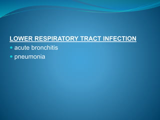
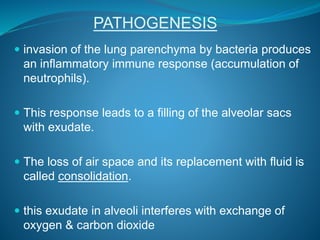
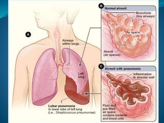
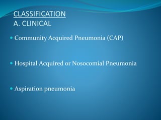
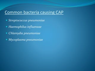
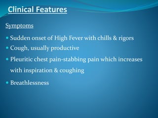
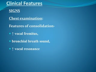
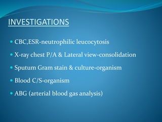
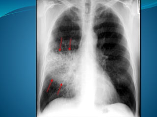
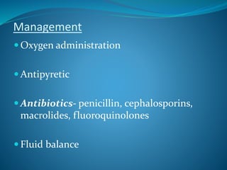
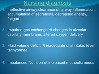
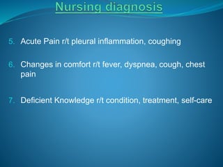
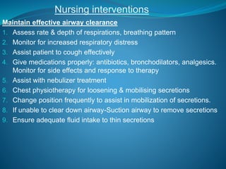
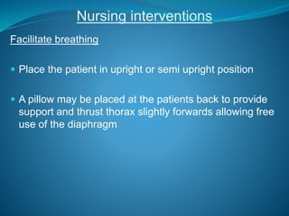
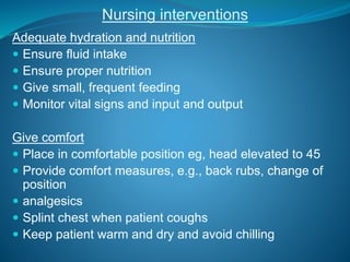
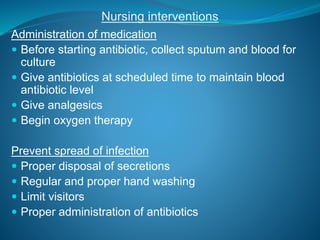
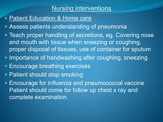
RESPIRATORY TRACT INFECTIONS
DR. RUBAYAT SHEIK GIASUDDIN
MBBS, MRCP(UK)
Assistant Professor of Medicine
RESPIRATORY TRACT
UPPER RESPIRATORY TRACT INFECTIONS
acute infection which involves the upper respiratory
tract: nose, sinuses, pharynx or larynx.
 Rhinitis
 Sinusitis
 Pharyngitis
 Laryngitis
LOWER RESPIRATORY TRACT INFECTION
 acute bronchitis
 pneumonia
Definition
 Pneumonia is an acute inflammation of the lung
parenchyma
Mode of transmission
Inhalation
Aspiration
Hematogenous
PATHOGENESIS
 invasion of the lung parenchyma by bacteria produces
an inflammatory immune response (accumulation of
neutrophils).
 This response leads to a filling of the alveolar sacs
with exudate.
 The loss of air space and its replacement with fluid is
called consolidation.
 this exudate in alveoli interferes with exchange of
oxygen & carbon dioxide
CLASSIFICATION
A. CLINICAL
 Community Acquired Pneumonia (CAP)
 Hospital Acquired or Nosocomial Pneumonia
 Aspiration pneumonia
Pathological Classification
B. Pathological Classification
 Lobar pneumonia: it is a type of pneumonia where
there is involvement of large & continuous area of
one or more lobes of lung
 Bronchopneumonia: It is a type
of pneumonia characterized by multiple patchy
involvement of one or more lobes.
RISK FACTORS THAT PREDISPOSE TO PNEUMONIA
 Cigarette smoking
 Upper respiratory tract infections
 Pre-existing lung disease e. COPD
 Immunosuppression-steroid therapy, Diabetes
 Old age
 Altered consciousness- stroke, Alcohol, seizures
Organisms causing CAP
 Bacteria
 Viruses
Common bacteria causing CAP
 Streptococcus pneumoniae
 Haemophilus influenzae
 Chlamydia pneumoniae
 Mycoplasma pneumoniae
•Pseudomonas aeruginosa
•E. Coli
•Proteus
•Klebsiella
Clinical Features
Symptoms
 Sudden onset of High Fever with chills & rigors
 Cough, usually productive
 Pleuritic chest pain-stabbing pain which increases
with inspiration & coughing
 Breathlessness
Clinical Features
SIGNS
Chest examination:
Features of consolidation-
 ↑ vocal fremitus,
 bronchial breath sound,
 ↑ vocal resonance
HOSPITAL ACQUIRED PNEUMONIA
 Hospital acquired Pneumonia or nosocomial
Pneumonia occur at least 2 days after admission to
hospital.
INVESTIGATIONS
 CBC,ESR-neutrophilic leucocytosis
 X-ray chest P/A & Lateral view-consolidation
 Sputum Gram stain & culture-organism
 Blood C/S-organism
 ABG (arterial blood gas analysis)
Management
 Oxygen administration
 Antipyretic
 Antibiotics- penicillin, cephalosporins,
macrolides, fluoroquinolones
 Fluid balance
COMPLICATIONS OF PNEUMONIA
 Pleurisy
 Para-pneumonic effusion
 Delayed resolution
 Lung abscess
 Empyema
 ARDS (acute respiratory distress syndrome)
 Multi-organ failure
 Metastatic abscess
1. Ineffective airway clearance r/t airway inflammation,
accumulation of secretions, decreased energy,
fatigue
2. Impaired gas exchange r/t changes in alveolar
capillary membrane, altered oxygen delivery
3. Fluid volume deficit r/t inadequate oral intake, fever,
tachypnoea.
4. Imbalanced Nutrition r/t increased metabolic needs
5. Acute Pain r/t pleural inflammation, coughing
6. Changes in comfort r/t fever, dyspnea, cough, chest
pain
7. Deficient Knowledge r/t condition, treatment, self-care
Nursing interventions
Maintain effective airway clearance
1. Assess rate & depth of respirations, breathing pattern
2. Monitor for increased respiratory distress
3. Assist patient to cough effectively
4. Give medications properly: antibiotics, bronchodilators, analgesics.
Monitor for side effects and response to therapy
5. Assist with nebulizer treatment
6. Chest physiotherapy for loosening & mobilising secretions
7. Change position frequently to assist in mobilization of secretions.
8. If unable to clear down airway-Suction airway to remove secretions
9. Ensure adequate fluid intake to thin secretions
Nursing interventions
Facilitate breathing
 Place the patient in upright or semi upright position
 A pillow may be placed at the patients back to provide
support and thrust thorax slightly forwards allowing free
use of the diaphragm
Adequate hydration and nutrition
 Ensure fluid intake
 Ensure proper nutrition
 Give small, frequent feeding
 Monitor vital signs and input and output
Give comfort
 Place in comfortable position eg, head elevated to 45
 Provide comfort measures, e.g., back rubs, change of
position
 analgesics
 Splint chest when patient coughs
 Keep patient warm and dry and avoid chilling
Nursing interventions
Administration of medication
 Before starting antibiotic, collect sputum and blood for
culture
 Give antibiotics at scheduled time to maintain blood
antibiotic level
 Give analgesics
 Begin oxygen therapy
Prevent spread of infection
 Proper disposal of secretions
 Regular and proper hand washing
 Limit visitors
 Proper administration of antibiotics
Nursing interventions
 Patient Education & Home care
 Assess patients understanding of pneumonia
 Teach proper handling of secretions, eg. Covering nose
and mouth with tissue when sneezing or coughing,
proper disposal of tissues, use of container for sputum
 Importance of handwashing after coughing, sneezing
 Encourage breathing exercises
 Patient should stop smoking
 Encourage for influenza and pneumococcal vaccine
Patient should come for follow up chest x ray and
complete examination.
Pneumonia

Pneumonia

  • 1.
    RESPIRATORY TRACT INFECTIONS DR.RUBAYAT SHEIK GIASUDDIN MBBS, MRCP(UK) Assistant Professor of Medicine
  • 2.
  • 3.
    UPPER RESPIRATORY TRACTINFECTIONS acute infection which involves the upper respiratory tract: nose, sinuses, pharynx or larynx.  Rhinitis  Sinusitis  Pharyngitis  Laryngitis
  • 4.
    LOWER RESPIRATORY TRACTINFECTION  acute bronchitis  pneumonia
  • 6.
    Definition  Pneumonia isan acute inflammation of the lung parenchyma
  • 7.
  • 8.
  • 9.
    PATHOGENESIS  invasion ofthe lung parenchyma by bacteria produces an inflammatory immune response (accumulation of neutrophils).  This response leads to a filling of the alveolar sacs with exudate.  The loss of air space and its replacement with fluid is called consolidation.  this exudate in alveoli interferes with exchange of oxygen & carbon dioxide
  • 13.
    CLASSIFICATION A. CLINICAL  CommunityAcquired Pneumonia (CAP)  Hospital Acquired or Nosocomial Pneumonia  Aspiration pneumonia
  • 14.
  • 15.
  • 16.
     Lobar pneumonia:it is a type of pneumonia where there is involvement of large & continuous area of one or more lobes of lung  Bronchopneumonia: It is a type of pneumonia characterized by multiple patchy involvement of one or more lobes.
  • 17.
    RISK FACTORS THATPREDISPOSE TO PNEUMONIA  Cigarette smoking  Upper respiratory tract infections  Pre-existing lung disease e. COPD  Immunosuppression-steroid therapy, Diabetes  Old age  Altered consciousness- stroke, Alcohol, seizures
  • 18.
    Organisms causing CAP Bacteria  Viruses
  • 19.
    Common bacteria causingCAP  Streptococcus pneumoniae  Haemophilus influenzae  Chlamydia pneumoniae  Mycoplasma pneumoniae
  • 20.
  • 21.
    Clinical Features Symptoms  Suddenonset of High Fever with chills & rigors  Cough, usually productive  Pleuritic chest pain-stabbing pain which increases with inspiration & coughing  Breathlessness
  • 22.
    Clinical Features SIGNS Chest examination: Featuresof consolidation-  ↑ vocal fremitus,  bronchial breath sound,  ↑ vocal resonance
  • 23.
    HOSPITAL ACQUIRED PNEUMONIA Hospital acquired Pneumonia or nosocomial Pneumonia occur at least 2 days after admission to hospital.
  • 24.
    INVESTIGATIONS  CBC,ESR-neutrophilic leucocytosis X-ray chest P/A & Lateral view-consolidation  Sputum Gram stain & culture-organism  Blood C/S-organism  ABG (arterial blood gas analysis)
  • 26.
    Management  Oxygen administration Antipyretic  Antibiotics- penicillin, cephalosporins, macrolides, fluoroquinolones  Fluid balance
  • 27.
    COMPLICATIONS OF PNEUMONIA Pleurisy  Para-pneumonic effusion  Delayed resolution  Lung abscess  Empyema  ARDS (acute respiratory distress syndrome)  Multi-organ failure  Metastatic abscess
  • 28.
    1. Ineffective airwayclearance r/t airway inflammation, accumulation of secretions, decreased energy, fatigue 2. Impaired gas exchange r/t changes in alveolar capillary membrane, altered oxygen delivery 3. Fluid volume deficit r/t inadequate oral intake, fever, tachypnoea. 4. Imbalanced Nutrition r/t increased metabolic needs
  • 29.
    5. Acute Painr/t pleural inflammation, coughing 6. Changes in comfort r/t fever, dyspnea, cough, chest pain 7. Deficient Knowledge r/t condition, treatment, self-care
  • 30.
    Nursing interventions Maintain effectiveairway clearance 1. Assess rate & depth of respirations, breathing pattern 2. Monitor for increased respiratory distress 3. Assist patient to cough effectively 4. Give medications properly: antibiotics, bronchodilators, analgesics. Monitor for side effects and response to therapy 5. Assist with nebulizer treatment 6. Chest physiotherapy for loosening & mobilising secretions 7. Change position frequently to assist in mobilization of secretions. 8. If unable to clear down airway-Suction airway to remove secretions 9. Ensure adequate fluid intake to thin secretions
  • 31.
    Nursing interventions Facilitate breathing Place the patient in upright or semi upright position  A pillow may be placed at the patients back to provide support and thrust thorax slightly forwards allowing free use of the diaphragm
  • 32.
    Adequate hydration andnutrition  Ensure fluid intake  Ensure proper nutrition  Give small, frequent feeding  Monitor vital signs and input and output Give comfort  Place in comfortable position eg, head elevated to 45  Provide comfort measures, e.g., back rubs, change of position  analgesics  Splint chest when patient coughs  Keep patient warm and dry and avoid chilling Nursing interventions
  • 33.
    Nursing interventions Administration ofmedication  Before starting antibiotic, collect sputum and blood for culture  Give antibiotics at scheduled time to maintain blood antibiotic level  Give analgesics  Begin oxygen therapy Prevent spread of infection  Proper disposal of secretions  Regular and proper hand washing  Limit visitors  Proper administration of antibiotics
  • 34.
    Nursing interventions  PatientEducation & Home care  Assess patients understanding of pneumonia  Teach proper handling of secretions, eg. Covering nose and mouth with tissue when sneezing or coughing, proper disposal of tissues, use of container for sputum  Importance of handwashing after coughing, sneezing  Encourage breathing exercises  Patient should stop smoking  Encourage for influenza and pneumococcal vaccine Patient should come for follow up chest x ray and complete examination.