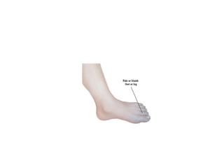
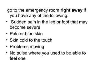
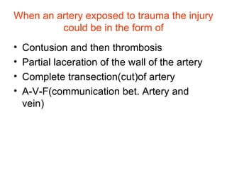
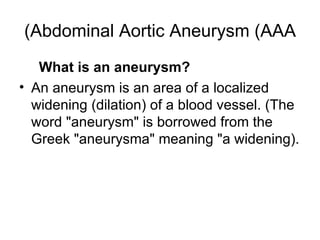
Artery Emergencies
Acute Arterial Occlusion Acute arterial occlusion   is serious .  It occurs when blood flow in a leg artery stops suddenly .  If blood flow to the toe, foot, or leg is completely blocked, the tissue begins to die .  This is called gangrene .  If this happens, medical care is needed right away to restore blood flow and save the leg . 
 
go to the emergency room   right away  if you have any of the following : Sudden pain in the leg or foot that may become severe Pale or blue skin Skin cold to the touch Problems moving No pulse where you used to be able to feel one
Causes of Acute Arterial Occlusion This problem is more likely to occur in patients with peripheral arterial disease  ( PAD ).  With PAD, leg arteries are narrowed .  This reduces blood flow to the legs and feet
How Is Acute Arterial Occlusion Diagnosed ? Tests on blood flow are done .  These tests may include : Ankle - brachial index  ( ABI ).   The blood pressure in the ankle is compared to the blood pressure in the arm .  What is a normal and an abnormal ABPI? An ABPI of greater than 0.9 (90%) is considered to be normal.  An ABPI of beween 0.5 and 0.9 (50% to 90%) is consistent with symptoms of  Intermittent  Claudication . An ABPI of less than 0.5 (50%) is usually associated with more severe symptoms, even  Critical  Ischaemia  . Duplex ultrasound .   Painless sound waves are used to create images of blood flow in the legs . Arteriography .   Contrast  ( x - ray fluid )  is injected into the artery .  This is done through a catheter  ( thin, flexible tube ).  The contrast makes blood vessels show up more clearly on x - rays .
How Is Acute Arterial Occlusion Treated ? Possible treatments for acute arterial occlusion include : Dissolving or removing a blood clot .   A catheter may be put into an artery in the groin to dissolve the clot .  The catheter is then used to deliver “clot - busting” medication, which dissolves the clot .  Or surgery may be done to remove the clot .  An incision is made in the artery at the blocked section .  The clot is then removed . Angioplasty .   A small, uninflated balloon is passed through a catheter to the narrowed part of the artery .  The balloon is then inflated to widen the artery .  The balloon is then deflated and removed . Stenting .   After angioplasty, a stent  ( tiny wire mesh tube )  may be placed in the artery to help hold it open .  The stent is also placed using a catheter . Endarterectomy .   An incision is made in the artery at the blocked section .  The material that blocks the artery is then removed from artery walls . Peripheral bypass surgery .   A natural or man - made graft is used to bypass the blocked section of the artery .
Peripheral Vascular Injuries   Peripheral vascular injuries may result from penetrating or blunt trauma to the extremities .  If not recognized and treated rapidly, injuries to major arteries, veins, and nerves may have disastrous consequences resulting in the loss of life and limb
Traumatic arterial injuries Arteries can be occluded as a result of changes :  In the lumen (thrombosis). In the wall (sub-intimal haematoma). In the surrounding tissues  (anterior tibial compartment).
Types of vascular injury * Contusion and thrombosis .  * Partial laceration of artery .  * Complete cut of artery . * Arterio-venous fistula .
Types of vascular injury Contusion   Puncture   Laceration   Transection   Clinical features Depends on site, mechanism and extent of injury   Signs classically divided into 'hard' and 'soft' sign
Hard signs of vascular injury Absent pulses   Bruit or palpable thrill   Active haemorrhage   Expanding haematoma   Distal ischaemia   Soft signs of vascular injury Haematoma   History of haemorrhage at seen of accident   Unexplained hypotension   Peripheral nerve deficit
Difinite or Hot signs Pulsatile or expanding haematoma Pulsatile bleeding  Presence of thrill or bruit End organ ischemia
Suggestive or Cold signs of arterial injury Unexplained shock Stable haematoma  Injury to an adjacent nerve Questionable history of arterial bleeding  proximity
When an artery exposed to trauma the injury could be in the form of Contusion and then thrombosis Partial laceration of the wall of the artery Complete transection(cut)of artery A-V-F(communication bet. Artery and vein)
Abdominal Aortic Aneurysm (AAA) What is an aneurysm ? An aneurysm is an area of a localized widening  ( dilation )  of a blood vessel . ( The word  " aneurysm "  is borrowed from the Greek  " aneurysma "  meaning  " a widening).
Where do aortic aneurysms tend to develop ? Aortic aneurysms can develop anywhere along the length of the aorta .  The majority, however, are located along the abdominal aorta .  Most  ( about 90% )  of abdominal aneurysms are located below the level of the renal arteries, the vessels that leave the aorta to go to the kidneys .  About two - thirds of abdominal aneurysms are not limited to just the aorta but extend from the aorta into one or both of the iliac arteries .
The most common symptoms of   abdominal aortic aneurysm       include general abdominal  ( belly )  pain or discomfort, which may come and go or be constant .  Other symptoms include : Pain in the chest ,  abdomen ,  lower back, or flank  ( over the   kidneys ),  possibly spreading to the groin, buttocks, or legs .  The pain may be deep, aching, gnawing, and / or throbbing, and may last for hours or days .  It is generally not affected by movement, although certain positions may be more comfortable than others .   A pulsating sensation in the abdomen .   A  " cold   foot "  or a black or blue painful toe can happen if an abdominal aortic aneurysm produces a   blood clot   that breaks off and blocks   blood   flow to the legs or feet .   Fever   or   weight loss ,  if it is an   inflammatory aortic aneurysm .
Surgery for Abdominal Aortic Aneurysm During surgery for abdominal aortic aneurysm  ( AAA ) , the weakened aortic wall is replaced with a hollow manmade tube  ( a   graft) Risks and Complications Infection Blood clots in legs Bleeding Kidney failure Pneumonia Injury to the colon’s blood supply Erectile dysfunction Spinal cord injury Heart attack, stroke, or death Reaching the Aneurysm The aorta can be reached through   open surgery .   Or a less invasive   endovascular procedure   may be done .
 

Surgery 6th year, Tutorial (Dr. Ahmed Al-Azzawi)

  • 1.
  • 2.
    Acute Arterial OcclusionAcute arterial occlusion is serious . It occurs when blood flow in a leg artery stops suddenly . If blood flow to the toe, foot, or leg is completely blocked, the tissue begins to die . This is called gangrene . If this happens, medical care is needed right away to restore blood flow and save the leg . 
  • 3.
  • 4.
    go to theemergency room right away if you have any of the following : Sudden pain in the leg or foot that may become severe Pale or blue skin Skin cold to the touch Problems moving No pulse where you used to be able to feel one
  • 5.
    Causes of AcuteArterial Occlusion This problem is more likely to occur in patients with peripheral arterial disease ( PAD ). With PAD, leg arteries are narrowed . This reduces blood flow to the legs and feet
  • 6.
    How Is AcuteArterial Occlusion Diagnosed ? Tests on blood flow are done . These tests may include : Ankle - brachial index ( ABI ).   The blood pressure in the ankle is compared to the blood pressure in the arm . What is a normal and an abnormal ABPI? An ABPI of greater than 0.9 (90%) is considered to be normal.  An ABPI of beween 0.5 and 0.9 (50% to 90%) is consistent with symptoms of Intermittent Claudication . An ABPI of less than 0.5 (50%) is usually associated with more severe symptoms, even Critical Ischaemia . Duplex ultrasound .   Painless sound waves are used to create images of blood flow in the legs . Arteriography .   Contrast ( x - ray fluid ) is injected into the artery . This is done through a catheter ( thin, flexible tube ). The contrast makes blood vessels show up more clearly on x - rays .
  • 7.
    How Is AcuteArterial Occlusion Treated ? Possible treatments for acute arterial occlusion include : Dissolving or removing a blood clot .   A catheter may be put into an artery in the groin to dissolve the clot . The catheter is then used to deliver “clot - busting” medication, which dissolves the clot . Or surgery may be done to remove the clot . An incision is made in the artery at the blocked section . The clot is then removed . Angioplasty .   A small, uninflated balloon is passed through a catheter to the narrowed part of the artery . The balloon is then inflated to widen the artery . The balloon is then deflated and removed . Stenting .   After angioplasty, a stent ( tiny wire mesh tube ) may be placed in the artery to help hold it open . The stent is also placed using a catheter . Endarterectomy .   An incision is made in the artery at the blocked section . The material that blocks the artery is then removed from artery walls . Peripheral bypass surgery .   A natural or man - made graft is used to bypass the blocked section of the artery .
  • 8.
    Peripheral Vascular Injuries  Peripheral vascular injuries may result from penetrating or blunt trauma to the extremities . If not recognized and treated rapidly, injuries to major arteries, veins, and nerves may have disastrous consequences resulting in the loss of life and limb
  • 9.
    Traumatic arterial injuriesArteries can be occluded as a result of changes : In the lumen (thrombosis). In the wall (sub-intimal haematoma). In the surrounding tissues (anterior tibial compartment).
  • 10.
    Types of vascularinjury * Contusion and thrombosis . * Partial laceration of artery . * Complete cut of artery . * Arterio-venous fistula .
  • 11.
    Types of vascularinjury Contusion Puncture Laceration Transection Clinical features Depends on site, mechanism and extent of injury Signs classically divided into 'hard' and 'soft' sign
  • 12.
    Hard signs ofvascular injury Absent pulses Bruit or palpable thrill Active haemorrhage Expanding haematoma Distal ischaemia Soft signs of vascular injury Haematoma History of haemorrhage at seen of accident Unexplained hypotension Peripheral nerve deficit
  • 13.
    Difinite or Hotsigns Pulsatile or expanding haematoma Pulsatile bleeding Presence of thrill or bruit End organ ischemia
  • 14.
    Suggestive or Coldsigns of arterial injury Unexplained shock Stable haematoma Injury to an adjacent nerve Questionable history of arterial bleeding proximity
  • 15.
    When an arteryexposed to trauma the injury could be in the form of Contusion and then thrombosis Partial laceration of the wall of the artery Complete transection(cut)of artery A-V-F(communication bet. Artery and vein)
  • 16.
    Abdominal Aortic Aneurysm(AAA) What is an aneurysm ? An aneurysm is an area of a localized widening ( dilation ) of a blood vessel . ( The word " aneurysm " is borrowed from the Greek " aneurysma " meaning " a widening).
  • 17.
    Where do aorticaneurysms tend to develop ? Aortic aneurysms can develop anywhere along the length of the aorta . The majority, however, are located along the abdominal aorta . Most ( about 90% ) of abdominal aneurysms are located below the level of the renal arteries, the vessels that leave the aorta to go to the kidneys . About two - thirds of abdominal aneurysms are not limited to just the aorta but extend from the aorta into one or both of the iliac arteries .
  • 18.
    The most commonsymptoms of abdominal aortic aneurysm include general abdominal ( belly ) pain or discomfort, which may come and go or be constant . Other symptoms include : Pain in the chest , abdomen , lower back, or flank ( over the kidneys ), possibly spreading to the groin, buttocks, or legs . The pain may be deep, aching, gnawing, and / or throbbing, and may last for hours or days . It is generally not affected by movement, although certain positions may be more comfortable than others . A pulsating sensation in the abdomen . A " cold foot " or a black or blue painful toe can happen if an abdominal aortic aneurysm produces a blood clot that breaks off and blocks blood flow to the legs or feet . Fever or weight loss , if it is an inflammatory aortic aneurysm .
  • 19.
    Surgery for AbdominalAortic Aneurysm During surgery for abdominal aortic aneurysm ( AAA ) , the weakened aortic wall is replaced with a hollow manmade tube ( a graft) Risks and Complications Infection Blood clots in legs Bleeding Kidney failure Pneumonia Injury to the colon’s blood supply Erectile dysfunction Spinal cord injury Heart attack, stroke, or death Reaching the Aneurysm The aorta can be reached through open surgery . Or a less invasive endovascular procedure may be done .
  • 20.