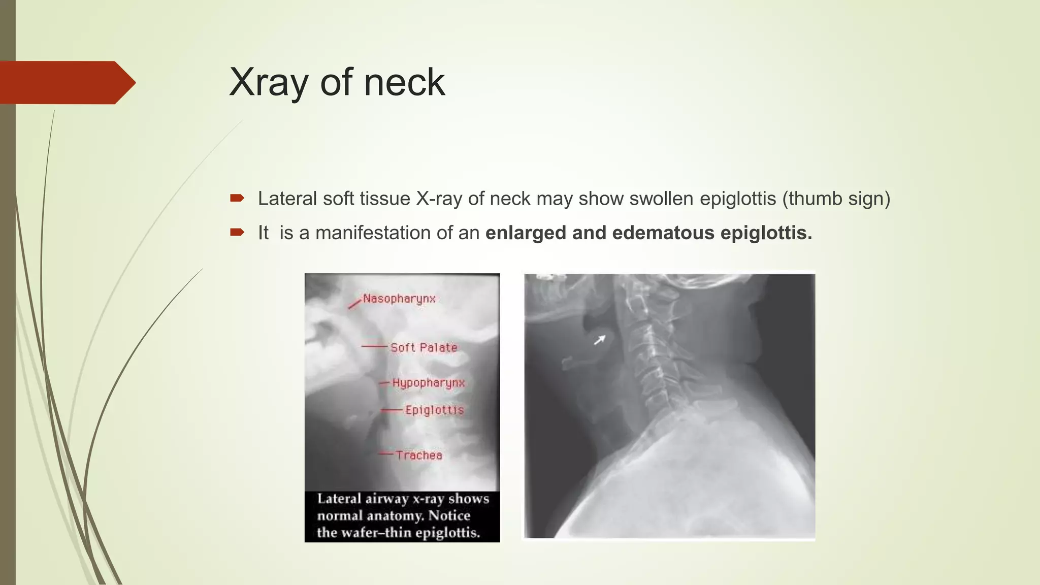
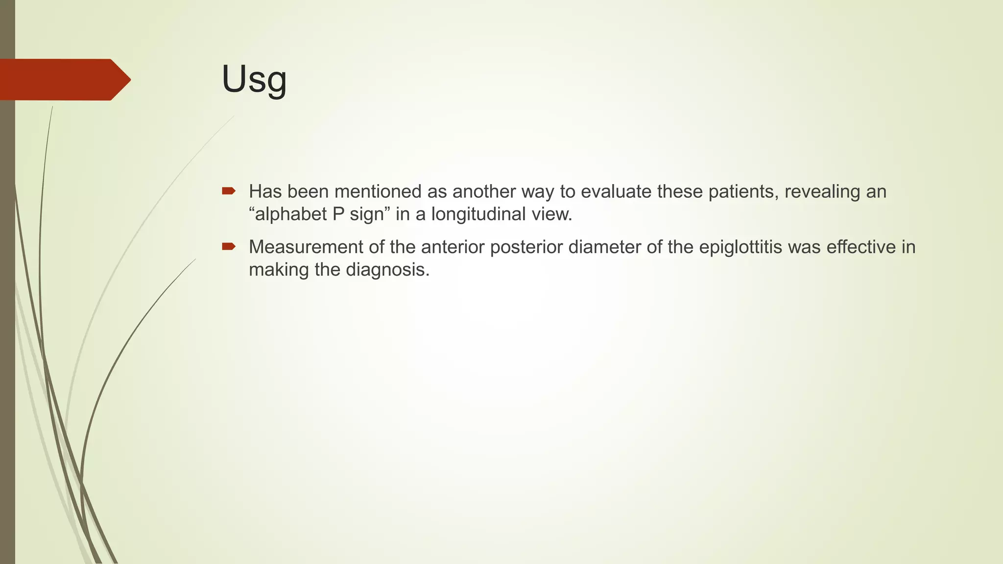
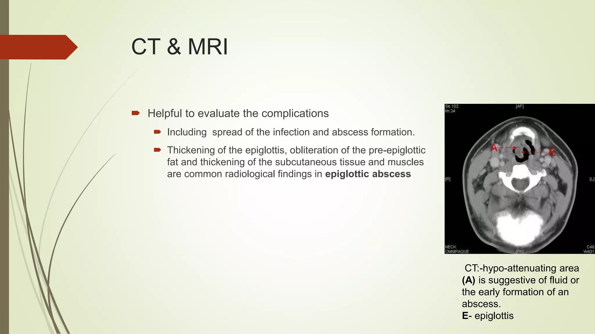
Acute Epiglottitis
Insp Dr Mahadev Deuja
Thursday, October 11, 2018
Outlines
 Introduction
 Anatomy
 Epidemiology
 Clinical presentation
 Evaluation
 Treatment
 Prognosis
 Prophylaxis
Introduction
 Epiglottitis is an inflammatory condition of the epiglottis and nearby structures like
the arytenoids, aryepiglottic folds, and vallecula.
 It is a life-threatening infection that causes profound swelling of the upper airways
which can lead to asphyxia and respiratory arrest
Anatomy
 Leaf like, yellow, elastic cartilage forming anterior
wall of laryngeal inlet.
 Attached to body of hyoid bone by hyoepiglottic
legament
Cont..
 Supraglottic structures
 Epiglottis
 Aryepiglottic folds and
 Arytenoids
Etiology
 Infectious (common)
 It can be bacterial or fungal in origin
 Haemophilus influenzae type B (HIB)
 Most common cause.
 This has decreased since the widespread use of immunization.
 However, even vaccinated children can develop epiglottitis due to non – type b H influenzae
 Other agents include Streptococcus pyrogenes, S. pneumoniae, and S. aureus
 In immunocompromised hosts, Pseudomonas aeruginosa and Candida
 Noninfectious
 Thermal inhalational injury or thermal ingestion
 Trauma to the upper airway, such as foreign bodies, and chemical irritation.
Epidemiology
 Addition of the HIB vaccine immunization has decreased the overall incidence of
epiglottitis in children.
 However, the incidence in adults has remained stable.
 Additionally, the age of children who have had epiglottitis has increased from 3
years old to about 6 to 12 years old
Clinical presentation
 Onset and progression of symptoms is rapid
(George Washington woke up with a sore throat and died the same night),
 Usually occurred within the last 12 to 24 hours.
 The patient will appear toxic.
 They will likely be sitting upright with their mouth open in
tripod position and possibly have a muffled voice.
 Drooling, dysphagia, and distress, or anxiety in children
(3 Ds) are common presenting symptoms
 Fever may go upto 40·c.
Cont..
 Swelling of the upper airway results in turbulent airflow during inspiration causing
stridor.
 Signs of severe upper airway obstruction such as
 Intercostal or suprasternal retractions
 Tachypnea
 cyanosis
(concerning for impending respiratory failure)
Examination
 Diagnosis is primarily one of clinical suspicion.
 An oropharyngeal exam is performed to evaluate a
suspected case of epiglottitis.
 Depressing the tongue with tongue depressor may
show red and swollen epiglottis.
 A flexible fiberoptic laryngoscopy
 May show edema and congestion of supraglottic
structures.
 Performed only in a very controlled setting such as the
operating room due to the risk of inducing
laryngospasm.
Xray of neck
 Lateral soft tissue X-ray of neck may show swollen epiglottis (thumb sign)
 It is a manifestation of an enlarged and edematous epiglottis.
Usg
 Has been mentioned as another way to evaluate these patients, revealing an
“alphabet P sign” in a longitudinal view.
 Measurement of the anterior posterior diameter of the epiglottitis was effective in
making the diagnosis.
Other investigations
 CBC with differential, blood culture.
 An epiglottal culture should only be obtained in patients with a secured ET tube.
CT & MRI
 Helpful to evaluate the complications
 Including spread of the infection and abscess formation.
 Thickening of the epiglottis, obliteration of the pre-epiglottic
fat and thickening of the subcutaneous tissue and muscles
are common radiological findings in epiglottic abscess
CT:-hypo-attenuating area
(A) is suggestive of fluid or
the early formation of an
abscess.
E- epiglottis
DD’S
 Infectious processes:
 Mononucleosis, diphtheria, pertussis, croup, tonsillitis
 Ludwig’s angina with retropharyngeal, Peripharyngeal and peritonsillar abscesses,
tracheobronchitis,
 subglottic laryngitis.
 Non-infectious diseases
 Allergic reactions, angioneurotic oedema, foreign body aspiration,
 Reflex laryngospasm, laryngeal trauma, tumours, hydrocarbon aspiration, systemic
lupus erythematosis and inhalation of toxic fumes or superheated steam
Complications
 In some cases, infection can spread to nearby parts of the body, including :
 Inner ear (otitis media)
 Brain (meningitis)
 Heart lining (pericarditis)
 Lungs (pneumonia)
Treatment/management
 Avoid agitating the patient, take a position in which he or she feels comfortable.
 The mainstay of treatment is to secure the airway.
 Unstable patients require immediate airway management
 Experienced providers should intubate these patients since their airways are regarded as difficult.
 An individual capable of performing a tracheotomy should be available if needed.
 Patients without signs of airway compromise, respiratory difficulty, stridor, or drooling, and who have
only mild swelling on laryngoscopy, may be managed without immediate airway intervention by close
monitoring in ICU
 Patient should be admitted to the ICU after the airway is secured.
 Corticosteroids (hydrocortisone or dexamethasone)
 To reduce edema
 To reduce ICU stay.
Treatment
 Empirically antimicrobials should be initiated.
 Ampicillin
 Third gen.cephalosporin-
 effective against H.influenzae
 given by parenteral route (i.m/i.v)
 Once culture and sensitivity results are available, the regimen should be adjusted.
 Adequate hydration - parenteral fluids
 Humidification and oxygen
Prophylaxis
 Close contacts of patients with H. influenzae should be prescribed rifampin
prophylaxis.
 Although H influenzae vaccine is available, it is not 100% effective
 Patients having recurrent episodes of acute epiglottitis warrant investigation of the
immune system.
Prognosis
 Good with appropriate and timely treatment.
 Most patients can be extubated within several days.
 However, unrecognized epiglottitis may rapidly lead to airway compromise and
resultant death.
 The risk of death is high due to sudden airway obstruction and difficulty intubating
patients with extensive swelling of supraglottic structures.
Take home message
 Epiglottitis is an inflammatory condition of the epiglottis and nearby structures.
 It is usually an infectious process of bacterial etiology directly or from bacteremia
 Diagnosis is primarily one of clinical suspicion.
 Lateral xray neck shows thumb sign.
 Airway management is the most urgent consideration.
Mcq’s
 Which is true of acute epiglottitis?
1. It is commonly seen in the elderly
2. It can be treated as an outpatient with antibiotics
3. It is a cause of stridor
4. Oral examination is necessary for these patients
What is the most common cause of epiglottitis?
1. E coli
2. Staphylococcus
3. H influenza
4. Streptococcus
 Which of the following symptoms associated with acute epiglottitis is
INCORRECT?
1. Sudden onset
2. Drooling
3. Better when lying flat
4. Fever
Refrences
 emedicine.com. Accessed October 9, 2018].
 Diseases of EAR, NOSE & THROAT. Elsevier India;
 https://knowledge.statpearls.com/chapter/0/21236?utm_source=pubmed
Thank you
Next presentation : Insp Dr Subash Chandra Gautam
AKI

Acute epiglottitis

  • 1.
    Acute Epiglottitis Insp DrMahadev Deuja Thursday, October 11, 2018
  • 2.
    Outlines  Introduction  Anatomy Epidemiology  Clinical presentation  Evaluation  Treatment  Prognosis  Prophylaxis
  • 3.
    Introduction  Epiglottitis isan inflammatory condition of the epiglottis and nearby structures like the arytenoids, aryepiglottic folds, and vallecula.  It is a life-threatening infection that causes profound swelling of the upper airways which can lead to asphyxia and respiratory arrest
  • 4.
    Anatomy  Leaf like,yellow, elastic cartilage forming anterior wall of laryngeal inlet.  Attached to body of hyoid bone by hyoepiglottic legament
  • 5.
    Cont..  Supraglottic structures Epiglottis  Aryepiglottic folds and  Arytenoids
  • 6.
    Etiology  Infectious (common) It can be bacterial or fungal in origin  Haemophilus influenzae type B (HIB)  Most common cause.  This has decreased since the widespread use of immunization.  However, even vaccinated children can develop epiglottitis due to non – type b H influenzae  Other agents include Streptococcus pyrogenes, S. pneumoniae, and S. aureus  In immunocompromised hosts, Pseudomonas aeruginosa and Candida  Noninfectious  Thermal inhalational injury or thermal ingestion  Trauma to the upper airway, such as foreign bodies, and chemical irritation.
  • 7.
    Epidemiology  Addition ofthe HIB vaccine immunization has decreased the overall incidence of epiglottitis in children.  However, the incidence in adults has remained stable.  Additionally, the age of children who have had epiglottitis has increased from 3 years old to about 6 to 12 years old
  • 8.
    Clinical presentation  Onsetand progression of symptoms is rapid (George Washington woke up with a sore throat and died the same night),  Usually occurred within the last 12 to 24 hours.  The patient will appear toxic.  They will likely be sitting upright with their mouth open in tripod position and possibly have a muffled voice.  Drooling, dysphagia, and distress, or anxiety in children (3 Ds) are common presenting symptoms  Fever may go upto 40·c.
  • 9.
    Cont..  Swelling ofthe upper airway results in turbulent airflow during inspiration causing stridor.  Signs of severe upper airway obstruction such as  Intercostal or suprasternal retractions  Tachypnea  cyanosis (concerning for impending respiratory failure)
  • 10.
    Examination  Diagnosis isprimarily one of clinical suspicion.  An oropharyngeal exam is performed to evaluate a suspected case of epiglottitis.  Depressing the tongue with tongue depressor may show red and swollen epiglottis.  A flexible fiberoptic laryngoscopy  May show edema and congestion of supraglottic structures.  Performed only in a very controlled setting such as the operating room due to the risk of inducing laryngospasm.
  • 11.
    Xray of neck Lateral soft tissue X-ray of neck may show swollen epiglottis (thumb sign)  It is a manifestation of an enlarged and edematous epiglottis.
  • 12.
    Usg  Has beenmentioned as another way to evaluate these patients, revealing an “alphabet P sign” in a longitudinal view.  Measurement of the anterior posterior diameter of the epiglottitis was effective in making the diagnosis.
  • 13.
    Other investigations  CBCwith differential, blood culture.  An epiglottal culture should only be obtained in patients with a secured ET tube.
  • 14.
    CT & MRI Helpful to evaluate the complications  Including spread of the infection and abscess formation.  Thickening of the epiglottis, obliteration of the pre-epiglottic fat and thickening of the subcutaneous tissue and muscles are common radiological findings in epiglottic abscess CT:-hypo-attenuating area (A) is suggestive of fluid or the early formation of an abscess. E- epiglottis
  • 15.
    DD’S  Infectious processes: Mononucleosis, diphtheria, pertussis, croup, tonsillitis  Ludwig’s angina with retropharyngeal, Peripharyngeal and peritonsillar abscesses, tracheobronchitis,  subglottic laryngitis.  Non-infectious diseases  Allergic reactions, angioneurotic oedema, foreign body aspiration,  Reflex laryngospasm, laryngeal trauma, tumours, hydrocarbon aspiration, systemic lupus erythematosis and inhalation of toxic fumes or superheated steam
  • 16.
    Complications  In somecases, infection can spread to nearby parts of the body, including :  Inner ear (otitis media)  Brain (meningitis)  Heart lining (pericarditis)  Lungs (pneumonia)
  • 17.
    Treatment/management  Avoid agitatingthe patient, take a position in which he or she feels comfortable.  The mainstay of treatment is to secure the airway.  Unstable patients require immediate airway management  Experienced providers should intubate these patients since their airways are regarded as difficult.  An individual capable of performing a tracheotomy should be available if needed.  Patients without signs of airway compromise, respiratory difficulty, stridor, or drooling, and who have only mild swelling on laryngoscopy, may be managed without immediate airway intervention by close monitoring in ICU  Patient should be admitted to the ICU after the airway is secured.  Corticosteroids (hydrocortisone or dexamethasone)  To reduce edema  To reduce ICU stay.
  • 18.
    Treatment  Empirically antimicrobialsshould be initiated.  Ampicillin  Third gen.cephalosporin-  effective against H.influenzae  given by parenteral route (i.m/i.v)  Once culture and sensitivity results are available, the regimen should be adjusted.  Adequate hydration - parenteral fluids  Humidification and oxygen
  • 19.
    Prophylaxis  Close contactsof patients with H. influenzae should be prescribed rifampin prophylaxis.  Although H influenzae vaccine is available, it is not 100% effective  Patients having recurrent episodes of acute epiglottitis warrant investigation of the immune system.
  • 20.
    Prognosis  Good withappropriate and timely treatment.  Most patients can be extubated within several days.  However, unrecognized epiglottitis may rapidly lead to airway compromise and resultant death.  The risk of death is high due to sudden airway obstruction and difficulty intubating patients with extensive swelling of supraglottic structures.
  • 21.
    Take home message Epiglottitis is an inflammatory condition of the epiglottis and nearby structures.  It is usually an infectious process of bacterial etiology directly or from bacteremia  Diagnosis is primarily one of clinical suspicion.  Lateral xray neck shows thumb sign.  Airway management is the most urgent consideration.
  • 22.
    Mcq’s  Which istrue of acute epiglottitis? 1. It is commonly seen in the elderly 2. It can be treated as an outpatient with antibiotics 3. It is a cause of stridor 4. Oral examination is necessary for these patients
  • 23.
    What is themost common cause of epiglottitis? 1. E coli 2. Staphylococcus 3. H influenza 4. Streptococcus
  • 24.
     Which ofthe following symptoms associated with acute epiglottitis is INCORRECT? 1. Sudden onset 2. Drooling 3. Better when lying flat 4. Fever
  • 25.
    Refrences  emedicine.com. AccessedOctober 9, 2018].  Diseases of EAR, NOSE & THROAT. Elsevier India;  https://knowledge.statpearls.com/chapter/0/21236?utm_source=pubmed
  • 26.
    Thank you Next presentation: Insp Dr Subash Chandra Gautam AKI