SlideShare a Scribd company logo
1 of 69
Scott brown learning 2020
Volume : 2 "Paediatrics
The ear skull base"
Chapter : 29.Acute laryngeal infections( p.no :325 to 332 )
30. Congenital disorders of larynx , trachea and bronchi
( p.no : 333 to 346 )
Acute laryngeal infections &
Congenital disorders of larynx ,
trachea and bronchi
Dr. Vinoth .S , postgraduate in department of ENT ,
Madurai medical college
Acute laryngeal infections – croup ( ALTB)
• Croup typically presents as hoarseness with distinctive barking cough
progressing to inspiratory or biphasic stridor.
• usually a preceding history of upper respiratory tract symptoms,
malaise and pyrexia.
• Children with croup do not typically drool or appear toxic, in contrast
to children presenting with epiglottitis.
• The symptoms are thought to be due to mucosal oedema of the
larynx and trachea following a viral illness.
caused by parainfluenza virus type I , parainfluenza virus type II,
respiratory syncytial virus (RSV) virus types A and B and rhinovirus.
• The diagnosis of croup is a clinical one and laboratorytests and
radiological tests are generally not needed
• Croup is usually self-limiting , is rare
• critical symptom of stridor is due to oedema in the subglottis, the
narrowest part of the paediatric
Chest x-ray :
• radiograph of the thoracic inlet will show characteristic narrowing of
the subglottis on an anteroposterior view(‘steeple’ or ‘pencil-tip’ sign)
Westley Croup Score :
A maximum score is 17, a score of
2–3 equates to mild croup
4–7 to moderate croup and
8 or more to severe croup
Treatment
• For mild croup, can be supportive humidified environment or mist
tent
Medical - Corticosteroids
• The mainstay of treatment for croup is corticosteroids.
• Corticosteroids have a systemic anti-inflammatory effect.
• There is a reduction in capillary endothelial permeability and
therefore in mucosal oedema, and stabilization of lysosomal
membranes, decreasing the inflammatory reaction.
• Topical corticosteroids have alpha-mediated vasoconstriction.
Nebulized epinephrine
• Nebulized epinephrine (1 mL of 1 in 1000 epinephrine diluted in 3 mL
of 0.9% saline) has an established role in the acute paediatric airway
in reducing mucosal oedema by an alpha-agonist effect causing
vasoconstriction and bronchodilation;
• a maximum effect is achieved within 30–60 minutes but there is no
lasting benefit beyond 2 hours.
Heliox
• HelioxR is a mixture of helium and oxygen generally supplied with a helium
: oxygen ratio of 70 : 30
• produce laminar flow in the presence of a partially obstructed airway than
air or oxygen.
• Inhaled helioxR delivered through a high-flow system should reduce the
work of breathing and result in larger tidal volumes and improved gas
exchange.
• Helium has a high thermal conductivity and so care is needed with
humidification in order to avoid hypothermia
BACTERIAL LARYNGOTRACHEOBRONCHITIS
also known as
• PSEUDOMEMBRANOUS CROUP,
• BACTERIAL TRACHEITIS,
• MEMBRANOUS LARYNGOTRACHEOBRONCHITIS,
• NEONATAL NECROTIZING TRACHEOBRONCHITIS
• a severe form of laryngotracheobronchitis characterized by the
presence of profuse mucopurulent secretions with sloughing of the
respiratory epithelium.
• Secretions are often adherent, are not effectively cleared by
coughing and may occlude the airway causing respiratory
compromise.
• Bacterial tracheitis must be differentiated from croup and other
causes of infectious upper airway obstruction in order to initiate
proper management.
• Classically the child with bacterial tracheitis appears toxic with high
fevers and worsening stridor that fails to respond to treatment with
steroids and nebulized epinephrine.
• It typically affects children older (mean age 4–8 years) than the usual
age group for croup.
• Bacterial tracheitis is a much less common condition than croup.
• However, there is increased susceptibility in children with Down
syndrome or immunodeficiency
• Diagnosis can only be confirmed on airway endoscopy; there is a
pseudomembrane in the subglottis and trachea and thick mucopus
and debris extending into the bronchi
• Direct laryngotracheobronchoscopy under general anaesthesia and
removal of all tracheal secretions, with pulmonary toilet, is
mandatory.
• to secure the airway by endotracheal intubation for a period of days.
• Staphylococcus aureus( is the pathogen most commonly isolated from
tracheal cultures) Haemophilus influenzae, Moraxella catarrhalis,
Streptococcus pneumoniae and Pseudomonas aeruginosa
• Viral cultures are also frequently positive, indicative that many cases
of bacterial tracheitis represent a secondary bacterial infection that
follows a viral respiratory tract infection.
• Most commonly influenza A is identified, although other viruses
including the H1N1 strain of influenza A and metapneumovirus have
also been isolated.2,
Complications
include
• airway stenosis,
• acute respiratory distress syndrome,
• respiratory failure,
• toxic shock syndrome,
• anoxic encephalopathy and
• death.
Diphtheria
• Diphtheria remains an important differential in the diagnosis of acute
laryngeal infection in children, particularly in those who have
travelled to endemic areas or have not been vaccinated
• The causative organisms in pathogenic diphtheria are the toxogenic
strains of Corynebacterium diphtheria and Corynebacterium ulcerans.
• The early clinical picture of upper respiratory tract symptoms is due
to the effects of the organism itself.
• Delayed effects are due to the release of exotoxin.
• Symptoms of diphtheria come on gradually and typically begin 2–5
days after exposure.
• Initial symptoms are of pharyngitis with sore throat and malaise.
• The child is feverish and on examination there is a typical appearance
of the pharyngeal tonsils with necrosis and the development of a
characteristic grey pseudomembrane over the surface.
• This consists of necrotic tissue, bacteria and a rich fibrinous exudate.
• Early removal causes bleeding but the pseudomembrane may
separate more easily in the later course of the disease.
• There may be a bull-neck appearance due to cellulitis and regional
lymphadenopathy
• Diphtheria infection may also involve the larynx.
• After initial symptoms of dysphagia and toxaemia, symptoms may progress to
inspiratory stridor and a barking cough;
• the cough is frequently paroxysmal and exhausting.
• Death may follow owing to acute airway obstruction or as a result of the later
effects of the exotoxin.
• The exotoxin can cause a toxic myocarditis in the second week of the disease and
this may be fatal.
• Peripheral neuritis may also occur, palatal paralysis being the most common
effect of peripheral neuropathy and presenting with nasal regurgitation of food
and hypernasal speech.
• Successful treatment depends on early diagnosis, and the timely administration
of high-dose benzylpenicillin and antitoxin (10 000–100 000 units, depending on
the severity of infection).
• Delaying the administration of the antitoxin is associated with increased mortality
as the antitoxin does not neutralize toxin that is already bound to tissues.
• The decision, therefore, to administer antitoxin should be made based on clinical
suspicion and should not await microbiological confirmation.
• Airway management consists of removal of the laryngeal membrane,
administration of oxygen and humidification, and endotracheal intubation or
tracheostomy if necessary.
• Systemic steroids may reduce the need for airway intervention.
• Bed rest is recommended until the danger of myocarditis is past.
It is of a toxic child with a short history of sore throat, inspiratory stridor,
muffled voice and drooling due to odynophagia and dysphagia.
Untreated results in progressive respiratory distress.
The child is febrile, tachypnoeic and classically will be sitting upright, with
the neck extended to optimize the airway, and using then arms to provide support
to the shoulder girdle to maximize the efficiency of the accessory muscles of
respiration.
It is commonest between the ages of 2 and 8 and there is an increased
prevalence in winter
Acute epiglottitis
• When acute epiglottitis is suspected, pharyngeal examination should
not be attempted, as simple manipulation with a tongue depressor
may precipitate acute airway obstruction,
• although the use of a fibreoptic or small rigid endoscope can assist
the diagnosis in patients with an atypical presentation.
• Direct examination of the airway should not be delayed, but should
be undertaken in a controlled setting such as an operating room or
paediatric intensive care unit by personnel skilled in airway
intervention;
• endoscopic evaluation will confirm gross erythema and oedema of
the supraglottic structures
• When the diagnosis is confirmed, the airway should be secured by
endotracheal intubation
• a soft-tissue lateral radiograph of the neck will typically show a
thickened oedematous epiglottis – the ‘thumb sign’
Etiology :
• acute epiglottitis has been a manifestation of invasive Hib infection
• meningococci,
• group A streptococci, pneumococci, Haemophilus parainfluenzae and
Staphylococcus aureus.
• Immunocompromised individuals are at increased risk of epiglottitis.
due to atypical organisms such as Herpes simplex type 1, Varicella
zoster, Parainfluenza or Candida albicans.
Treatment :
• Treatment is with intravenous antibiotics; ampicillin resistance due to beta-
lactamase production is now over 50% in Haemophilus influenzae, so empirical
treatment with third-generation cephalosporins for 5–7 days is advised.
• Rifampicin prophylaxis has been recommended to eradicate the carrier state for
the index case as well as household and school contacts.
• Haemophilus influenzae type b immunization
AIRWAY MANAGEMENT
• Endotracheal intubation is now considered preferable to tracheostomy
• If endotracheal intubation is to be undertaken, it should be performed in an
operating room or paediatric intensive care unit with personnel and equipment
available and ready to undertake tracheostomy if the airway cannot be secured.
• humidified air should be administered to discourage the development of
tenacious secretions
Nasotracheal intubation
• When a prolonged period of intubation is anticipated, the
nasotracheal rather than the orotracheal route is preferred
• In acute epiglottitis, visualization of the epiglottis with a flexible
nasendoscope can be useful in monitoring resolution of
inflammation.
• in laryngotracheobronchitis, whether viral or bacterial, the airway
can be monitored using endoscopy until a reduction in tracheal
inflammation shows that it is safe to extubate.
• Systemic steroids administered 6 hours prior to removal of the tube
will minimize post-intubation oedema, and nebulized epinephrine can
assist with airway patency after extubation.
• For acute epiglottitis the period of intubation is usually less than 48
hours but for croup and bacterial laryngotracheobronchitis it may be
up to a week.
• Tracheostomy is no longer considered the first choice to secure the
airway in acute laryngeal infection.
Congenital conditions of
larynx
Congenital conditions of larynx -Laryngomalacia
• Laryngomalacia is characterized by partial or complete collapse of the
supraglottic structures on inspiration
• is the most common congenital cause of stridor
• Signs- the epiglottis is long and curled (omega-shaped)
the aryepiglottic folds are short and tightly tethered to the epiglottis;
there may also be redundant mucosa and submucosa of
the aryepiglottic folds medially resulted in a tall,
narrow supraglottis with a deep interarytenoid cleft
- epiglottis is soft and may curl and collapse and the
mucosa may prolapse into the airway .
- due to neuromuscular immaturity and consequent
incoordination of arytenoid movements
• high-pitched, fluttering inspiratory stridor is usually present at, or
shortly after, birth , increases during activity and decreases at sleep
The severity of the stridor increases during the first 9 months
of life, and disappears at two years of age ,
rarely may persist into late childhood.
• The diagnosis can be confirmed by flexible fibre-optic laryngoscopy
• Treatment :
• for mild, no intervention is needed and the parents can be reassured
• In severe laryngomalacia, there is serious respiratory obstruction with
substantial sternal and intercostal recession together with feeding
difficulties results in failure to thrive
• May associated with congenital cyanotic heart disease
• Children who show signs of failure to thrive should undergo an endoscopic
aryepiglottoplasty (sometimes termed a supraglottoplasty)
• In this procedure, each aryepiglottic fold is first divided to release it from the
edge of the epiglottis, and any redundant mucosa and submucosal tissue are
then excised from over the arytenoids, together, if necessary, with part or all of
the cuneiform cartilages.
• The ‘bridge’ of mucosa between the arytenoids is carefully preserved to prevent
interarytenoid scarring.
Vocal cord paralysis
• Vocal cord paralysis is the second most common congenital anomaly
of the larynx after laryngomalacia.
• patients may have other, coexisting airway pathology
• Diagnosed by flexible fibre-optic laryngoscopy
Laryngeal ultrasound
• Unilateral vocal cord paralysis is usually not congenital, most cases being acquired
as a result of surgical injury to the left recurrent laryngeal nerve, often following
correction of a congenital cardiac anomaly.
• Patients present with mild stridor, dysphonia and sometimes aspiration.
• If the vocal cord lies in an intermediate position, surgical intervention is not
usually necessary, and the voice can be expected to improveby compensation.
• If the vocal cord lies in a more abducted position, the dysphonia may be more
pronounced and aspiration more likely.
• Vocal cord medialization procedures such as thyroplasty and augmentation
injection can improve the dysphonia and aspiration, but these may result in
worsening of the stridor.
• Bilateral vocal cord palsy is usually a congenital abductor paralysis.
• The vocal cords lie in the paramedian position with consequent
inspiratory stridor,
• and a tracheostomy is necessary for airway management
• Presented with the Arnold-Chiari malformation
• If it have no movement ,then an endoscopic laser cordotomy or
arytenoidectomy should be considered at the age of 11
Laryngocoeles :
• Laryngeal cysts divided into laryngocoeles and saccular cysts.
• laryngocoele is an air-filled dilatation of the laryngeal ventricle which
communicates with the laryngeal lumen.
• uncommon lesion which usually occurs in middle age but may rarely be seen in
infancy,
• can produce respiratory distress which typically becomes worse on crying due to
increased distension of the laryngocoele with air.
• a laryngocoele may obstruct and fill with mucus or become infected
(laryngopyocoele), thus becoming indistinguishable from a saccular cyst
• A also represents an abnormal dilatation or herniation of the saccule
of the ventricle of the larynx;
• it differs from a laryngocoele in that there is no opening into the
larynx and it is filled with mucus instead of air.
• result from a developmental failure to maintain patency of the
orifice between the saccule and the ventricle, and may be of anterior
or lateral type.
Saccular cyst
The anterior saccular cyst extends medially
and posteriorly from the saccule and so
protrudes into the laryngeal airway
between the true and false vocal cords.
The lateral saccular cyst is most common in
infants and
expands posterosuperiorly into the false
cord and
aryepiglottic fold
• Laryngeal cysts are classified as internal or type 1 if contained entirely within the
laryngeal framework, and external or type 2 if it pierces the thyrohyoid
membrane.
• Diagnosis is confirmed by endoscopy, except in an external laryngocoele where
no abnormality may be seen except on imaging.
• Saccular cysts are best treated at the initial endoscopy by wide endoscopic
marsupialization
• If the cyst recurs, then the procedure of choice is a lateral cervical approach
extending through the thyrohyoid membrane at the superior margin of the ala of
the thyroid cartilage, with subperichondrial resection of a portion of the upper
part of the ala. Through this ‘window’ the cyst can be completely excised, using
short-term intubation to secure the airway post-operatively.
Vascular malformations
• include lymphatic malformations, venous malformations, av malformations
• Lymphatic malformations (also termed lymphagiomas or cystic hygromas) are
cystic malformations that result from abnormal development of the lymphatic
vessels.
• In the head neck they may be macrocystic (usually infrahyoid), microcystic
(usually suprahyoid) or a combination of the two.
• Occasionally, a microcystic lymphatic malformation may extend into the tongue
base, valleculae and supraglottis, and airway obstruction may result.
• If the lymphatic malformation is very extensive, a tracheostomy may be required,
• Surgical modality is to debulk the lesion by endoscopic vaporization using a CO2
laser or radiofrequency ablation.
Bifid epiglottis
• is a rare laryngeal anomaly in which the epiglottis fails to fuse in the
midline and thus has a cleft extending down to its tubercle.
• It may be seen as a feature of Pallister–Hall syndrome
• presents with feeding difficulties due to aspiration and with stridor
because of collapse and enfolding of the two halves of the epiglottis.
• Endoscopy establishes the diagnosis,
• treatment options include amputation of the epiglottis and
tracheostomy.
Glottis - Laryngeal webs
• Failure of complete canalization of the larynx during embryogenesis
may result in a glottic or, very rarely, a supraglottic web.
• Mostly involves anterior glottis , associated with congenital vocal cord
paralysis .
• Due to delayed maturation in the vagal nuclei
• glottic enlargement surgery in order to avoid a tracheostomy
• an endoscopic laser cordotomy or arytenoidectomy if presented with
stridor
Laryngeal atresia
• Laryngeal atresia is incompatible with life unless there is an
associated tracheo-oesophageal fistula (TOF)
• emergency tracheostomy is performed in the delivery room.
• now recognized antenatally on ultrasound imaging and managed with
ex utero intrapartum treatment (EXIT) procedure,
• whereby a tracheostomy is undertaken following elective Caesarean
section with the neonate still on placental circulation.
Congenital subglottic stenosis
• due to defective canalization of the cricoid cartilage and/or conus elasticus,
resulting in either gross thickening of the anterior lamina of the abnormal cricoid
• a small, elliptical, thickened cricoid with excessive submucosal soft tissue
• there may be anterior fusion of the vocal cords, forming a web with subglottic
extension, as seen in 22q11 deletion syndromes.
• Subglottic stenosis is said to be the third most common congenital anomaly of
the larynx
• may be a combined congenital plus acquired stenosis for intubation
The Myer-Cotton grading system
• Grade I : 0–50% obstruction
• • Grade II: 51–70% obstruction
• • Grade III: 71–99% obstruction
• • Grade IV: 100% obstruction.
• If the airway is not severely compromised, surgery may not be
required
• a strict contraindication to dilatation or laser resection:
• any type of endoscopic treatment is liable to worsen the initial
condition, and attempted dilatation is inevitably ineffective as the
thickened ring of cricoid cartilage cannot be expanded
Milder degrees of stenosis present as
inspiratory or biphasic stridor as the
child becomes older and more active, or
as recurrent ‘croup’ owing to
superimposed oedema from upper
respiratory tract infections
• If the airway is severely compromised, a tracheostomy is needed.
• The surgical options have evolved from the classical castellated
laryngotracheoplasty designed by Evans and Todd
• The LTR involves augmentation of the laryngotracheal complex by
anterior and/or posterior midline incision of the cricoid with insertion
of costal cartilage grafts to expand the airway
Treatment :
• Grade I subglottic stenosis usually requires no surgical intervention.
• Grade II stenosis can be reconstructed by means of an LTR with
anterior cartilage grafting +/− posterior cricoid split.
• Mild grade III stenosis needs an anterior graft with posterior cricoid
split +/− posterior cartilage grafting.
• Severe grade III stenosis (a pinhole airway) requires both anterior and
posterior grafts.
• Grade IV stenosis demands anterior and posterior grafts with
prolonged stenting
Subglottic haemangioma
• Infantile subglottic haemangioma causes gradually worsening
inspiratory or biphasic stridor presenting in the first few weeks of life
• Associated with cutaneous haemangioma
• The natural history is typically a proliferative phase lasting 6–12
months followed by complete involution over 1–5 years
• At endoscopy the typical appearance is of a compressible, pear-
shaped red swelling in the subglottis on one side, left more
commonly than right
• Subglottic haemangioma is life-threatening
because of its situation in the narrowest part
of the airway and therefore requires rapid
intervention.
• Tracheostomy will maintain the airway
until involution
• radiotherapy, CO2 laser ablation, systemic steroids,
intralesional steroid injection followed by intubation, and interferon
alpha-2a
• propranolol is an effective treatment for cutaneous haemangioma
and subglottic haemangioma
• Submucous resection remains a valid alternative for non-
circumferential haemangiomas and those not involving the vocal
cords
Laryngeal and laryngotracheooesophageal
cleft
• Posterior laryngeal clefts result from failure of the posterior cricoid
lamina to fuse with incomplete development of the tracheo-
oesophageal septum.
• Benjamin and Inglis classification
- type I cleft extends down to the level ofthe vocal cords;
-type II cleft extends below the vocal cords into the cricoid
- type III cleft extends down into the cervical trachea
-type IV cleft extends into the thoracic trachea and may even reach
the carina
• With Opitz-Frias syndrome, TOF, Pallister–Hall syndrome
Symptoms :
• Type I clefts present with cyanotic attacks on feeding and recurrent chest
infections
• Type II and III clefts produce dramatic aspiration with recurrent
pneumonia, sometimes with stridor and an abnormal cry.
• Type IV clefts cause severe aspiration, cyanosis and incipient
cardiorespiratory failure.
• Suspension microlaryngoscopy to probe the arytenoids
• A plain chest X-ray may show changes secondary to recurrent aspiration
pneumonitis
• Videofluoroscopic contrast swallow studies may not differentiate laryngeal
incompetence from neuromuscular incoordination
Treatment :
• A short type I cleft with no aspiration requires no treatment. Minimal aspiration
may be managed by thickening the feeds
• A very short type II cleft may also be repaired endoscopically,
• a long type II or a type III cleft needs to be approached anteriorly through an
extended laryngofissure with a low tracheostomy
• type IV clefts may be managed by an anterior approach through a cervical
incision, if necessary pulling the trachea up into the neck to reach the lower end
of the cleft.
• Longer clefts will require a lateral cervical approach in combination with a
thoracotomy, or preferably an anterior cervicothoracic approach via a median
sternotomy with repair on extracorporeal membrane oxygenation (ECMO) or
cardiopulmonary bypass.
• Mortality remains significant, being approximately 14% overall
37 rising to 66% for type IV laryngotracheooesophageal clefts and
up to 100% for full-length clefts ending at the carina
Agenesis
• Tracheal agenesis may be complete (full-length) or partial, but in
either case there is no continuity between the larynx and the bronchi
• Occasionally, short-term survival may be possible if there is a
broncho-oesophageal fistula, which can permit some airflow into the
lungs, but surgical efforts to use the oesophagus as a tracheal
replacement have not been successful and long-term survival has not
proved possible
Stenosis
• Occasionally, a membranous web may be encountered in
the trachea with a normal underlying cartilaginous
ring structure. Such cases usually fare well with endoscopic
rupture and dilatation.
• Thicker congenital fibrous stenoses may be amenable to radial KTP laser incision
and balloon dilatation, followed by application of mitomycin C to reduce the risk
of restenosis42 or tracheal resection with end-to-end anastomosis.
• A short stenosis may also be associated with one or two absent tracheal rings,
although the most common finding in congenital tracheal stenosisis (CTS) is a
segment of complete tracheal cartilaginous rings with an airway lumen as narrow
as 2 mm
• Children with congenital tracheal stenosis usually present with
respiratory distress
• Symptoms may become apparent in the neonatal period with
biphasic stridor, respiratory distress, tracheal tug and episodes of
cyanosis in the first year of life
• Endoscopy remains the ‘gold standard’ investigation for a child with
stridor
• Bronchoscopy and bronchography (B&B) demonstrate the the size of the tracheal
lumen and outline the trachea and bronchi distal to the stenosis, which may be
too narrow to permit passage of a bronchoscope and also provides a dynamic
assessment of any malacic segments.
• Optical coherence tomography (OCT) can be combined with B&B to confirm the
presence of complete rings if there is any doubt.
• Contrast CT and an echocardiogram are essential in view of the high incidence of
associated anomalies of the heart and great vessels.
• CT is particularly helpful in assessing the vascular anatomy and its relationship to
the trachea and 3D reconstructions allow better surgical planning
• Mild cases with minimal symptoms may require no intervention after
the diagnostic endoscopy,
• Very narrow, short segments can be excised with end-to-end
anastomosis , Augmentation tracheoplasty with costal cartilage
grafting or a pericardial flap or free patch can be tried
• Successful endoscopic balloon dilatation
• Absorbable PDS stents offer an advantage over metallic stents and are
used to treat malacia, particularly bronchomalacia
Tracheomalacia and bronchomalacia
• Tracheomalacia is a condition in which there is reduced stiffness of
the tracheal wall, resulting in abnormal collapse of the trachea during
expiration
• produce symptoms of airway obstruction
• Pathologically, the striking finding is an
increased muscle- to-cartilage ratio
seen on the transverse section of the
trachea
• Tracheomalacia is traditionally classified as primary (idiopathic), due to an
intrinsic abnormality in the wall of the airway, or
secondary, due to another associated anomaly or to external compression.
• high-pitched expiratory noise stridor becomes apparent during the first few
weeks of life with a harsh, barking cough
• Characteristically, the stridor becomes much worse when the child is active,
feeding, upset, coughing or crying,
• it may be associated with cyanotic attacks which are sometimes sufficiently
severe to be termed ‘dying spells’.
• There may be a prolonged expiratory phase to respiration, possibly with faint
expiratory stridor
• A barium swallow may identify a TOF and can also demonstrate the airway
collapse on lateral screening
• CT angiogram is extremely useful for identifying the cause of extrinsic
compression of the trachea by normal or abnormal vessels, including vascular
rings.
• Contrast bronchography may be the most useful radiological investigation:
Treatment :
• Mild tracheobronchomalacia (less than 75% collapse) requires no
intervention, and the stridor can be expected to resolve
spontaneously by around the age of 2 years.
• If it is secondary to a vascular anomaly, this should be corrected an
extended tracheostomy tube will effectively support a midtracheal
malacic segment, but this is not a satisfactory solution for lower-end
tracheal or for bronchial collapse include internal or external stenting
of the trachea, segmental resection and cartilage grafting
Anomalous bifurcations
• The right upper lobe bronchus may take origin from the right lateral
wall of the trachea above the carina
• is termed a tracheal bronchus or
suis bronchus(PIG BRONCHUS)
• asymptomatic, incidental finding,
but may sometimes be associated
with tracheal stenosis
Tracheo-oesophageal fistula
• It is a fairly common congenital malformation of the neonatal air and food
passages, which usually occurs in association with oesophageal atresia.
• Eighty-seven per cent of cases have oesophageal atresia with a TOF
communicating between the distal oesophagus and the mid- to lower trachea or
a main bronchus
• m/c present to intensivist TOF without atresia (‘H-type fistula’)
the least symptomatic of the group, with no swallowing difficulty, but small
amounts of fluid pass through the fistula into the trachea and produce symptoms
and signs of recurrent minor aspiration.
barium swallow confirms the diagnosis
treatment by ligation and division of fistula
Vascular compressions
• Vascular ring :
• common is double aortic arch mostly involving ascending aorta
• Presents earlier in life with airway obstructions
• Barium swallow shows double impression upon the column of
contrast
• Surgical treatment involves diving the lesser component of the ring
• Post op tracheomalacia will persists for few monthds
• Vascular sling :
• Common is aberrant innominate artery crosses the anterior surface of trachea
above the carina
• Presents with less severe airway obstruction
• Expiratory stridor , cough ,recurrent URTI
• Bronchoscopy shows sloping pulsatile compression of the trachea 2 cm above the
carina
• Upward compression with bronchoscopes compress the artery against the
sternum thus obliterating the radial pulse
• Treatment by aortopexy

More Related Content

What's hot

Otitis media with effusion
Otitis media with effusionOtitis media with effusion
Otitis media with effusionshriyashsinha
 
Ménière's disease it’s definition ,etiopathogenesis and management
Ménière's disease it’s definition ,etiopathogenesis and managementMénière's disease it’s definition ,etiopathogenesis and management
Ménière's disease it’s definition ,etiopathogenesis and managementsritama1988
 
The formation and management of middle ear granulation
The formation and management of middle ear granulationThe formation and management of middle ear granulation
The formation and management of middle ear granulationDaria Otgonbayar
 
Diseases of the external ear
Diseases of the external earDiseases of the external ear
Diseases of the external earAjay Manickam
 
Chronic suppurative otitis media (csom)
Chronic suppurative otitis media (csom)Chronic suppurative otitis media (csom)
Chronic suppurative otitis media (csom)Aditi Kataria
 
Csom, cholesteatoma
Csom, cholesteatomaCsom, cholesteatoma
Csom, cholesteatomafarranajwa
 
Diseases of middle ear;csom(safe&unsafe)&cholesteatoma dr.davis thoma...
Diseases of middle ear;csom(safe&unsafe)&cholesteatoma dr.davis thoma...Diseases of middle ear;csom(safe&unsafe)&cholesteatoma dr.davis thoma...
Diseases of middle ear;csom(safe&unsafe)&cholesteatoma dr.davis thoma...ophthalmgmcri
 
Tumours of Ear
Tumours of EarTumours of Ear
Tumours of EarAnwaaar
 

What's hot (20)

Otitis media with effusion
Otitis media with effusionOtitis media with effusion
Otitis media with effusion
 
choanal atresia
choanal atresiachoanal atresia
choanal atresia
 
Meniere’s disease
Meniere’s diseaseMeniere’s disease
Meniere’s disease
 
Ototoxicity
Ototoxicity Ototoxicity
Ototoxicity
 
Meniere’s disease
Meniere’s diseaseMeniere’s disease
Meniere’s disease
 
Ménière's disease it’s definition ,etiopathogenesis and management
Ménière's disease it’s definition ,etiopathogenesis and managementMénière's disease it’s definition ,etiopathogenesis and management
Ménière's disease it’s definition ,etiopathogenesis and management
 
The formation and management of middle ear granulation
The formation and management of middle ear granulationThe formation and management of middle ear granulation
The formation and management of middle ear granulation
 
The formation and management of middle ear granulation
The formation and management of middle ear granulationThe formation and management of middle ear granulation
The formation and management of middle ear granulation
 
Diseases of the external ear
Diseases of the external earDiseases of the external ear
Diseases of the external ear
 
05 ome
05 ome05 ome
05 ome
 
Laryngocele
LaryngoceleLaryngocele
Laryngocele
 
Chronic suppurative otitis media (csom)
Chronic suppurative otitis media (csom)Chronic suppurative otitis media (csom)
Chronic suppurative otitis media (csom)
 
Treatment of sinusitis
Treatment of sinusitisTreatment of sinusitis
Treatment of sinusitis
 
Meniere’s disease
Meniere’s diseaseMeniere’s disease
Meniere’s disease
 
Csom, cholesteatoma
Csom, cholesteatomaCsom, cholesteatoma
Csom, cholesteatoma
 
Endoscopic ear sugery
Endoscopic ear sugeryEndoscopic ear sugery
Endoscopic ear sugery
 
Otittis media
Otittis mediaOtittis media
Otittis media
 
Sinunasal malignacy
Sinunasal malignacySinunasal malignacy
Sinunasal malignacy
 
Diseases of middle ear;csom(safe&unsafe)&cholesteatoma dr.davis thoma...
Diseases of middle ear;csom(safe&unsafe)&cholesteatoma dr.davis thoma...Diseases of middle ear;csom(safe&unsafe)&cholesteatoma dr.davis thoma...
Diseases of middle ear;csom(safe&unsafe)&cholesteatoma dr.davis thoma...
 
Tumours of Ear
Tumours of EarTumours of Ear
Tumours of Ear
 

Similar to Acute larynx infections , congenital cause copy

Respiratory Infections in Children.pptx
Respiratory Infections in Children.pptxRespiratory Infections in Children.pptx
Respiratory Infections in Children.pptxShilpasree Saha
 
Acute inflammations-of-larynx
Acute inflammations-of-larynxAcute inflammations-of-larynx
Acute inflammations-of-larynxsunitisingh6
 
Wheezing and noisy breathing seminar
Wheezing and noisy breathing seminarWheezing and noisy breathing seminar
Wheezing and noisy breathing seminarNaqib Bajuri
 
acute laryngotracheobronchitis
acute laryngotracheobronchitisacute laryngotracheobronchitis
acute laryngotracheobronchitisSaima Mustafa
 
Upper respiratory infections
Upper respiratory infectionsUpper respiratory infections
Upper respiratory infectionsJOFREY MTEWELE
 
upper air way obstruction
upper air way obstruction upper air way obstruction
upper air way obstruction Lulwah Althumali
 
Pediatric Pneumonia.pptx
Pediatric Pneumonia.pptxPediatric Pneumonia.pptx
Pediatric Pneumonia.pptxLydiahkawira1
 
stridor and wheeze-1-1.pptx
stridor and wheeze-1-1.pptxstridor and wheeze-1-1.pptx
stridor and wheeze-1-1.pptxsarath267362
 
Acute inflammatory upper airway obstruction
Acute inflammatory upper airway obstructionAcute inflammatory upper airway obstruction
Acute inflammatory upper airway obstructionS. Ismat
 
Acute respiratory infections in children
Acute respiratory infections in childrenAcute respiratory infections in children
Acute respiratory infections in childrenLaith Ali
 
PNEUMONIA.pptx
PNEUMONIA.pptxPNEUMONIA.pptx
PNEUMONIA.pptxSAMOEINESH
 
ACUTE & CHRONIC RHINOSINUSITIS
ACUTE & CHRONIC RHINOSINUSITISACUTE & CHRONIC RHINOSINUSITIS
ACUTE & CHRONIC RHINOSINUSITISDr Harjitpal Singh
 

Similar to Acute larynx infections , congenital cause copy (20)

Laryngeal infections
Laryngeal infectionsLaryngeal infections
Laryngeal infections
 
Respiratory Infections in Children.pptx
Respiratory Infections in Children.pptxRespiratory Infections in Children.pptx
Respiratory Infections in Children.pptx
 
Acute inflammations-of-larynx
Acute inflammations-of-larynxAcute inflammations-of-larynx
Acute inflammations-of-larynx
 
Wheezing and noisy breathing seminar
Wheezing and noisy breathing seminarWheezing and noisy breathing seminar
Wheezing and noisy breathing seminar
 
acute laryngotracheobronchitis
acute laryngotracheobronchitisacute laryngotracheobronchitis
acute laryngotracheobronchitis
 
Upper respiratory infections
Upper respiratory infectionsUpper respiratory infections
Upper respiratory infections
 
UNIT 4.ppt
UNIT 4.pptUNIT 4.ppt
UNIT 4.ppt
 
upper air way obstruction
upper air way obstruction upper air way obstruction
upper air way obstruction
 
Pediatric Pneumonia.pptx
Pediatric Pneumonia.pptxPediatric Pneumonia.pptx
Pediatric Pneumonia.pptx
 
stridor and wheeze-1-1.pptx
stridor and wheeze-1-1.pptxstridor and wheeze-1-1.pptx
stridor and wheeze-1-1.pptx
 
Acute inflammatory upper airway obstruction
Acute inflammatory upper airway obstructionAcute inflammatory upper airway obstruction
Acute inflammatory upper airway obstruction
 
PNEUMONIA.pptx
PNEUMONIA.pptxPNEUMONIA.pptx
PNEUMONIA.pptx
 
Pneumonia
PneumoniaPneumonia
Pneumonia
 
Acute infections of the larynx
Acute infections of the larynxAcute infections of the larynx
Acute infections of the larynx
 
Pneu
PneuPneu
Pneu
 
Unit II
Unit IIUnit II
Unit II
 
Acute respiratory infections in children
Acute respiratory infections in childrenAcute respiratory infections in children
Acute respiratory infections in children
 
Pneumonia Lecture.pptx
Pneumonia Lecture.pptxPneumonia Lecture.pptx
Pneumonia Lecture.pptx
 
PNEUMONIA.pptx
PNEUMONIA.pptxPNEUMONIA.pptx
PNEUMONIA.pptx
 
ACUTE & CHRONIC RHINOSINUSITIS
ACUTE & CHRONIC RHINOSINUSITISACUTE & CHRONIC RHINOSINUSITIS
ACUTE & CHRONIC RHINOSINUSITIS
 

More from Arul Lakshmanaperumal

More from Arul Lakshmanaperumal (19)

Tinnitus
Tinnitus Tinnitus
Tinnitus
 
Hearing tests in children
Hearing tests in children Hearing tests in children
Hearing tests in children
 
Acquired laryngotracheal stenosis
Acquired laryngotracheal stenosisAcquired laryngotracheal stenosis
Acquired laryngotracheal stenosis
 
Vestibular system
Vestibular system Vestibular system
Vestibular system
 
Pediatric trachostomy
Pediatric trachostomyPediatric trachostomy
Pediatric trachostomy
 
Cervicofacial infection
Cervicofacial infection Cervicofacial infection
Cervicofacial infection
 
Perinatal airway management haemangiomas and vascular malformations
Perinatal   airway   management  haemangiomas   and   vascular   malformationsPerinatal   airway   management  haemangiomas   and   vascular   malformations
Perinatal airway management haemangiomas and vascular malformations
 
Lacrimal disorders in children
Lacrimal disorders in children Lacrimal disorders in children
Lacrimal disorders in children
 
Examination of ear.
Examination of ear. Examination of ear.
Examination of ear.
 
Acute otitis media final
Acute otitis media finalAcute otitis media final
Acute otitis media final
 
Vesti. migraine, neuronitis, ssc dehiscence
Vesti. migraine, neuronitis, ssc dehiscenceVesti. migraine, neuronitis, ssc dehiscence
Vesti. migraine, neuronitis, ssc dehiscence
 
Inner ear scott brown full
Inner ear scott brown fullInner ear scott brown full
Inner ear scott brown full
 
Reflux and eosinophilic oesophagitis final
Reflux and eosinophilic oesophagitis finalReflux and eosinophilic oesophagitis final
Reflux and eosinophilic oesophagitis final
 
Stridor
Stridor Stridor
Stridor
 
Pediatric crs complications final
Pediatric crs complications finalPediatric crs complications final
Pediatric crs complications final
 
Peadiatric stridor
Peadiatric stridorPeadiatric stridor
Peadiatric stridor
 
Tinnitus final
Tinnitus finalTinnitus final
Tinnitus final
 
Anatomy & embryology of external & middle ear
Anatomy & embryology of external & middle earAnatomy & embryology of external & middle ear
Anatomy & embryology of external & middle ear
 
Exam of nose pg
Exam of nose pgExam of nose pg
Exam of nose pg
 

Recently uploaded

VIP Call Girls Lucknow Nandini 7001305949 Independent Escort Service Lucknow
VIP Call Girls Lucknow Nandini 7001305949 Independent Escort Service LucknowVIP Call Girls Lucknow Nandini 7001305949 Independent Escort Service Lucknow
VIP Call Girls Lucknow Nandini 7001305949 Independent Escort Service Lucknownarwatsonia7
 
Low Rate Call Girls Pune Esha 9907093804 Short 1500 Night 6000 Best call girl...
Low Rate Call Girls Pune Esha 9907093804 Short 1500 Night 6000 Best call girl...Low Rate Call Girls Pune Esha 9907093804 Short 1500 Night 6000 Best call girl...
Low Rate Call Girls Pune Esha 9907093804 Short 1500 Night 6000 Best call girl...Miss joya
 
Hemostasis Physiology and Clinical correlations by Dr Faiza.pdf
Hemostasis Physiology and Clinical correlations by Dr Faiza.pdfHemostasis Physiology and Clinical correlations by Dr Faiza.pdf
Hemostasis Physiology and Clinical correlations by Dr Faiza.pdfMedicoseAcademics
 
High Profile Call Girls Jaipur Vani 8445551418 Independent Escort Service Jaipur
High Profile Call Girls Jaipur Vani 8445551418 Independent Escort Service JaipurHigh Profile Call Girls Jaipur Vani 8445551418 Independent Escort Service Jaipur
High Profile Call Girls Jaipur Vani 8445551418 Independent Escort Service Jaipurparulsinha
 
Call Girl Lucknow Mallika 7001305949 Independent Escort Service Lucknow
Call Girl Lucknow Mallika 7001305949 Independent Escort Service LucknowCall Girl Lucknow Mallika 7001305949 Independent Escort Service Lucknow
Call Girl Lucknow Mallika 7001305949 Independent Escort Service Lucknownarwatsonia7
 
Call Girls Service Nandiambakkam | 7001305949 At Low Cost Cash Payment Booking
Call Girls Service Nandiambakkam | 7001305949 At Low Cost Cash Payment BookingCall Girls Service Nandiambakkam | 7001305949 At Low Cost Cash Payment Booking
Call Girls Service Nandiambakkam | 7001305949 At Low Cost Cash Payment BookingNehru place Escorts
 
Artifacts in Nuclear Medicine with Identifying and resolving artifacts.
Artifacts in Nuclear Medicine with Identifying and resolving artifacts.Artifacts in Nuclear Medicine with Identifying and resolving artifacts.
Artifacts in Nuclear Medicine with Identifying and resolving artifacts.MiadAlsulami
 
Kolkata Call Girls Services 9907093804 @24x7 High Class Babes Here Call Now
Kolkata Call Girls Services 9907093804 @24x7 High Class Babes Here Call NowKolkata Call Girls Services 9907093804 @24x7 High Class Babes Here Call Now
Kolkata Call Girls Services 9907093804 @24x7 High Class Babes Here Call NowNehru place Escorts
 
Call Girls Hosur Just Call 7001305949 Top Class Call Girl Service Available
Call Girls Hosur Just Call 7001305949 Top Class Call Girl Service AvailableCall Girls Hosur Just Call 7001305949 Top Class Call Girl Service Available
Call Girls Hosur Just Call 7001305949 Top Class Call Girl Service Availablenarwatsonia7
 
Call Girls Frazer Town Just Call 7001305949 Top Class Call Girl Service Avail...
Call Girls Frazer Town Just Call 7001305949 Top Class Call Girl Service Avail...Call Girls Frazer Town Just Call 7001305949 Top Class Call Girl Service Avail...
Call Girls Frazer Town Just Call 7001305949 Top Class Call Girl Service Avail...narwatsonia7
 
College Call Girls Vyasarpadi Whatsapp 7001305949 Independent Escort Service
College Call Girls Vyasarpadi Whatsapp 7001305949 Independent Escort ServiceCollege Call Girls Vyasarpadi Whatsapp 7001305949 Independent Escort Service
College Call Girls Vyasarpadi Whatsapp 7001305949 Independent Escort ServiceNehru place Escorts
 
Call Girls Whitefield Just Call 7001305949 Top Class Call Girl Service Available
Call Girls Whitefield Just Call 7001305949 Top Class Call Girl Service AvailableCall Girls Whitefield Just Call 7001305949 Top Class Call Girl Service Available
Call Girls Whitefield Just Call 7001305949 Top Class Call Girl Service Availablenarwatsonia7
 
Call Girls Budhwar Peth 7001305949 All Area Service COD available Any Time
Call Girls Budhwar Peth 7001305949 All Area Service COD available Any TimeCall Girls Budhwar Peth 7001305949 All Area Service COD available Any Time
Call Girls Budhwar Peth 7001305949 All Area Service COD available Any Timevijaych2041
 
Call Girls Jayanagar Just Call 7001305949 Top Class Call Girl Service Available
Call Girls Jayanagar Just Call 7001305949 Top Class Call Girl Service AvailableCall Girls Jayanagar Just Call 7001305949 Top Class Call Girl Service Available
Call Girls Jayanagar Just Call 7001305949 Top Class Call Girl Service Availablenarwatsonia7
 
Asthma Review - GINA guidelines summary 2024
Asthma Review - GINA guidelines summary 2024Asthma Review - GINA guidelines summary 2024
Asthma Review - GINA guidelines summary 2024Gabriel Guevara MD
 
VIP Call Girls Pune Vrinda 9907093804 Short 1500 Night 6000 Best call girls S...
VIP Call Girls Pune Vrinda 9907093804 Short 1500 Night 6000 Best call girls S...VIP Call Girls Pune Vrinda 9907093804 Short 1500 Night 6000 Best call girls S...
VIP Call Girls Pune Vrinda 9907093804 Short 1500 Night 6000 Best call girls S...Miss joya
 
Mumbai Call Girls Service 9910780858 Real Russian Girls Looking Models
Mumbai Call Girls Service 9910780858 Real Russian Girls Looking ModelsMumbai Call Girls Service 9910780858 Real Russian Girls Looking Models
Mumbai Call Girls Service 9910780858 Real Russian Girls Looking Modelssonalikaur4
 
Call Girls ITPL Just Call 7001305949 Top Class Call Girl Service Available
Call Girls ITPL Just Call 7001305949 Top Class Call Girl Service AvailableCall Girls ITPL Just Call 7001305949 Top Class Call Girl Service Available
Call Girls ITPL Just Call 7001305949 Top Class Call Girl Service Availablenarwatsonia7
 
Call Girls Jp Nagar Just Call 7001305949 Top Class Call Girl Service Available
Call Girls Jp Nagar Just Call 7001305949 Top Class Call Girl Service AvailableCall Girls Jp Nagar Just Call 7001305949 Top Class Call Girl Service Available
Call Girls Jp Nagar Just Call 7001305949 Top Class Call Girl Service Availablenarwatsonia7
 

Recently uploaded (20)

VIP Call Girls Lucknow Nandini 7001305949 Independent Escort Service Lucknow
VIP Call Girls Lucknow Nandini 7001305949 Independent Escort Service LucknowVIP Call Girls Lucknow Nandini 7001305949 Independent Escort Service Lucknow
VIP Call Girls Lucknow Nandini 7001305949 Independent Escort Service Lucknow
 
Low Rate Call Girls Pune Esha 9907093804 Short 1500 Night 6000 Best call girl...
Low Rate Call Girls Pune Esha 9907093804 Short 1500 Night 6000 Best call girl...Low Rate Call Girls Pune Esha 9907093804 Short 1500 Night 6000 Best call girl...
Low Rate Call Girls Pune Esha 9907093804 Short 1500 Night 6000 Best call girl...
 
Russian Call Girls in Delhi Tanvi ➡️ 9711199012 💋📞 Independent Escort Service...
Russian Call Girls in Delhi Tanvi ➡️ 9711199012 💋📞 Independent Escort Service...Russian Call Girls in Delhi Tanvi ➡️ 9711199012 💋📞 Independent Escort Service...
Russian Call Girls in Delhi Tanvi ➡️ 9711199012 💋📞 Independent Escort Service...
 
Hemostasis Physiology and Clinical correlations by Dr Faiza.pdf
Hemostasis Physiology and Clinical correlations by Dr Faiza.pdfHemostasis Physiology and Clinical correlations by Dr Faiza.pdf
Hemostasis Physiology and Clinical correlations by Dr Faiza.pdf
 
High Profile Call Girls Jaipur Vani 8445551418 Independent Escort Service Jaipur
High Profile Call Girls Jaipur Vani 8445551418 Independent Escort Service JaipurHigh Profile Call Girls Jaipur Vani 8445551418 Independent Escort Service Jaipur
High Profile Call Girls Jaipur Vani 8445551418 Independent Escort Service Jaipur
 
Call Girl Lucknow Mallika 7001305949 Independent Escort Service Lucknow
Call Girl Lucknow Mallika 7001305949 Independent Escort Service LucknowCall Girl Lucknow Mallika 7001305949 Independent Escort Service Lucknow
Call Girl Lucknow Mallika 7001305949 Independent Escort Service Lucknow
 
Call Girls Service Nandiambakkam | 7001305949 At Low Cost Cash Payment Booking
Call Girls Service Nandiambakkam | 7001305949 At Low Cost Cash Payment BookingCall Girls Service Nandiambakkam | 7001305949 At Low Cost Cash Payment Booking
Call Girls Service Nandiambakkam | 7001305949 At Low Cost Cash Payment Booking
 
Artifacts in Nuclear Medicine with Identifying and resolving artifacts.
Artifacts in Nuclear Medicine with Identifying and resolving artifacts.Artifacts in Nuclear Medicine with Identifying and resolving artifacts.
Artifacts in Nuclear Medicine with Identifying and resolving artifacts.
 
Kolkata Call Girls Services 9907093804 @24x7 High Class Babes Here Call Now
Kolkata Call Girls Services 9907093804 @24x7 High Class Babes Here Call NowKolkata Call Girls Services 9907093804 @24x7 High Class Babes Here Call Now
Kolkata Call Girls Services 9907093804 @24x7 High Class Babes Here Call Now
 
Call Girls Hosur Just Call 7001305949 Top Class Call Girl Service Available
Call Girls Hosur Just Call 7001305949 Top Class Call Girl Service AvailableCall Girls Hosur Just Call 7001305949 Top Class Call Girl Service Available
Call Girls Hosur Just Call 7001305949 Top Class Call Girl Service Available
 
Call Girls Frazer Town Just Call 7001305949 Top Class Call Girl Service Avail...
Call Girls Frazer Town Just Call 7001305949 Top Class Call Girl Service Avail...Call Girls Frazer Town Just Call 7001305949 Top Class Call Girl Service Avail...
Call Girls Frazer Town Just Call 7001305949 Top Class Call Girl Service Avail...
 
College Call Girls Vyasarpadi Whatsapp 7001305949 Independent Escort Service
College Call Girls Vyasarpadi Whatsapp 7001305949 Independent Escort ServiceCollege Call Girls Vyasarpadi Whatsapp 7001305949 Independent Escort Service
College Call Girls Vyasarpadi Whatsapp 7001305949 Independent Escort Service
 
Call Girls Whitefield Just Call 7001305949 Top Class Call Girl Service Available
Call Girls Whitefield Just Call 7001305949 Top Class Call Girl Service AvailableCall Girls Whitefield Just Call 7001305949 Top Class Call Girl Service Available
Call Girls Whitefield Just Call 7001305949 Top Class Call Girl Service Available
 
Call Girls Budhwar Peth 7001305949 All Area Service COD available Any Time
Call Girls Budhwar Peth 7001305949 All Area Service COD available Any TimeCall Girls Budhwar Peth 7001305949 All Area Service COD available Any Time
Call Girls Budhwar Peth 7001305949 All Area Service COD available Any Time
 
Call Girls Jayanagar Just Call 7001305949 Top Class Call Girl Service Available
Call Girls Jayanagar Just Call 7001305949 Top Class Call Girl Service AvailableCall Girls Jayanagar Just Call 7001305949 Top Class Call Girl Service Available
Call Girls Jayanagar Just Call 7001305949 Top Class Call Girl Service Available
 
Asthma Review - GINA guidelines summary 2024
Asthma Review - GINA guidelines summary 2024Asthma Review - GINA guidelines summary 2024
Asthma Review - GINA guidelines summary 2024
 
VIP Call Girls Pune Vrinda 9907093804 Short 1500 Night 6000 Best call girls S...
VIP Call Girls Pune Vrinda 9907093804 Short 1500 Night 6000 Best call girls S...VIP Call Girls Pune Vrinda 9907093804 Short 1500 Night 6000 Best call girls S...
VIP Call Girls Pune Vrinda 9907093804 Short 1500 Night 6000 Best call girls S...
 
Mumbai Call Girls Service 9910780858 Real Russian Girls Looking Models
Mumbai Call Girls Service 9910780858 Real Russian Girls Looking ModelsMumbai Call Girls Service 9910780858 Real Russian Girls Looking Models
Mumbai Call Girls Service 9910780858 Real Russian Girls Looking Models
 
Call Girls ITPL Just Call 7001305949 Top Class Call Girl Service Available
Call Girls ITPL Just Call 7001305949 Top Class Call Girl Service AvailableCall Girls ITPL Just Call 7001305949 Top Class Call Girl Service Available
Call Girls ITPL Just Call 7001305949 Top Class Call Girl Service Available
 
Call Girls Jp Nagar Just Call 7001305949 Top Class Call Girl Service Available
Call Girls Jp Nagar Just Call 7001305949 Top Class Call Girl Service AvailableCall Girls Jp Nagar Just Call 7001305949 Top Class Call Girl Service Available
Call Girls Jp Nagar Just Call 7001305949 Top Class Call Girl Service Available
 

Acute larynx infections , congenital cause copy

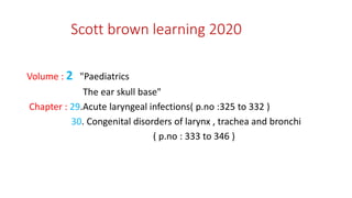
  • 1. Scott brown learning 2020 Volume : 2 "Paediatrics The ear skull base" Chapter : 29.Acute laryngeal infections( p.no :325 to 332 ) 30. Congenital disorders of larynx , trachea and bronchi ( p.no : 333 to 346 )
  • 2. Acute laryngeal infections & Congenital disorders of larynx , trachea and bronchi Dr. Vinoth .S , postgraduate in department of ENT , Madurai medical college
  • 3. Acute laryngeal infections – croup ( ALTB) • Croup typically presents as hoarseness with distinctive barking cough progressing to inspiratory or biphasic stridor. • usually a preceding history of upper respiratory tract symptoms, malaise and pyrexia. • Children with croup do not typically drool or appear toxic, in contrast to children presenting with epiglottitis. • The symptoms are thought to be due to mucosal oedema of the larynx and trachea following a viral illness.
  • 4. caused by parainfluenza virus type I , parainfluenza virus type II, respiratory syncytial virus (RSV) virus types A and B and rhinovirus. • The diagnosis of croup is a clinical one and laboratorytests and radiological tests are generally not needed • Croup is usually self-limiting , is rare • critical symptom of stridor is due to oedema in the subglottis, the narrowest part of the paediatric
  • 5. Chest x-ray : • radiograph of the thoracic inlet will show characteristic narrowing of the subglottis on an anteroposterior view(‘steeple’ or ‘pencil-tip’ sign)
  • 6. Westley Croup Score : A maximum score is 17, a score of 2–3 equates to mild croup 4–7 to moderate croup and 8 or more to severe croup
  • 7. Treatment • For mild croup, can be supportive humidified environment or mist tent Medical - Corticosteroids • The mainstay of treatment for croup is corticosteroids. • Corticosteroids have a systemic anti-inflammatory effect. • There is a reduction in capillary endothelial permeability and therefore in mucosal oedema, and stabilization of lysosomal membranes, decreasing the inflammatory reaction. • Topical corticosteroids have alpha-mediated vasoconstriction.
  • 8. Nebulized epinephrine • Nebulized epinephrine (1 mL of 1 in 1000 epinephrine diluted in 3 mL of 0.9% saline) has an established role in the acute paediatric airway in reducing mucosal oedema by an alpha-agonist effect causing vasoconstriction and bronchodilation; • a maximum effect is achieved within 30–60 minutes but there is no lasting benefit beyond 2 hours.
  • 9. Heliox • HelioxR is a mixture of helium and oxygen generally supplied with a helium : oxygen ratio of 70 : 30 • produce laminar flow in the presence of a partially obstructed airway than air or oxygen. • Inhaled helioxR delivered through a high-flow system should reduce the work of breathing and result in larger tidal volumes and improved gas exchange. • Helium has a high thermal conductivity and so care is needed with humidification in order to avoid hypothermia
  • 10. BACTERIAL LARYNGOTRACHEOBRONCHITIS also known as • PSEUDOMEMBRANOUS CROUP, • BACTERIAL TRACHEITIS, • MEMBRANOUS LARYNGOTRACHEOBRONCHITIS, • NEONATAL NECROTIZING TRACHEOBRONCHITIS
  • 11. • a severe form of laryngotracheobronchitis characterized by the presence of profuse mucopurulent secretions with sloughing of the respiratory epithelium. • Secretions are often adherent, are not effectively cleared by coughing and may occlude the airway causing respiratory compromise. • Bacterial tracheitis must be differentiated from croup and other causes of infectious upper airway obstruction in order to initiate proper management.
  • 12. • Classically the child with bacterial tracheitis appears toxic with high fevers and worsening stridor that fails to respond to treatment with steroids and nebulized epinephrine. • It typically affects children older (mean age 4–8 years) than the usual age group for croup. • Bacterial tracheitis is a much less common condition than croup. • However, there is increased susceptibility in children with Down syndrome or immunodeficiency
  • 13. • Diagnosis can only be confirmed on airway endoscopy; there is a pseudomembrane in the subglottis and trachea and thick mucopus and debris extending into the bronchi • Direct laryngotracheobronchoscopy under general anaesthesia and removal of all tracheal secretions, with pulmonary toilet, is mandatory. • to secure the airway by endotracheal intubation for a period of days.
  • 14. • Staphylococcus aureus( is the pathogen most commonly isolated from tracheal cultures) Haemophilus influenzae, Moraxella catarrhalis, Streptococcus pneumoniae and Pseudomonas aeruginosa • Viral cultures are also frequently positive, indicative that many cases of bacterial tracheitis represent a secondary bacterial infection that follows a viral respiratory tract infection. • Most commonly influenza A is identified, although other viruses including the H1N1 strain of influenza A and metapneumovirus have also been isolated.2,
  • 15. Complications include • airway stenosis, • acute respiratory distress syndrome, • respiratory failure, • toxic shock syndrome, • anoxic encephalopathy and • death.
  • 16. Diphtheria • Diphtheria remains an important differential in the diagnosis of acute laryngeal infection in children, particularly in those who have travelled to endemic areas or have not been vaccinated • The causative organisms in pathogenic diphtheria are the toxogenic strains of Corynebacterium diphtheria and Corynebacterium ulcerans. • The early clinical picture of upper respiratory tract symptoms is due to the effects of the organism itself. • Delayed effects are due to the release of exotoxin.
  • 17. • Symptoms of diphtheria come on gradually and typically begin 2–5 days after exposure. • Initial symptoms are of pharyngitis with sore throat and malaise. • The child is feverish and on examination there is a typical appearance of the pharyngeal tonsils with necrosis and the development of a characteristic grey pseudomembrane over the surface. • This consists of necrotic tissue, bacteria and a rich fibrinous exudate. • Early removal causes bleeding but the pseudomembrane may separate more easily in the later course of the disease. • There may be a bull-neck appearance due to cellulitis and regional lymphadenopathy
  • 18. • Diphtheria infection may also involve the larynx. • After initial symptoms of dysphagia and toxaemia, symptoms may progress to inspiratory stridor and a barking cough; • the cough is frequently paroxysmal and exhausting. • Death may follow owing to acute airway obstruction or as a result of the later effects of the exotoxin. • The exotoxin can cause a toxic myocarditis in the second week of the disease and this may be fatal. • Peripheral neuritis may also occur, palatal paralysis being the most common effect of peripheral neuropathy and presenting with nasal regurgitation of food and hypernasal speech.
  • 19. • Successful treatment depends on early diagnosis, and the timely administration of high-dose benzylpenicillin and antitoxin (10 000–100 000 units, depending on the severity of infection). • Delaying the administration of the antitoxin is associated with increased mortality as the antitoxin does not neutralize toxin that is already bound to tissues. • The decision, therefore, to administer antitoxin should be made based on clinical suspicion and should not await microbiological confirmation. • Airway management consists of removal of the laryngeal membrane, administration of oxygen and humidification, and endotracheal intubation or tracheostomy if necessary. • Systemic steroids may reduce the need for airway intervention. • Bed rest is recommended until the danger of myocarditis is past.
  • 20. It is of a toxic child with a short history of sore throat, inspiratory stridor, muffled voice and drooling due to odynophagia and dysphagia. Untreated results in progressive respiratory distress. The child is febrile, tachypnoeic and classically will be sitting upright, with the neck extended to optimize the airway, and using then arms to provide support to the shoulder girdle to maximize the efficiency of the accessory muscles of respiration. It is commonest between the ages of 2 and 8 and there is an increased prevalence in winter
  • 21. Acute epiglottitis • When acute epiglottitis is suspected, pharyngeal examination should not be attempted, as simple manipulation with a tongue depressor may precipitate acute airway obstruction, • although the use of a fibreoptic or small rigid endoscope can assist the diagnosis in patients with an atypical presentation. • Direct examination of the airway should not be delayed, but should be undertaken in a controlled setting such as an operating room or paediatric intensive care unit by personnel skilled in airway intervention;
  • 22. • endoscopic evaluation will confirm gross erythema and oedema of the supraglottic structures • When the diagnosis is confirmed, the airway should be secured by endotracheal intubation • a soft-tissue lateral radiograph of the neck will typically show a thickened oedematous epiglottis – the ‘thumb sign’
  • 23. Etiology : • acute epiglottitis has been a manifestation of invasive Hib infection • meningococci, • group A streptococci, pneumococci, Haemophilus parainfluenzae and Staphylococcus aureus. • Immunocompromised individuals are at increased risk of epiglottitis. due to atypical organisms such as Herpes simplex type 1, Varicella zoster, Parainfluenza or Candida albicans.
  • 24. Treatment : • Treatment is with intravenous antibiotics; ampicillin resistance due to beta- lactamase production is now over 50% in Haemophilus influenzae, so empirical treatment with third-generation cephalosporins for 5–7 days is advised. • Rifampicin prophylaxis has been recommended to eradicate the carrier state for the index case as well as household and school contacts. • Haemophilus influenzae type b immunization
  • 25. AIRWAY MANAGEMENT • Endotracheal intubation is now considered preferable to tracheostomy • If endotracheal intubation is to be undertaken, it should be performed in an operating room or paediatric intensive care unit with personnel and equipment available and ready to undertake tracheostomy if the airway cannot be secured. • humidified air should be administered to discourage the development of tenacious secretions
  • 26. Nasotracheal intubation • When a prolonged period of intubation is anticipated, the nasotracheal rather than the orotracheal route is preferred • In acute epiglottitis, visualization of the epiglottis with a flexible nasendoscope can be useful in monitoring resolution of inflammation. • in laryngotracheobronchitis, whether viral or bacterial, the airway can be monitored using endoscopy until a reduction in tracheal inflammation shows that it is safe to extubate.
  • 27. • Systemic steroids administered 6 hours prior to removal of the tube will minimize post-intubation oedema, and nebulized epinephrine can assist with airway patency after extubation. • For acute epiglottitis the period of intubation is usually less than 48 hours but for croup and bacterial laryngotracheobronchitis it may be up to a week. • Tracheostomy is no longer considered the first choice to secure the airway in acute laryngeal infection.
  • 29. Congenital conditions of larynx -Laryngomalacia • Laryngomalacia is characterized by partial or complete collapse of the supraglottic structures on inspiration • is the most common congenital cause of stridor • Signs- the epiglottis is long and curled (omega-shaped) the aryepiglottic folds are short and tightly tethered to the epiglottis; there may also be redundant mucosa and submucosa of the aryepiglottic folds medially resulted in a tall, narrow supraglottis with a deep interarytenoid cleft
  • 30. - epiglottis is soft and may curl and collapse and the mucosa may prolapse into the airway . - due to neuromuscular immaturity and consequent incoordination of arytenoid movements
  • 31. • high-pitched, fluttering inspiratory stridor is usually present at, or shortly after, birth , increases during activity and decreases at sleep The severity of the stridor increases during the first 9 months of life, and disappears at two years of age , rarely may persist into late childhood.
  • 32. • The diagnosis can be confirmed by flexible fibre-optic laryngoscopy • Treatment : • for mild, no intervention is needed and the parents can be reassured • In severe laryngomalacia, there is serious respiratory obstruction with substantial sternal and intercostal recession together with feeding difficulties results in failure to thrive • May associated with congenital cyanotic heart disease
  • 33. • Children who show signs of failure to thrive should undergo an endoscopic aryepiglottoplasty (sometimes termed a supraglottoplasty) • In this procedure, each aryepiglottic fold is first divided to release it from the edge of the epiglottis, and any redundant mucosa and submucosal tissue are then excised from over the arytenoids, together, if necessary, with part or all of the cuneiform cartilages. • The ‘bridge’ of mucosa between the arytenoids is carefully preserved to prevent interarytenoid scarring.
  • 34. Vocal cord paralysis • Vocal cord paralysis is the second most common congenital anomaly of the larynx after laryngomalacia. • patients may have other, coexisting airway pathology • Diagnosed by flexible fibre-optic laryngoscopy Laryngeal ultrasound
  • 35. • Unilateral vocal cord paralysis is usually not congenital, most cases being acquired as a result of surgical injury to the left recurrent laryngeal nerve, often following correction of a congenital cardiac anomaly. • Patients present with mild stridor, dysphonia and sometimes aspiration. • If the vocal cord lies in an intermediate position, surgical intervention is not usually necessary, and the voice can be expected to improveby compensation. • If the vocal cord lies in a more abducted position, the dysphonia may be more pronounced and aspiration more likely. • Vocal cord medialization procedures such as thyroplasty and augmentation injection can improve the dysphonia and aspiration, but these may result in worsening of the stridor.
  • 36. • Bilateral vocal cord palsy is usually a congenital abductor paralysis. • The vocal cords lie in the paramedian position with consequent inspiratory stridor, • and a tracheostomy is necessary for airway management • Presented with the Arnold-Chiari malformation • If it have no movement ,then an endoscopic laser cordotomy or arytenoidectomy should be considered at the age of 11
  • 37. Laryngocoeles : • Laryngeal cysts divided into laryngocoeles and saccular cysts. • laryngocoele is an air-filled dilatation of the laryngeal ventricle which communicates with the laryngeal lumen. • uncommon lesion which usually occurs in middle age but may rarely be seen in infancy, • can produce respiratory distress which typically becomes worse on crying due to increased distension of the laryngocoele with air. • a laryngocoele may obstruct and fill with mucus or become infected (laryngopyocoele), thus becoming indistinguishable from a saccular cyst
  • 38. • A also represents an abnormal dilatation or herniation of the saccule of the ventricle of the larynx; • it differs from a laryngocoele in that there is no opening into the larynx and it is filled with mucus instead of air. • result from a developmental failure to maintain patency of the orifice between the saccule and the ventricle, and may be of anterior or lateral type. Saccular cyst
  • 39. The anterior saccular cyst extends medially and posteriorly from the saccule and so protrudes into the laryngeal airway between the true and false vocal cords. The lateral saccular cyst is most common in infants and expands posterosuperiorly into the false cord and aryepiglottic fold
  • 40. • Laryngeal cysts are classified as internal or type 1 if contained entirely within the laryngeal framework, and external or type 2 if it pierces the thyrohyoid membrane. • Diagnosis is confirmed by endoscopy, except in an external laryngocoele where no abnormality may be seen except on imaging. • Saccular cysts are best treated at the initial endoscopy by wide endoscopic marsupialization • If the cyst recurs, then the procedure of choice is a lateral cervical approach extending through the thyrohyoid membrane at the superior margin of the ala of the thyroid cartilage, with subperichondrial resection of a portion of the upper part of the ala. Through this ‘window’ the cyst can be completely excised, using short-term intubation to secure the airway post-operatively.
  • 41. Vascular malformations • include lymphatic malformations, venous malformations, av malformations • Lymphatic malformations (also termed lymphagiomas or cystic hygromas) are cystic malformations that result from abnormal development of the lymphatic vessels. • In the head neck they may be macrocystic (usually infrahyoid), microcystic (usually suprahyoid) or a combination of the two. • Occasionally, a microcystic lymphatic malformation may extend into the tongue base, valleculae and supraglottis, and airway obstruction may result. • If the lymphatic malformation is very extensive, a tracheostomy may be required, • Surgical modality is to debulk the lesion by endoscopic vaporization using a CO2 laser or radiofrequency ablation.
  • 42. Bifid epiglottis • is a rare laryngeal anomaly in which the epiglottis fails to fuse in the midline and thus has a cleft extending down to its tubercle. • It may be seen as a feature of Pallister–Hall syndrome • presents with feeding difficulties due to aspiration and with stridor because of collapse and enfolding of the two halves of the epiglottis. • Endoscopy establishes the diagnosis, • treatment options include amputation of the epiglottis and tracheostomy.
  • 43. Glottis - Laryngeal webs • Failure of complete canalization of the larynx during embryogenesis may result in a glottic or, very rarely, a supraglottic web. • Mostly involves anterior glottis , associated with congenital vocal cord paralysis . • Due to delayed maturation in the vagal nuclei • glottic enlargement surgery in order to avoid a tracheostomy • an endoscopic laser cordotomy or arytenoidectomy if presented with stridor
  • 44. Laryngeal atresia • Laryngeal atresia is incompatible with life unless there is an associated tracheo-oesophageal fistula (TOF) • emergency tracheostomy is performed in the delivery room. • now recognized antenatally on ultrasound imaging and managed with ex utero intrapartum treatment (EXIT) procedure, • whereby a tracheostomy is undertaken following elective Caesarean section with the neonate still on placental circulation.
  • 45. Congenital subglottic stenosis • due to defective canalization of the cricoid cartilage and/or conus elasticus, resulting in either gross thickening of the anterior lamina of the abnormal cricoid • a small, elliptical, thickened cricoid with excessive submucosal soft tissue • there may be anterior fusion of the vocal cords, forming a web with subglottic extension, as seen in 22q11 deletion syndromes. • Subglottic stenosis is said to be the third most common congenital anomaly of the larynx • may be a combined congenital plus acquired stenosis for intubation
  • 46. The Myer-Cotton grading system • Grade I : 0–50% obstruction • • Grade II: 51–70% obstruction • • Grade III: 71–99% obstruction • • Grade IV: 100% obstruction. • If the airway is not severely compromised, surgery may not be required • a strict contraindication to dilatation or laser resection: • any type of endoscopic treatment is liable to worsen the initial condition, and attempted dilatation is inevitably ineffective as the thickened ring of cricoid cartilage cannot be expanded
  • 47. Milder degrees of stenosis present as inspiratory or biphasic stridor as the child becomes older and more active, or as recurrent ‘croup’ owing to superimposed oedema from upper respiratory tract infections
  • 48. • If the airway is severely compromised, a tracheostomy is needed. • The surgical options have evolved from the classical castellated laryngotracheoplasty designed by Evans and Todd • The LTR involves augmentation of the laryngotracheal complex by anterior and/or posterior midline incision of the cricoid with insertion of costal cartilage grafts to expand the airway
  • 49. Treatment : • Grade I subglottic stenosis usually requires no surgical intervention. • Grade II stenosis can be reconstructed by means of an LTR with anterior cartilage grafting +/− posterior cricoid split. • Mild grade III stenosis needs an anterior graft with posterior cricoid split +/− posterior cartilage grafting. • Severe grade III stenosis (a pinhole airway) requires both anterior and posterior grafts. • Grade IV stenosis demands anterior and posterior grafts with prolonged stenting
  • 50. Subglottic haemangioma • Infantile subglottic haemangioma causes gradually worsening inspiratory or biphasic stridor presenting in the first few weeks of life • Associated with cutaneous haemangioma • The natural history is typically a proliferative phase lasting 6–12 months followed by complete involution over 1–5 years • At endoscopy the typical appearance is of a compressible, pear- shaped red swelling in the subglottis on one side, left more commonly than right
  • 51. • Subglottic haemangioma is life-threatening because of its situation in the narrowest part of the airway and therefore requires rapid intervention. • Tracheostomy will maintain the airway until involution • radiotherapy, CO2 laser ablation, systemic steroids, intralesional steroid injection followed by intubation, and interferon alpha-2a
  • 52. • propranolol is an effective treatment for cutaneous haemangioma and subglottic haemangioma • Submucous resection remains a valid alternative for non- circumferential haemangiomas and those not involving the vocal cords
  • 53. Laryngeal and laryngotracheooesophageal cleft • Posterior laryngeal clefts result from failure of the posterior cricoid lamina to fuse with incomplete development of the tracheo- oesophageal septum. • Benjamin and Inglis classification - type I cleft extends down to the level ofthe vocal cords; -type II cleft extends below the vocal cords into the cricoid - type III cleft extends down into the cervical trachea -type IV cleft extends into the thoracic trachea and may even reach the carina • With Opitz-Frias syndrome, TOF, Pallister–Hall syndrome
  • 54. Symptoms : • Type I clefts present with cyanotic attacks on feeding and recurrent chest infections • Type II and III clefts produce dramatic aspiration with recurrent pneumonia, sometimes with stridor and an abnormal cry. • Type IV clefts cause severe aspiration, cyanosis and incipient cardiorespiratory failure. • Suspension microlaryngoscopy to probe the arytenoids • A plain chest X-ray may show changes secondary to recurrent aspiration pneumonitis • Videofluoroscopic contrast swallow studies may not differentiate laryngeal incompetence from neuromuscular incoordination
  • 55. Treatment : • A short type I cleft with no aspiration requires no treatment. Minimal aspiration may be managed by thickening the feeds • A very short type II cleft may also be repaired endoscopically, • a long type II or a type III cleft needs to be approached anteriorly through an extended laryngofissure with a low tracheostomy • type IV clefts may be managed by an anterior approach through a cervical incision, if necessary pulling the trachea up into the neck to reach the lower end of the cleft. • Longer clefts will require a lateral cervical approach in combination with a thoracotomy, or preferably an anterior cervicothoracic approach via a median sternotomy with repair on extracorporeal membrane oxygenation (ECMO) or cardiopulmonary bypass.
  • 56. • Mortality remains significant, being approximately 14% overall 37 rising to 66% for type IV laryngotracheooesophageal clefts and up to 100% for full-length clefts ending at the carina
  • 57. Agenesis • Tracheal agenesis may be complete (full-length) or partial, but in either case there is no continuity between the larynx and the bronchi • Occasionally, short-term survival may be possible if there is a broncho-oesophageal fistula, which can permit some airflow into the lungs, but surgical efforts to use the oesophagus as a tracheal replacement have not been successful and long-term survival has not proved possible
  • 58. Stenosis • Occasionally, a membranous web may be encountered in the trachea with a normal underlying cartilaginous ring structure. Such cases usually fare well with endoscopic rupture and dilatation. • Thicker congenital fibrous stenoses may be amenable to radial KTP laser incision and balloon dilatation, followed by application of mitomycin C to reduce the risk of restenosis42 or tracheal resection with end-to-end anastomosis. • A short stenosis may also be associated with one or two absent tracheal rings, although the most common finding in congenital tracheal stenosisis (CTS) is a segment of complete tracheal cartilaginous rings with an airway lumen as narrow as 2 mm
  • 59. • Children with congenital tracheal stenosis usually present with respiratory distress • Symptoms may become apparent in the neonatal period with biphasic stridor, respiratory distress, tracheal tug and episodes of cyanosis in the first year of life • Endoscopy remains the ‘gold standard’ investigation for a child with stridor
  • 60. • Bronchoscopy and bronchography (B&B) demonstrate the the size of the tracheal lumen and outline the trachea and bronchi distal to the stenosis, which may be too narrow to permit passage of a bronchoscope and also provides a dynamic assessment of any malacic segments. • Optical coherence tomography (OCT) can be combined with B&B to confirm the presence of complete rings if there is any doubt. • Contrast CT and an echocardiogram are essential in view of the high incidence of associated anomalies of the heart and great vessels. • CT is particularly helpful in assessing the vascular anatomy and its relationship to the trachea and 3D reconstructions allow better surgical planning
  • 61. • Mild cases with minimal symptoms may require no intervention after the diagnostic endoscopy, • Very narrow, short segments can be excised with end-to-end anastomosis , Augmentation tracheoplasty with costal cartilage grafting or a pericardial flap or free patch can be tried • Successful endoscopic balloon dilatation • Absorbable PDS stents offer an advantage over metallic stents and are used to treat malacia, particularly bronchomalacia
  • 62. Tracheomalacia and bronchomalacia • Tracheomalacia is a condition in which there is reduced stiffness of the tracheal wall, resulting in abnormal collapse of the trachea during expiration • produce symptoms of airway obstruction • Pathologically, the striking finding is an increased muscle- to-cartilage ratio seen on the transverse section of the trachea
  • 63. • Tracheomalacia is traditionally classified as primary (idiopathic), due to an intrinsic abnormality in the wall of the airway, or secondary, due to another associated anomaly or to external compression. • high-pitched expiratory noise stridor becomes apparent during the first few weeks of life with a harsh, barking cough • Characteristically, the stridor becomes much worse when the child is active, feeding, upset, coughing or crying, • it may be associated with cyanotic attacks which are sometimes sufficiently severe to be termed ‘dying spells’.
  • 64. • There may be a prolonged expiratory phase to respiration, possibly with faint expiratory stridor • A barium swallow may identify a TOF and can also demonstrate the airway collapse on lateral screening • CT angiogram is extremely useful for identifying the cause of extrinsic compression of the trachea by normal or abnormal vessels, including vascular rings. • Contrast bronchography may be the most useful radiological investigation:
  • 65. Treatment : • Mild tracheobronchomalacia (less than 75% collapse) requires no intervention, and the stridor can be expected to resolve spontaneously by around the age of 2 years. • If it is secondary to a vascular anomaly, this should be corrected an extended tracheostomy tube will effectively support a midtracheal malacic segment, but this is not a satisfactory solution for lower-end tracheal or for bronchial collapse include internal or external stenting of the trachea, segmental resection and cartilage grafting
  • 66. Anomalous bifurcations • The right upper lobe bronchus may take origin from the right lateral wall of the trachea above the carina • is termed a tracheal bronchus or suis bronchus(PIG BRONCHUS) • asymptomatic, incidental finding, but may sometimes be associated with tracheal stenosis
  • 67. Tracheo-oesophageal fistula • It is a fairly common congenital malformation of the neonatal air and food passages, which usually occurs in association with oesophageal atresia. • Eighty-seven per cent of cases have oesophageal atresia with a TOF communicating between the distal oesophagus and the mid- to lower trachea or a main bronchus • m/c present to intensivist TOF without atresia (‘H-type fistula’) the least symptomatic of the group, with no swallowing difficulty, but small amounts of fluid pass through the fistula into the trachea and produce symptoms and signs of recurrent minor aspiration. barium swallow confirms the diagnosis treatment by ligation and division of fistula
  • 68. Vascular compressions • Vascular ring : • common is double aortic arch mostly involving ascending aorta • Presents earlier in life with airway obstructions • Barium swallow shows double impression upon the column of contrast • Surgical treatment involves diving the lesser component of the ring • Post op tracheomalacia will persists for few monthds
  • 69. • Vascular sling : • Common is aberrant innominate artery crosses the anterior surface of trachea above the carina • Presents with less severe airway obstruction • Expiratory stridor , cough ,recurrent URTI • Bronchoscopy shows sloping pulsatile compression of the trachea 2 cm above the carina • Upward compression with bronchoscopes compress the artery against the sternum thus obliterating the radial pulse • Treatment by aortopexy