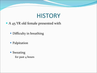
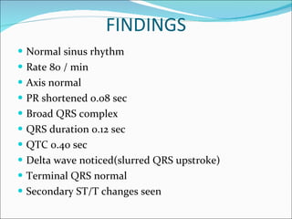
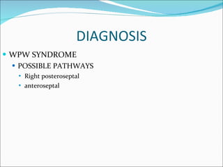
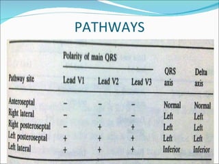
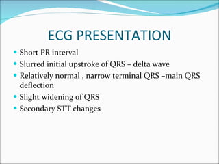
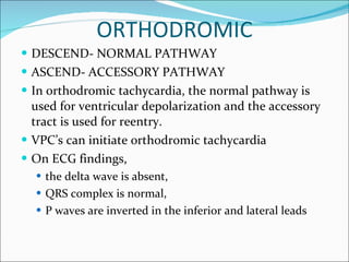
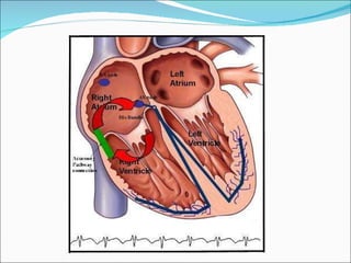
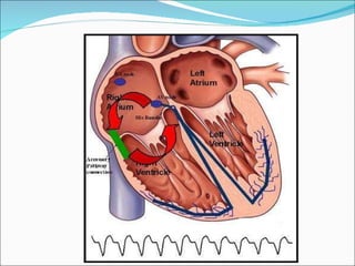
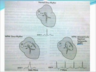
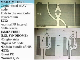
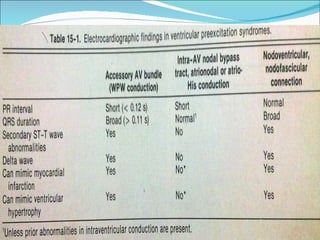
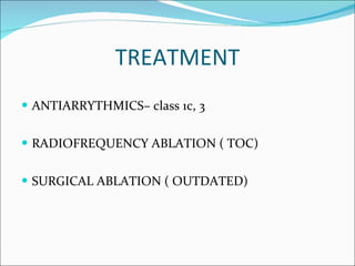
PROF .Dr G.SUNDARAMURTY ‘S UNIT M6 S.DHANRAJ Ist YEAR PG
HISTORY A 45 YR old female presented with Difficulty in breathing Palpitation Sweating for past 4 hours
ECG
FINDINGS Normal sinus rhythm Rate 80 / min Axis normal PR shortened 0.08 sec Broad QRS complex QRS duration 0.12 sec QTC 0.40 sec Delta wave noticed(slurred QRS upstroke) Terminal QRS normal Secondary ST/T changes seen
DIAGNOSIS WPW SYNDROME POSSIBLE PATHWAYS Right posteroseptal anteroseptal
PATHWAYS
HISTORY Named after  three scientists  WOLFF  PARKINSON WHITE In the year 1930
DEFINITION WPW is a electrocardiographic syndrome it is the expression of anomalous atrio ventricular  conduction pathway congenital in origin This pathway forms a bypass which enables supraventricular impulse to bypass AV node , bundle of HIS and distal conducting system and so activate or pre exite the ventricles  This anomalous bypass, most commonly bundle of kent situated any where along AV node
ECG PRESENTATION Short PR interval Slurred initial upstroke of QRS – delta wave Relatively normal , narrow terminal QRS –main QRS deflection Slight widening of QRS  Secondary STT changes
CARDIAC ACTIVATION PHASE 1  Atrial activation- normal  PHASE 2: Ventricular pre-exitation sinus activation occurs through both normal , anomalous pathway anomalous pathway lacks AV nodal conduction delay so  sinus impulse conducted at a rapid rate this enables ventricles to be activated or pre exited- short PR interval , delta wave Further activation through normal pathway PHASE 3: Narrow terminal QRS
 
OLD CLASSIFICATION Type A In this type of WPW syndrome, the delta wave and QRS complex are predominantly upright in the precordial leads. The dominant R wave in lead V1 may be misinterpreted as right bundle branch block. Type B The delta wave and QRS complex are predominantly negative in leads V1 and V2 and positive in the other precordial leads, resembling left bundle branch block.
PATHWAY
 
ORTHODROMIC DESCEND- NORMAL PATHWAY ASCEND- ACCESSORY PATHWAY In orthodromic tachycardia, the normal pathway is used for ventricular depolarization and the accessory tract is used for reentry.  VPC’s can initiate orthodromic tachycardia On ECG findings,  the delta wave is absent,  QRS complex is normal,  P waves are inverted in the inferior and lateral leads
 
ANTIDROMIC LESS COMMON PATHWAY. DESCEND- ACCESSORY PATHWAY. ASCEND – NORMAL PATHWAY On ECG findings,  the QRS is wide, which is an exaggeration of the delta wave during sinus rhythm (ie, wide-QRS tachycardia).  Such tachycardias are difficult to differentiate from ventricular tachycardia
 
 
PATHWAYS
FIBRES KENT PATHWAY : ATRIO-VENTRICULAR JAMES PATHWAY : ATRIO-HIS MAHAIM PATHWAY: HISO- VENTRICULAR
MAHAIM FIBRE: Orgin- distal to AV node Ends in the venricular myocardium ECG : normal PR interval delta waves JAMES FIBRE (LGL SYNDROME) Origin- atria Bypass AV node Ends in bundle of HIS ECG : Short PR Normal QRS
 
COMPLICATION Tachyarrhythmia Syncopal attacks Sudden cardiac death Complications of drug therapy (eg, proarrhythmia, organ toxicity) Complications associated with invasive procedures and surgery Recurrence
TREATMENT ANTIARRYTHMICS– class 1c, 3 RADIOFREQUENCY ABLATION ( TOC) SURGICAL ABLATION ( OUTDATED)
CAUTION Usual presentation is SVT Sudden cadiac death possible Digoxin, beta blockers,verapamil are contraindicated Underlying Ebstein’s anomaly, hypertrophic cardiomyopathy should be evaluated
THANK YOU

ECG: WPW Syndrome

  • 1.
    PROF .Dr G.SUNDARAMURTY‘S UNIT M6 S.DHANRAJ Ist YEAR PG
  • 2.
    HISTORY A 45YR old female presented with Difficulty in breathing Palpitation Sweating for past 4 hours
  • 3.
  • 4.
    FINDINGS Normal sinusrhythm Rate 80 / min Axis normal PR shortened 0.08 sec Broad QRS complex QRS duration 0.12 sec QTC 0.40 sec Delta wave noticed(slurred QRS upstroke) Terminal QRS normal Secondary ST/T changes seen
  • 5.
    DIAGNOSIS WPW SYNDROMEPOSSIBLE PATHWAYS Right posteroseptal anteroseptal
  • 6.
  • 7.
    HISTORY Named after three scientists WOLFF PARKINSON WHITE In the year 1930
  • 8.
    DEFINITION WPW isa electrocardiographic syndrome it is the expression of anomalous atrio ventricular conduction pathway congenital in origin This pathway forms a bypass which enables supraventricular impulse to bypass AV node , bundle of HIS and distal conducting system and so activate or pre exite the ventricles This anomalous bypass, most commonly bundle of kent situated any where along AV node
  • 9.
    ECG PRESENTATION ShortPR interval Slurred initial upstroke of QRS – delta wave Relatively normal , narrow terminal QRS –main QRS deflection Slight widening of QRS Secondary STT changes
  • 10.
    CARDIAC ACTIVATION PHASE1 Atrial activation- normal PHASE 2: Ventricular pre-exitation sinus activation occurs through both normal , anomalous pathway anomalous pathway lacks AV nodal conduction delay so sinus impulse conducted at a rapid rate this enables ventricles to be activated or pre exited- short PR interval , delta wave Further activation through normal pathway PHASE 3: Narrow terminal QRS
  • 11.
  • 12.
    OLD CLASSIFICATION TypeA In this type of WPW syndrome, the delta wave and QRS complex are predominantly upright in the precordial leads. The dominant R wave in lead V1 may be misinterpreted as right bundle branch block. Type B The delta wave and QRS complex are predominantly negative in leads V1 and V2 and positive in the other precordial leads, resembling left bundle branch block.
  • 13.
  • 14.
  • 15.
    ORTHODROMIC DESCEND- NORMALPATHWAY ASCEND- ACCESSORY PATHWAY In orthodromic tachycardia, the normal pathway is used for ventricular depolarization and the accessory tract is used for reentry. VPC’s can initiate orthodromic tachycardia On ECG findings, the delta wave is absent, QRS complex is normal, P waves are inverted in the inferior and lateral leads
  • 16.
  • 17.
    ANTIDROMIC LESS COMMONPATHWAY. DESCEND- ACCESSORY PATHWAY. ASCEND – NORMAL PATHWAY On ECG findings, the QRS is wide, which is an exaggeration of the delta wave during sinus rhythm (ie, wide-QRS tachycardia). Such tachycardias are difficult to differentiate from ventricular tachycardia
  • 18.
  • 19.
  • 20.
  • 21.
    FIBRES KENT PATHWAY: ATRIO-VENTRICULAR JAMES PATHWAY : ATRIO-HIS MAHAIM PATHWAY: HISO- VENTRICULAR
  • 22.
    MAHAIM FIBRE: Orgin-distal to AV node Ends in the venricular myocardium ECG : normal PR interval delta waves JAMES FIBRE (LGL SYNDROME) Origin- atria Bypass AV node Ends in bundle of HIS ECG : Short PR Normal QRS
  • 23.
  • 24.
    COMPLICATION Tachyarrhythmia Syncopalattacks Sudden cardiac death Complications of drug therapy (eg, proarrhythmia, organ toxicity) Complications associated with invasive procedures and surgery Recurrence
  • 25.
    TREATMENT ANTIARRYTHMICS– class1c, 3 RADIOFREQUENCY ABLATION ( TOC) SURGICAL ABLATION ( OUTDATED)
  • 26.
    CAUTION Usual presentationis SVT Sudden cadiac death possible Digoxin, beta blockers,verapamil are contraindicated Underlying Ebstein’s anomaly, hypertrophic cardiomyopathy should be evaluated
  • 27.