2. Definition
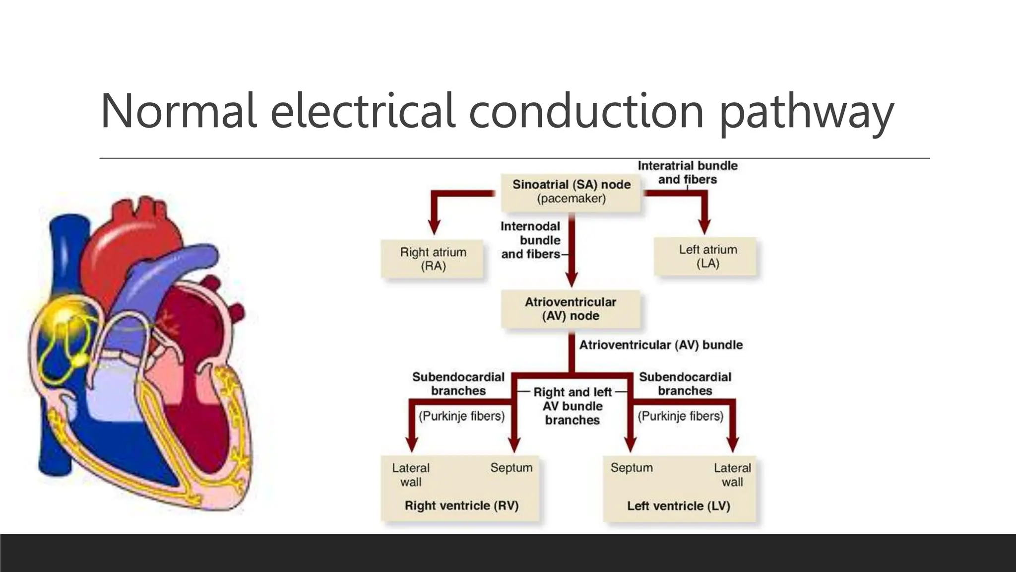
Ventricular preexcitation syndrome refers to abnormal accessory
pathways in the heart's electrical conduction system. These pathways
create extra routes for electrical impulses, bypassing the normal pathway.
This leads to premature activation of the ventricles and an altered ECG
pattern. It can cause symptoms like palpitations and irregular heartbeat.
Common forms include Wolff-Parkinson-White syndrome,Lown-Ganong-
Levine syndrome and Mahaim syndrome
3. Prevalence and incidence
The prevalence and incidence of ventricular preexcitation syndrome, specifically referring to Wolff-Parkinson-
White (WPW) syndrome, can vary across different populations. However, estimates suggest that WPW
syndrome is relatively rare, with a prevalence ranging from 0.1% to 0.3% in the general population.
The incidence of WPW syndrome, referring to new cases occurring within a specific time period, is estimated to
be around 1.5 to 3 cases per 1,000 person-years. This incidence rate can also vary based on factors such as age,
gender, and underlying cardiac conditions.
It is worth noting that Lown-Ganong-Levine syndrome, another form of ventricular preexcitation, is less
common than WPW syndrome, and its prevalence and incidence are even more infrequent compared to WPW
syndrome.
5. Accessory pathways
•They are also known as bypass tracts
•Abnormal conduction pathways formed during cardiac development
•Impulse conduction:
a) Anterograde (Normal forward propagation of electrical impulses through the
heart's conduction system A->V)
b) Retrograde (Backward propagation of electrical impulses within the heart's
conduction system V->A)
c) Both Directions
7. Underlying causes and risk factors
1.Congenital: Ventricular preexcitation syndromes are often congenital, meaning they are present at
birth. Abnormal development of the electrical conduction system during fetal development can lead to
the formation of accessory pathways.
2.Structural Heart Abnormalities: Certain structural heart abnormalities, such as Ebstein's anomaly or
hypertrophic cardiomyopathy, may increase the likelihood of developing ventricular preexcitation
syndrome.
3.Genetic Factors: There is evidence of a genetic predisposition to ventricular preexcitation syndromes.
Mutations in certain genes involved in cardiac conduction system development and function, such as
PRKAG2 and SCN5A, have been linked to an increased risk of developing WPW syndrome.
4.Age and Gender: Ventricular preexcitation syndromes, especially WPW syndrome, tend to be more
common in younger individuals. The condition may present earlier in life but can be present across all
age groups. In terms of gender, WPW syndrome is slightly more common in males than females.
5.Other Cardiac Conditions: Certain cardiac conditions, such as mitral valve prolapse, atrial septal
defect, or coronary artery disease, may be associated with an increased risk of ventricular preexcitation
syndrome.
8. Classification
Type
Conduction
pathway
PR interval QRS Interval Delta wave
Wolff-Parkinson-
White
Bundle of Kent
(Atria to
ventricles)
Short Long Yes
Lown-Ganong-
Levine
James Bundle
(Atria to Bundle
of His)
Short Normal No
Mahaim Mahaim Fibres Normal Long Normal
9. Wolff-Parkinson-White syndrome
•Combination of the presence of congenital
accessory pathway and episodes of
tachyarrhythmia
•Bundle of Kent Facilitates wave Depolarization
into the ventricles that Bypass the AV node
•Can Lead to Rapidly conducted supraventricular
tachycardias and reentrant tachycardias
10. WPW ECG Features
•PR Interval <120ms
•Delta Wave
•QRS Prolongation (>110ms)
•ST Segment and T wave Discordant changes
•Pseudo-Infarction pattern is seen in up to 70% of patient
•WPW Types
1) Type A: +ve Delta wave in all precordial leads with R/S >1 in V1 (Left sided
accessory pathway)
2) Type B: -ve Delta wave in Leads V1 and V2 with dominant S in V1 (Right
sided accessory pathway)
15. Mahaim Type Preexitation syndrome
•Accessory pathway arising from the lateral right atrium
extending to the body of the RV(Right Bundle Branch)
•ECG Features
1. Wide QRS interval
2. Sinus Rhythm ECG may be Normal
3. May result in variation in Ventricular Morphology
4. Re entry Tachycardia typically has LBBB Morphology
17. Management and Treatment
•Observation: Asymptomatic patients with ventricular preexcitation may not require immediate
treatment. Regular monitoring and periodic follow-up visits with a cardiologist may be
recommended to assess symptom progression or the development of arrhythmias.
•Medications: Certain medications can help manage symptoms and prevent arrhythmias
associated with ventricular preexcitation. These may include antiarrhythmic drugs, such as
beta-blockers or calcium channel blockers, to slow down the heart rate and prevent abnormal
rhythms.
•Catheter Ablation: Catheter ablation is a commonly performed procedure for ventricular
preexcitation syndrome. It involves the use of a specialized catheter to identify and eliminate
the abnormal accessory pathway causing the preexcitation. Radiofrequency energy or
cryoablation is applied to destroy the accessory pathway and restore normal electrical
conduction
18. Management and Treatment
•Risk Stratification: In individuals at higher risk of developing arrhythmias or complications,
risk stratification is important. This involves assessing the risk based on factors such as the
presence of symptoms, characteristics of the accessory pathway, and other associated cardiac
conditions. High-risk individuals may require closer monitoring or consideration for catheter
ablation.
•Education and Lifestyle Modifications: Patient education plays a crucial role in the
management of ventricular preexcitation syndrome. Patients should be aware of their condition,
recognize symptoms of arrhythmias, and understand when to seek medical attention. Lifestyle
modifications, such as avoiding excessive caffeine, alcohol, or stimulants, may be
recommended to minimize triggers for arrhythmias.
•Emergency Preparedness: Individuals with ventricular preexcitation syndrome should be
educated about the signs and symptoms of serious arrhythmias and sudden cardiac arrest.
They may be advised to have an emergency plan in place, including knowing how to perform
cardiopulmonary resuscitation (CPR) and the availability of automatic external defibrillators