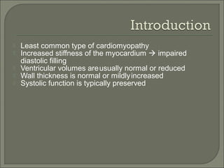
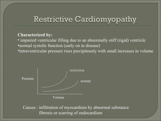
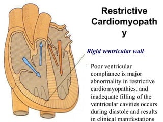
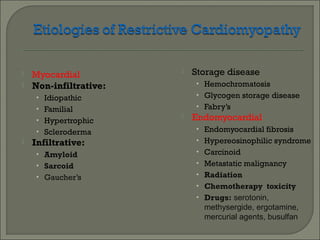
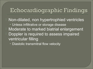
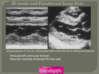
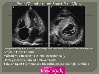
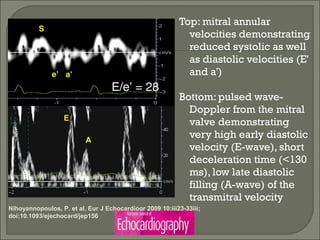
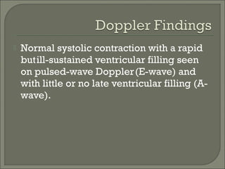
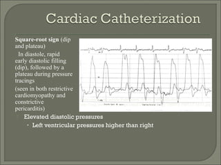
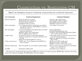
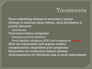
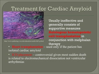
DR. MD. Saiful Islam
MD (cardiology) Final part student
Department of Cardiology
DMCH
Definition:
 It is a heterogenous group of disease of
myocardium,
 associated with mechanical or electrical
dysfunction,
 which is usually but not invariably exhibits
inappropriate ventricular hypertrophy or
dilation
 & are due to variety of etiology that
frequently are genetic.
 Primary
(those resulting from
genetic abnormalities of
cardiac muscle)
• Dilated
• Hypertrophic
• Restrictive
 Secondary
(those resulting from
infections, metabolic
and nutritional diseases,
endocrine disorders,
neuromuscular diseases,
blood diseases, tumors)
Br Heart J 1980; 44:672-673
Cardiomyopathies
WHO Classification
1. Dilated
• Enlarged
• Systolic dysfunction
2. Hypertrophic
• Thickened
• Diastolic dysfunction
3. Restrictive
• Diastolic dysfunction
3. Arrhythmogenic RV dysplasia
• Fibrofatty replacement
3. Unclassified
• Fibroelastosis
• LV noncompaction
Circ 93:841, 1996
Progressive fibro-fatty replacement of the
right ventricle
Arrhythmogenic right ventricular
cardiomyopathy
Restricted filling and reduced diastolic size
of either or both ventricles with normal or
near-normal systolic function.
Restrictive cardiomyopathy
Left and/or right ventricular hypertrophy,
often asymmetrical, which usually involves
the interventricular septum.
Hypertrophic cardiomyopathy
Dilatation and impaired contraction of the
left or both ventricles.
Dilated cardiomyopathy
 Introduction
 Symptoms
 Causes
 Myocardial
 Endomyocardial
 Diagnosis:
 ECG
 Echocardiography
 invasive hemodynamics
 cardiac MRI
 Treatment options
 Least common type of cardiomyopathy
 Increased stiffness of the myocardium  impaired
diastolic filling
 Ventricular volumes areusually normal or reduced
 Wall thickness is normal or mildlyincreased
 Systolic function is typically preserved
Characterized by:
• impaired ventricular filling due to an abnormally stiff (rigid) ventricle
•normal systolic function (early on in disease)
•intraventricular pressure rises precipitously with small increases in volume
Pressure
Volume
Causes : infiltration of myocardium by abnormal substance
fibrosis or scarring of endocardium
normal
restriction
Restrictive
Cardiomyopath
y
 Poor ventricular
compliance is major
abnormality in restrictive
cardiomyopathies, and
inadequate filling of the
ventricular cavities occurs
during diastole and results
in clinical manifestations
Rigid ventricular wall
 Myocardial
 Non-infiltrative:
• Idiopathic
• Familial
• Hypertrophic
• Scleroderma
 Infiltrative:
• Amyloid
• Sarcoid
• Gaucher’s
 Storage disease
• Hemochromatosis
• Glycogen storage disease
• Fabry’s
 Endomyocardial
• Endomyocardial fibrosis
• Hypereosinophilic syndrome
• Carcinoid
• Metastatic malignancy
• Radiation
• Chemotherapy toxicity
• Drugs: serotonin,
methysergide, ergotamine,
mercurial agents, busulfan
Secondary restrictive cardiomyopathy caused by cardiac amyloidosis.
William J. McKenna et al. Circ Res. 2017;121:722-730
Copyright © American Heart Association, Inc. All rights reserved.
Conduction System Disease
Ventricular Arrhythmias
(Sudden Cardiac Death)
Endemic in parts of Africa, India, South and Central America,
Asia
15-25% of cardiac deaths in equatorial Africa
hypereosinophilic syndrome (Loffler’s endocarditis)
Thickening of basal inferior wall
endocardial deposition of thrombus
apical obliteration
mitral regurgitation
80-90% die within 1-2 years
Rigid
myocardium
Diastolic
Ventricular
pressure
Ventricular
filling
Venous
congestion
CO
Jugular venous distention
Hepatomegaly
Ascites
Weakness
Fatigue
 Volume overload
 Fatigue
 Dyspnea
 Orthopnea
 Noctural dyspnea
 Arrhythmia  palpitations, syncope, exercise
intolerance
 Reduced cardiac output
 Exercise intolerance
 Cognitive difficulties
 Angina, Dyspnea,syncope-on exertion
 Sudden cardiac death
17
 Pulse-Tachycardia , Bradycardia, Irregular,
Weak peripheral pulse.
 BP-Low
 Jugular venous pressure- Raised
 S3 and/or S4
 Pulmonarycrackles
 Inspiratory increase in venous pressure
(Kussmaul’s sign)
 Findings of Rt. Heart Failure may predominate
i.e. edema, hepatomegaly
ECG
Echo cardiography
MRI
Cardiac catherization
Laboratory diagnosis of amyloid protein – free
light chains, paraprotein
Endomyocardial biopsy
 Large P waves indicating biatrial
enlargement
 Conduction delays -High-grade AV block
(sarcoidosis, amyloid > hemochromatosis)
 Various ST and T segment changes
 Ventricular tachycardias
• Especially in sarcoidosis
 Atrial tachyarrhythmias, including atrial
fibrillation
 In amyloid, classically – low QRS voltage
 Non-dilated, non hypertrophied ventricles
• Unless infiltrative or storage disease
 Moderate to marked biatrial enlargement
 Doppler is required to assess impaired
ventricular filling
• Diastolic transmitral flow velocity
Reduced left ventricular function
Note the markedly thickened RV free wall
Nihoyannopoulos, P. et al. Eur J Echocardiogr 2009 10:iii23-33iii; doi:10.1093/ejechocard/jep156
Amyloid Heart Disease
Marked wall thickness (15 mm) concentrically
Homogeneous texture of both ventricles
Thickening of the mitral and tricuspid leaflets and right ventricle
Top: mitral annular
velocities demonstrating
reduced systolic as well
as diastolic velocities (E'
and a')
Bottom: pulsed wave-
Doppler from the mitral
valve demonstrating
very high early diastolic
velocity (E-wave), short
deceleration time (<130
ms), low late diastolic
filling (A-wave) of the
transmitral velocity
Nihoyannopoulos, P. et al. Eur J Echocardiogr 2009 10:iii23-33iii;
doi:10.1093/ejechocard/jep156
 Normal systolic contraction with a rapid
butill-sustained ventricular filling seen
on pulsed-wave Doppler(E-wave) and
with little or no late ventricular filling (A-
wave).
 Elevated diastolic pressures
• Left ventricular pressures higher than right
Square-root sign (dip
and plateau)
In diastole, rapid
early diastolic filling
(dip), followed by a
plateau during pressure
tracings
(seen in both restrictive
cardiomyopathy and
constrictive
pericarditis)
From emedicine article Pulmonary Artery Catheterization
http://img.medscape.com/pi/emed/ckb/cardiology/150072-160317-3323.jpg
 High diagnostic accuracy for constrictive
pericarditis, which can present similar to
restrictive cardiomyopathy
• Important to distinguish from restrictive
cardiomyopathy as definitive surgical therapy
available for constrictive pericarditis
 Gold standard for noninvasive diagnosis
of cardiac hemochromatosis
 Acute dilated cardiomyopathy with refractory heart
failure symptoms
 Rapidly progressive ventricular dysfunction in an
unexplained cardiomyopathy of recent onset
 New onset cardiomyopathy with recurrent ventricular
tachycardia or high grade heart block
 Heart failure in the setting of fever, rash, and peripheral
eosinophilia
 cardiomyopathy in setting of systemic diseases known
to affect the myocardium (systemic lupus erythematosus,
polymyositis, sarcoidosis)
Wu LA, et al. Mayo Clin Proc 2001;76:1030-8
 The principal purpose of right ventricular
endomyocardial biopsy has been to differentiate
patients with myocarditis from those with IDC.
 The usual rate of detection is approximately 10
percent.
 Biopsy should currently be considered for patients
participating in clinical trials and those with myocardial
dysfunction and a treatable systemic disease known to
affect the myocardium, such as amyloidosis, sarcoidosis
or eosinophilia.
RIGHT
VENTRICUL
AR BIOPSY
TECHNIQUE
ENDOMYOCARDIAL BIOPSY IN DILATED CARDIOMYOPATHY
 Constrictive pericarditis
 Diastolic left ventricular dysfunction(due
to ischemic or hypertensive heart
disease)
 Apical hypertrophic cardiomyopathy
 Treat underlying disease in secondary causes
 Attempt to maintain sinus rhythm, atrial fibrillation is
poorly tolerated
• Amiodarone
 Treat heart failure symptoms
• Diuretics and ACE inhibitors
• Avoid digitalis, nifedipine, ACE-I and verapamil in Amyloid
 Most are irreversible and require cardiac
transplantation, regardless poor prognosis
 Pacemaker for conduction system disease
 Anticoagulation for thrombus (esp in atrial appendages
 Usually ineffective and
generally consists of
supportive measures
 Autologous hematopoietic
cell transplantation in
conjunction with melphalan
therapy
• Heart transplantation – used only if the patient has
isolated cardiac amyloid
• ICD placement – controversial given most sudden death
is related to electromechanical dissociation not ventricular
arrhythmias
http://www.pathology.vcu.edu/education/cardio/lab3.g.html
 Goal is to control inflammation and fibrosis
 Glucocorticoids – thought to halt or slow process
of inflammation and fibrosis
• Dose unclear
• Relapses common after taper
 Chloroquine, hydroxychloroquine,
cyclosporine, and methotrexate – can be used for
patients that are resistant to steroids
 ICD placement – 30-65% of deaths in patient’s with
cardiac sarcoid are due to ventricular arrhythmias
or conduction block
 Hemachromatosis
• Treatment with serial phlebotomy
 Endo myocardial fibrosis
• Poor prognosis with medical therapy (HF
therapy beta blockers, diuresis) or prednisone if
acute carditis
• Endomyocardial resection with valve
replacement or repair
 Fabry’s – no cure
• Treatment with recombinant a-galactosidase A
(alpha-Gal A), likely require dialysis
 Restrictive cardiomyopathy is
uncommon, however mortality is high
 Systolic function is typically preserved
 Many etiologies including both
myocardial and endomyocardial causes
 Echo reveals impaired filling
 Cardiac cath shows ‘square-root sign’
 MRI can be useful in distinguishing from
constrictive pericarditis

Restrictive cardiomyopathy

  • 1.
    DR. MD. SaifulIslam MD (cardiology) Final part student Department of Cardiology DMCH
  • 2.
    Definition:  It isa heterogenous group of disease of myocardium,  associated with mechanical or electrical dysfunction,  which is usually but not invariably exhibits inappropriate ventricular hypertrophy or dilation  & are due to variety of etiology that frequently are genetic.
  • 3.
     Primary (those resultingfrom genetic abnormalities of cardiac muscle) • Dilated • Hypertrophic • Restrictive  Secondary (those resulting from infections, metabolic and nutritional diseases, endocrine disorders, neuromuscular diseases, blood diseases, tumors) Br Heart J 1980; 44:672-673 Cardiomyopathies
  • 4.
    WHO Classification 1. Dilated •Enlarged • Systolic dysfunction 2. Hypertrophic • Thickened • Diastolic dysfunction 3. Restrictive • Diastolic dysfunction 3. Arrhythmogenic RV dysplasia • Fibrofatty replacement 3. Unclassified • Fibroelastosis • LV noncompaction Circ 93:841, 1996
  • 6.
    Progressive fibro-fatty replacementof the right ventricle Arrhythmogenic right ventricular cardiomyopathy Restricted filling and reduced diastolic size of either or both ventricles with normal or near-normal systolic function. Restrictive cardiomyopathy Left and/or right ventricular hypertrophy, often asymmetrical, which usually involves the interventricular septum. Hypertrophic cardiomyopathy Dilatation and impaired contraction of the left or both ventricles. Dilated cardiomyopathy
  • 7.
     Introduction  Symptoms Causes  Myocardial  Endomyocardial  Diagnosis:  ECG  Echocardiography  invasive hemodynamics  cardiac MRI  Treatment options
  • 8.
     Least commontype of cardiomyopathy  Increased stiffness of the myocardium  impaired diastolic filling  Ventricular volumes areusually normal or reduced  Wall thickness is normal or mildlyincreased  Systolic function is typically preserved
  • 9.
    Characterized by: • impairedventricular filling due to an abnormally stiff (rigid) ventricle •normal systolic function (early on in disease) •intraventricular pressure rises precipitously with small increases in volume Pressure Volume Causes : infiltration of myocardium by abnormal substance fibrosis or scarring of endocardium normal restriction
  • 10.
    Restrictive Cardiomyopath y  Poor ventricular complianceis major abnormality in restrictive cardiomyopathies, and inadequate filling of the ventricular cavities occurs during diastole and results in clinical manifestations Rigid ventricular wall
  • 11.
     Myocardial  Non-infiltrative: •Idiopathic • Familial • Hypertrophic • Scleroderma  Infiltrative: • Amyloid • Sarcoid • Gaucher’s  Storage disease • Hemochromatosis • Glycogen storage disease • Fabry’s  Endomyocardial • Endomyocardial fibrosis • Hypereosinophilic syndrome • Carcinoid • Metastatic malignancy • Radiation • Chemotherapy toxicity • Drugs: serotonin, methysergide, ergotamine, mercurial agents, busulfan
  • 12.
    Secondary restrictive cardiomyopathycaused by cardiac amyloidosis. William J. McKenna et al. Circ Res. 2017;121:722-730 Copyright © American Heart Association, Inc. All rights reserved.
  • 13.
    Conduction System Disease VentricularArrhythmias (Sudden Cardiac Death)
  • 14.
    Endemic in partsof Africa, India, South and Central America, Asia 15-25% of cardiac deaths in equatorial Africa hypereosinophilic syndrome (Loffler’s endocarditis) Thickening of basal inferior wall endocardial deposition of thrombus apical obliteration mitral regurgitation 80-90% die within 1-2 years
  • 15.
  • 16.
     Volume overload Fatigue  Dyspnea  Orthopnea  Noctural dyspnea  Arrhythmia  palpitations, syncope, exercise intolerance  Reduced cardiac output  Exercise intolerance  Cognitive difficulties  Angina, Dyspnea,syncope-on exertion  Sudden cardiac death
  • 17.
    17  Pulse-Tachycardia ,Bradycardia, Irregular, Weak peripheral pulse.  BP-Low  Jugular venous pressure- Raised  S3 and/or S4  Pulmonarycrackles  Inspiratory increase in venous pressure (Kussmaul’s sign)  Findings of Rt. Heart Failure may predominate i.e. edema, hepatomegaly
  • 18.
    ECG Echo cardiography MRI Cardiac catherization Laboratorydiagnosis of amyloid protein – free light chains, paraprotein Endomyocardial biopsy
  • 19.
     Large Pwaves indicating biatrial enlargement  Conduction delays -High-grade AV block (sarcoidosis, amyloid > hemochromatosis)  Various ST and T segment changes  Ventricular tachycardias • Especially in sarcoidosis  Atrial tachyarrhythmias, including atrial fibrillation  In amyloid, classically – low QRS voltage
  • 21.
     Non-dilated, nonhypertrophied ventricles • Unless infiltrative or storage disease  Moderate to marked biatrial enlargement  Doppler is required to assess impaired ventricular filling • Diastolic transmitral flow velocity
  • 22.
    Reduced left ventricularfunction Note the markedly thickened RV free wall Nihoyannopoulos, P. et al. Eur J Echocardiogr 2009 10:iii23-33iii; doi:10.1093/ejechocard/jep156
  • 23.
    Nihoyannopoulos, P. etal. Eur J Echocardiogr 2009 10:iii23-33iii; doi:10.1093/ejechocard/jep156 Amyloid Heart Disease Marked wall thickness (15 mm) concentrically Homogeneous texture of both ventricles Thickening of the mitral and tricuspid leaflets and right ventricle
  • 24.
    Top: mitral annular velocitiesdemonstrating reduced systolic as well as diastolic velocities (E' and a') Bottom: pulsed wave- Doppler from the mitral valve demonstrating very high early diastolic velocity (E-wave), short deceleration time (<130 ms), low late diastolic filling (A-wave) of the transmitral velocity Nihoyannopoulos, P. et al. Eur J Echocardiogr 2009 10:iii23-33iii; doi:10.1093/ejechocard/jep156
  • 25.
     Normal systoliccontraction with a rapid butill-sustained ventricular filling seen on pulsed-wave Doppler(E-wave) and with little or no late ventricular filling (A- wave).
  • 26.
     Elevated diastolicpressures • Left ventricular pressures higher than right Square-root sign (dip and plateau) In diastole, rapid early diastolic filling (dip), followed by a plateau during pressure tracings (seen in both restrictive cardiomyopathy and constrictive pericarditis) From emedicine article Pulmonary Artery Catheterization http://img.medscape.com/pi/emed/ckb/cardiology/150072-160317-3323.jpg
  • 27.
     High diagnosticaccuracy for constrictive pericarditis, which can present similar to restrictive cardiomyopathy • Important to distinguish from restrictive cardiomyopathy as definitive surgical therapy available for constrictive pericarditis  Gold standard for noninvasive diagnosis of cardiac hemochromatosis
  • 28.
     Acute dilatedcardiomyopathy with refractory heart failure symptoms  Rapidly progressive ventricular dysfunction in an unexplained cardiomyopathy of recent onset  New onset cardiomyopathy with recurrent ventricular tachycardia or high grade heart block  Heart failure in the setting of fever, rash, and peripheral eosinophilia  cardiomyopathy in setting of systemic diseases known to affect the myocardium (systemic lupus erythematosus, polymyositis, sarcoidosis) Wu LA, et al. Mayo Clin Proc 2001;76:1030-8
  • 29.
     The principalpurpose of right ventricular endomyocardial biopsy has been to differentiate patients with myocarditis from those with IDC.  The usual rate of detection is approximately 10 percent.  Biopsy should currently be considered for patients participating in clinical trials and those with myocardial dysfunction and a treatable systemic disease known to affect the myocardium, such as amyloidosis, sarcoidosis or eosinophilia.
  • 30.
  • 31.
     Constrictive pericarditis Diastolic left ventricular dysfunction(due to ischemic or hypertensive heart disease)  Apical hypertrophic cardiomyopathy
  • 33.
     Treat underlyingdisease in secondary causes  Attempt to maintain sinus rhythm, atrial fibrillation is poorly tolerated • Amiodarone  Treat heart failure symptoms • Diuretics and ACE inhibitors • Avoid digitalis, nifedipine, ACE-I and verapamil in Amyloid  Most are irreversible and require cardiac transplantation, regardless poor prognosis  Pacemaker for conduction system disease  Anticoagulation for thrombus (esp in atrial appendages
  • 34.
     Usually ineffectiveand generally consists of supportive measures  Autologous hematopoietic cell transplantation in conjunction with melphalan therapy • Heart transplantation – used only if the patient has isolated cardiac amyloid • ICD placement – controversial given most sudden death is related to electromechanical dissociation not ventricular arrhythmias http://www.pathology.vcu.edu/education/cardio/lab3.g.html
  • 35.
     Goal isto control inflammation and fibrosis  Glucocorticoids – thought to halt or slow process of inflammation and fibrosis • Dose unclear • Relapses common after taper  Chloroquine, hydroxychloroquine, cyclosporine, and methotrexate – can be used for patients that are resistant to steroids  ICD placement – 30-65% of deaths in patient’s with cardiac sarcoid are due to ventricular arrhythmias or conduction block
  • 36.
     Hemachromatosis • Treatmentwith serial phlebotomy  Endo myocardial fibrosis • Poor prognosis with medical therapy (HF therapy beta blockers, diuresis) or prednisone if acute carditis • Endomyocardial resection with valve replacement or repair  Fabry’s – no cure • Treatment with recombinant a-galactosidase A (alpha-Gal A), likely require dialysis
  • 37.
     Restrictive cardiomyopathyis uncommon, however mortality is high  Systolic function is typically preserved  Many etiologies including both myocardial and endomyocardial causes  Echo reveals impaired filling  Cardiac cath shows ‘square-root sign’  MRI can be useful in distinguishing from constrictive pericarditis

Editor's Notes

  • #13 Secondary restrictive cardiomyopathy caused by cardiac amyloidosis.A, Macroscopic specimen sectioned to reveal the left atrium and left ventricle. The walls are stiff, the mitral valve normal, and the left atrium dilated. B, Congo Red histochemical staining reveals amyloid interstitial deposits. C, The same as (B) at polarized light, revealing green apple–colored deposits.