10.0000@jamanetwork.com@jamainternalmedicine@article abstract@2774115
•
0 likes•7 views

sd wsw

Recommended
Recommended
More Related Content
What's hot
What's hot (20)
Similar to 10.0000@jamanetwork.com@jamainternalmedicine@article abstract@2774115
Similar to 10.0000@jamanetwork.com@jamainternalmedicine@article abstract@2774115 (20)
Recently uploaded
Recently uploaded (20)
10.0000@jamanetwork.com@jamainternalmedicine@article abstract@2774115
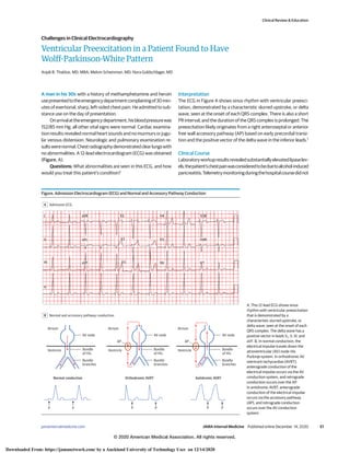
- 1. Ventricular Preexcitation in a Patient Found to Have Wolff-Parkinson-White Pattern Anjali B. Thakkar, MD, MBA; Melvin Scheinman, MD; Nora Goldschlager, MD A man in his 30s with a history of methamphetamine and heroin usepresentedtotheemergencydepartmentcomplainingof30min- utes of exertional, sharp, left-sided chest pain. He admitted to sub- stance use on the day of presentation. Onarrivalattheemergencydepartment,hisbloodpressurewas 152/85 mm Hg; all other vital signs were normal. Cardiac examina- tion results revealed normal heart sounds and no murmurs or jugu- lar venous distension. Neurologic and pulmonary examination re- sultswerenormal.Chestradiographydemonstratedclearlungswith no abnormalities. A 12-lead electrocardiogram (ECG) was obtained (Figure, A). Questions: What abnormalities are seen in this ECG, and how would you treat this patient’s condition? Interpretation The ECG in Figure A shows sinus rhythm with ventricular preexci- tation, demonstrated by a characteristic slurred upstroke, or delta wave, seen at the onset of each QRS complex. There is also a short PR interval, and the duration of the QRS complex is prolonged. The preexcitation likely originates from a right anteroseptal or anterior free wall accessory pathway (AP) based on early precordial transi- tion and the positive vector of the delta wave in the inferior leads.1 Clinical Course Laboratoryworkupresultsrevealedsubstantiallyelevatedlipaselev- els;thepatient’schestpainwasconsideredtobeduetoalcohol-induced pancreatitis.Telemetrymonitoringduringthehospitalcoursedidnot Figure. Admission Electrocardiogram (ECG) and Normal and Accessory Pathway Conduction Admission ECG A aVR V1 V4 V3R V2 V3 V5 V6 V4R V7 I II aVL III II aVF Normal and accessory pathway conduction B Atrium Atrium Atrium Ventricle Ventricle Ventricle Bundle of His Bundle of His AV node AV node Bundle branches Bundle of His AV node Bundle branches Bundle branches Orthodromic AVRT Normal conduction Antidromic AVRT P P AP AP P P P P A, This 12-lead ECG shows sinus rhythm with ventricular preexcitation that is demonstrated by a characteristic slurred upstroke, or delta wave, seen at the onset of each QRS complex. The delta wave has a positive vector in leads V1, II, III, and aVF. B, In normal conduction, the electrical impulse travels down the atrioventricular (AV) node-His Purkinje system. In orthodromic AV reentrant tachycardias (AVRT), anterograde conduction of the electrical impulse occurs via the AV conduction system, and retrograde conduction occurs over the AP. In antidromic AVRT, anterograde conduction of the electrical impulse occurs via the accessory pathway (AP), and retrograde conduction occurs over the AV conduction system. Clinical Review & Education Challenges in Clinical Electrocardiography jamainternalmedicine.com (Reprinted) JAMA Internal Medicine Published online December 14, 2020 E1 © 2020 American Medical Association. All rights reserved. Downloaded From: https://jamanetwork.com/ by a Auckland University of Technology User on 12/14/2020
- 2. reveal evidence of arrhythmias. The patient left the hospital against medical advice before additional workup could be performed. Discussion Wolff-Parkinson-White (WPW) is an uncommon syndrome result- ing from myocardial remnant tissue connecting atria and ven- tricles, allowing for anomalous ventricular activation (preexcita- tion)fromtheatriumtotheventricle,bypassingtheatrioventricular (AV) node-His axis. The degree of conduction via the AP can vary from entirely over the AV node-His-Purkinje system (no preexcita- tion) to entirely over the AP (maximal preexcitation). Wolff- Parkinson-White is most commonly diagnosed on routine ECG in asymptomaticindividualsforwhom3characteristicfeaturesarepre- sent:aslurredupstroke(deltawave)oftheQRScomplex(whichcan bepositiveornegative),widenedQRScomplex,andshortPRinter- val. When the WPW ECG pattern is seen in the absence of sympto- matic arrhythmias, it is known as WPW pattern; in the presence of symptomatic arrhythmias, it is known as WPW syndrome. The prevalence of WPW in multiple community-based popula- tionstudiesisestimatedtobebetween1to3per1000individuals.2 Autosomal dominant familial forms of WPW syndrome have been identified; the prevalence is 5.5 in 1000 first-degree relatives of in- dividuals with WPW.2,3 Electrophysiologicstudieshaveelucidatedthemechanismsun- derlying symptoms in patients with WPW. Symptomatic arrhyth- mias are due either to tachycardias that use the AP to initiate and sustain a reentrant circuit, or tachycardias in which the AP serves as a bystander route of arrhythmia conduction. Tachycardias that use the AP as a critical component of the tachycardia circuit are AV reentrant tachycardias (AVRT); tachycardias in which the AP is a bystander to other atrial arrhythmias include atrial tachycardia, AV nodal reentrant tachycardia, and atrial flutter or fibrillation. The presenceofanterogradeconductionresultsintheclassicdeltawave finding on ECG; pathways with retrograde-only properties, known as concealed pathways, will not manifest a delta wave.4 In AVRT, the circuit consists of the normal AV conduction sys- tem and an AP, both of which link the atria and ventricles (Figure, B). The electrical impulse can conduct either via the AV conduction sys- tem or the AP. When anterograde conduction occurs via the AV con- duction system and retrograde conduction occurs over the AP, the tachycardia is known as orthodromic AVRT and is associated with anarrowQRScomplex,orbundlebranchblockmorphologyintheset- ting of aberrant ventricular conduction. In orthodromic AVRT, the tachycardia can be initiated with an atrial premature depolarization thatblocksintheAP,conductsoverthenode,andthenreturnstothe atrium via the AP. Orthodromic AVRT is the most common arrhyth- mia observed in WPW syndrome because the AV nodal refractory period is shorter than that of the AP. In contrast, in up to 8% of cases, anterograde conduction occurs over the AP with retrograde nodal conduction, which is associated with a broad QRS because of maximalpreexcitation;thistachycardiaisknownasantidromicAVRT.5 AsymptomaticindividualswhoarefoundtohaveWPWpattern,as inthispatient,shouldbeevaluatedfortheriskofsuddencardiacarrest. While electrophysiologic study is the criterion standard for risk strati- fication,ECGduringrestandexercisestresscanbehelpful.Whenstress testresultsshowalossofpreexcitationatlowheartrates,thissuggests amorebenignpathway.Thisisbecausepreexcitedatrialtachyarrhyth- mias,likeatrialfibrillation,wouldbeunabletoconductrapidlyenough viatheaccessorypathwaytoprecipitatelife-threateningarrhythmias. Ifpreexcitationpersistsduringastresstest,invasiveelectrophysiologic studyiswarrantedtoidentifythelocationandelectrophysiologicprop- ertiesoftheAPandtheneedforcatheterablation. Take-Home Points • Wolff-Parkinson-Whiteisanuncommonsyndromeresultingfrommyo- cardialremnanttissueconnectingatriaandventricles,allowingforven- tricularactivationseparatefromtheAVnode-HisPurkinjesyndrome. • The WPW pattern is manifest by 3 characteristic ECG features: a delta wave, widened QRS complex, and short PR interval. • InpatientswithWPWsyndrome,symptomaticarrhythmiasaredue either to tachycardias that use the AP as a critical part of the reen- trant circuit, or tachycardias in which the AP acts as a bystander. • AsymptomaticindividualsfoundtohaveWPWpatternshouldundergo riskstratificationwithECGduringrestandexercisestresstoassessfor lossofpreexcitation;ifpreexcitationpersists,electrophysiologicstudy iswarrantedtodeterminetheneedforcatheterablation. ARTICLE INFORMATION Author Affiliations: Department of Medicine, University of California, San Francisco, San Francisco (Thakkar); Division of Cardiology, Department of Medicine, University of California San Francisco, San Francisco (Scheinman, Goldschlager); Division of Cardiology, Department of Medicine, Zuckerberg San Francisco General Hospital and Trauma Center, San Francisco, California (Goldschlager); Section Editor, JAMA Internal Medicine (Goldschlager). Corresponding Author: Anjali B. Thakkar, MD, MBA, Department of Medicine, University of California, San Francisco, 505 Parnassus Ave, Room M-1483, San Francisco, CA 94143-0119 (anjali.thakkar@ucsf.edu). Published Online: December 14, 2020. doi:10.1001/jamainternmed.2020.6847 Conflict of Interest Disclosures: None reported. Disclaimer: Dr Goldschlager is a Section Editor of the Challenges in Electrocardiography section of JAMA Internal Medicine, but she was not involved in any of the decisions regarding review of the manuscript or its acceptance. REFERENCES 1. Fitzpatrick AP, Gonzales RP, Lesh MD, Modin GW, Lee RJ, Scheinman MM. New algorithm for the localization of accessory atrioventricular connections using a baseline electrocardiogram. J Am Coll Cardiol. 1994;23(1):107-116. doi:10.1016/ 0735-1097(94)90508-8 2. Cohen MI, Triedman JK, Cannon BC, et al; Pediatric and Congenital Electrophysiology Society (PACES); Heart Rhythm Society (HRS); American College of Cardiology Foundation (ACCF); American Heart Association (AHA); American Academy of Pediatrics (AAP); Canadian Heart Rhythm Society (CHRS). PACES/HRS expert consensus statement on the management of the asymptomatic young patient with a Wolff-Parkinson-White (WPW, ventricular preexcitation) electrocardiographic pattern: developed in partnership between the Pediatric and Congenital Electrophysiology Society (PACES) and the Heart Rhythm Society (HRS). endorsed by the governing bodies of PACES, HRS, the American College of Cardiology Foundation (ACCF), the American Heart Association (AHA), the American Academy of Pediatrics (AAP), and the Canadian Heart Rhythm Society (CHRS). Heart Rhythm. 2012;9(6):1006-1024. doi:10.1016/j. hrthm.2012.03.050 3. Vidaillet HJJ Jr, Pressley JC, Henke E, Harrell FEJ Jr, German LD. Familial occurrence of accessory atrioventricular pathways (preexcitation syndrome). N Engl J Med. 1987;317(2):65-69. doi:10.1056/NEJM198707093170201 4. Narula OS. Wolff-Parkinson-White syndrome. a review. Circulation. 1973;47(4):872-887. doi:10. 1161/01.CIR.47.4.872 5. Brembilla-Perrot B, Pauriah M, Sellal J-M, et al. Incidence and prognostic significance of spontaneous and inducible antidromic tachycardia. Europace. 2013;15(6):871-876. doi:10.1093/europace/ eus354 Clinical Review & Education Challenges in Clinical Electrocardiography E2 JAMA Internal Medicine Published online December 14, 2020 (Reprinted) jamainternalmedicine.com © 2020 American Medical Association. All rights reserved. Downloaded From: https://jamanetwork.com/ by a Auckland University of Technology User on 12/14/2020