Downloaded 236 times


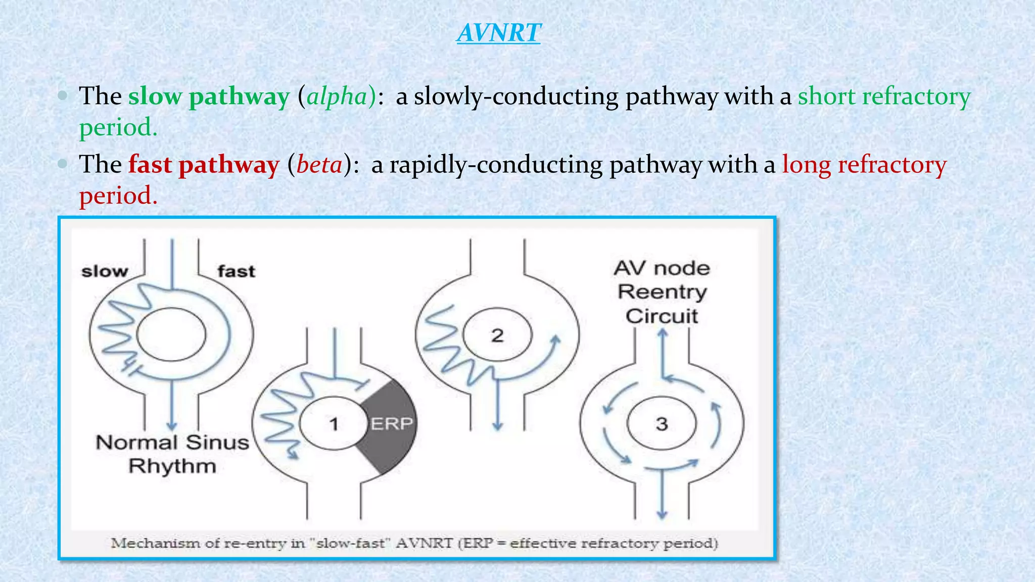


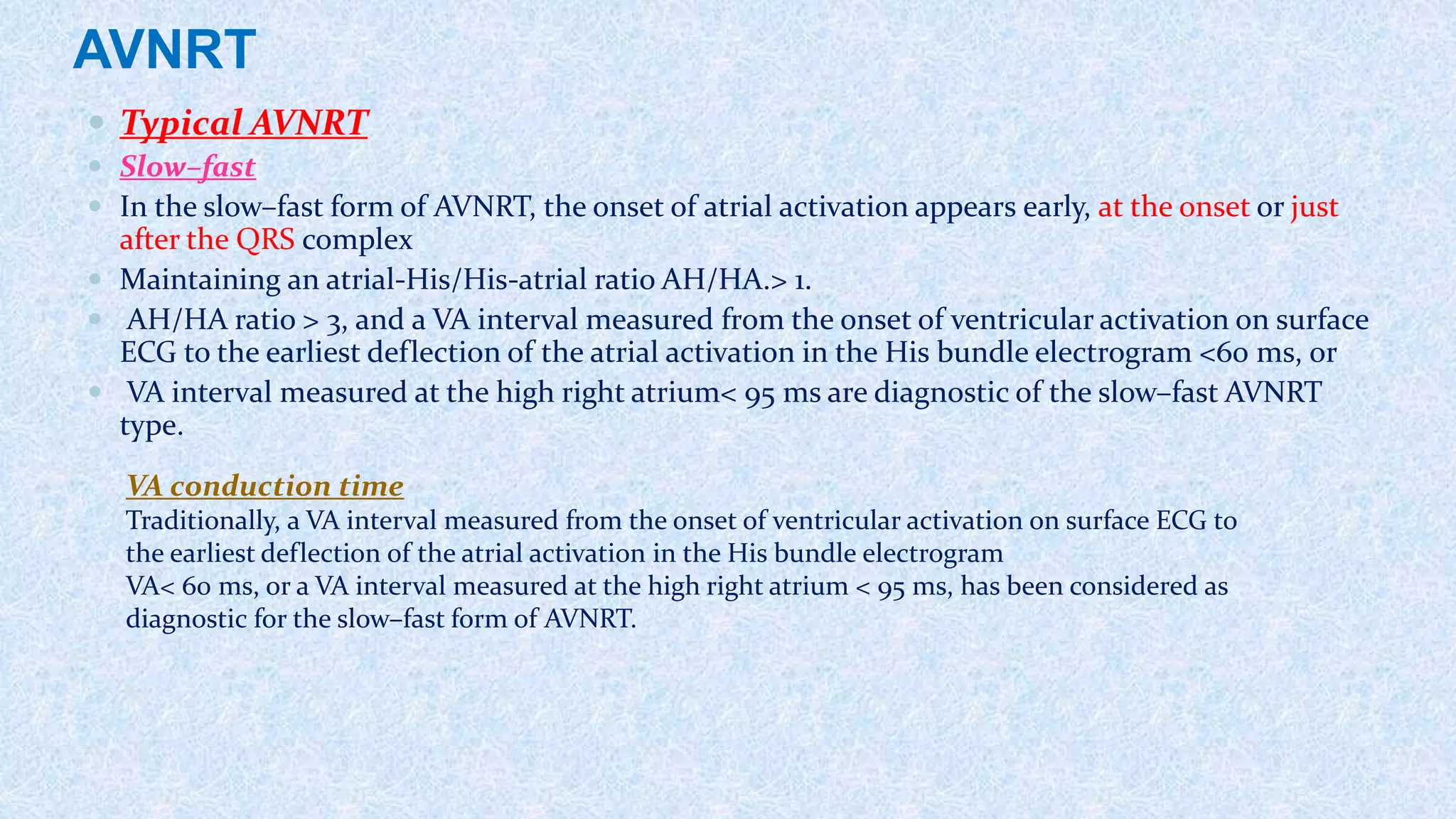


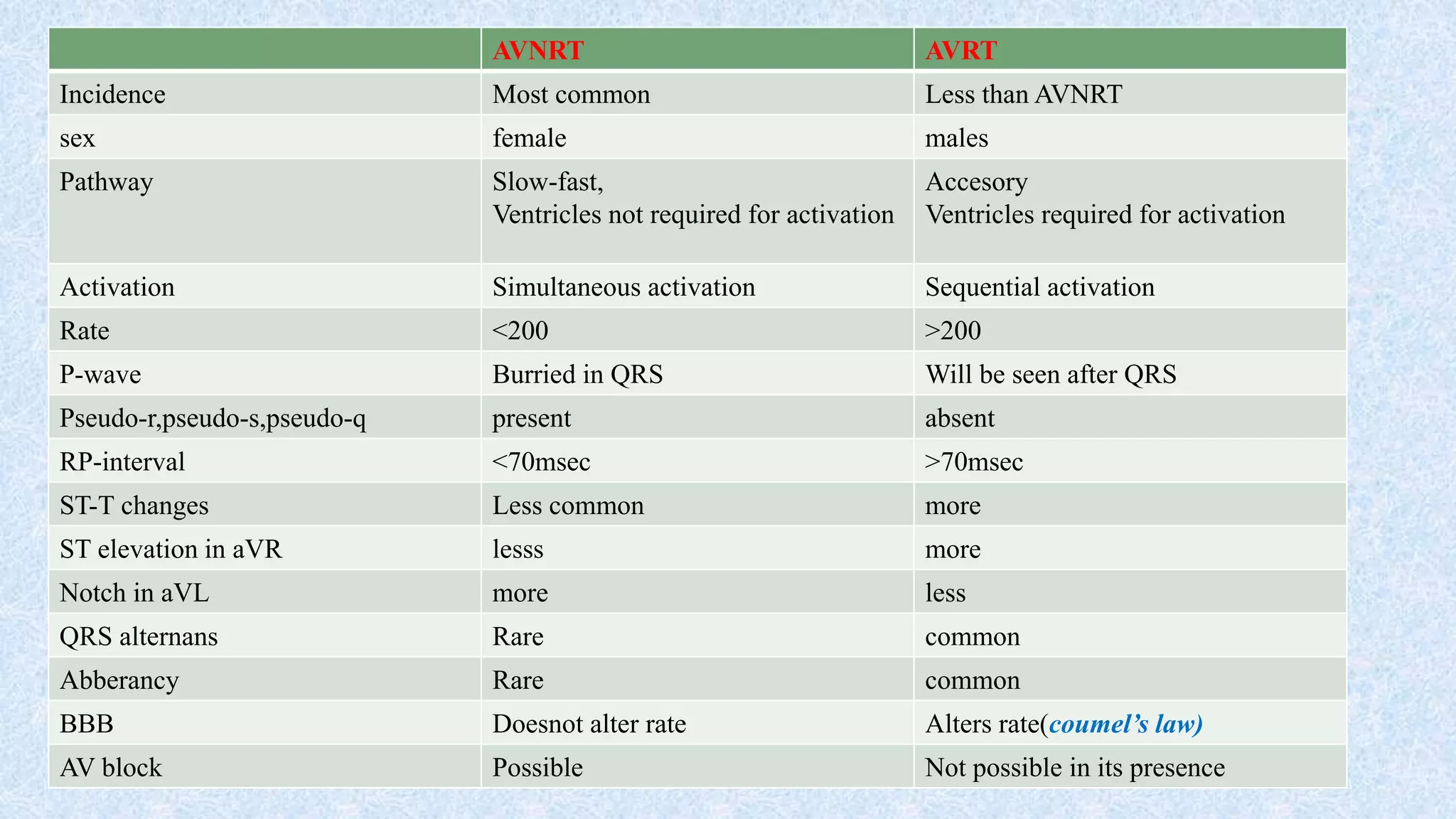


1. AVNRT and AVRT are types of supraventricular tachycardia involving abnormal pathways for electrical conduction between the atria and ventricles. 2. AVNRT is caused by a reentry circuit within the AV node, while AVRT involves an accessory pathway bypassing the AV node. 3. There are different subtypes of AVNRT and AVRT depending on which pathways are involved in the antegrade and retrograde directions. Typical AVNRT involves a slow-fast pathway while typical AVRT involves orthodromic conduction over an accessory pathway.


