Download as PPSX, PPTX

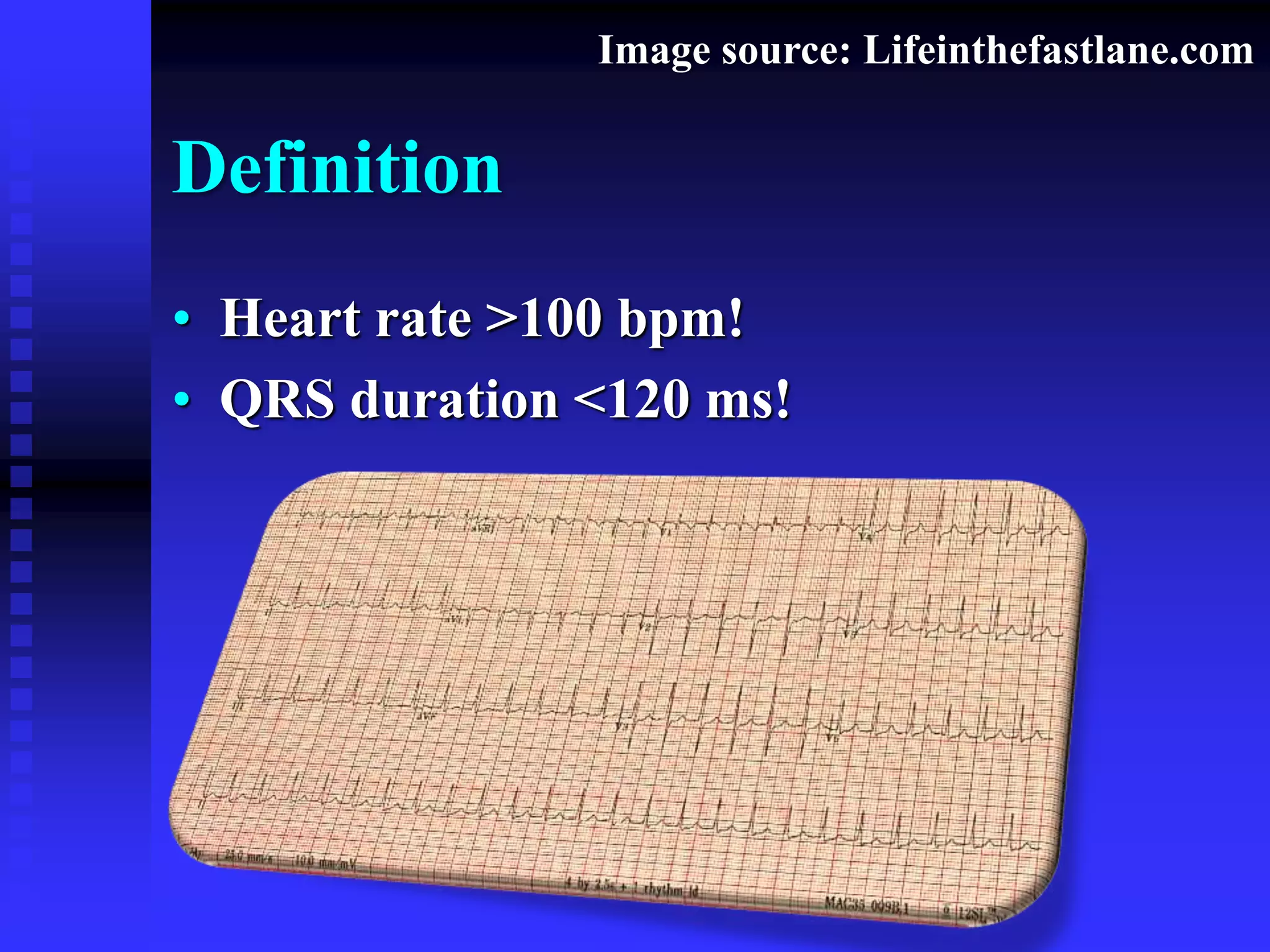

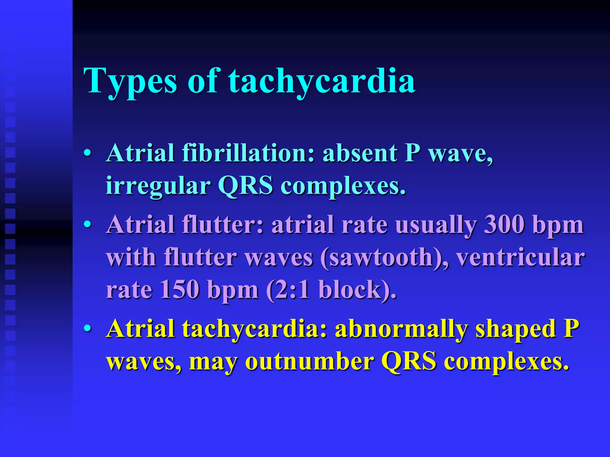

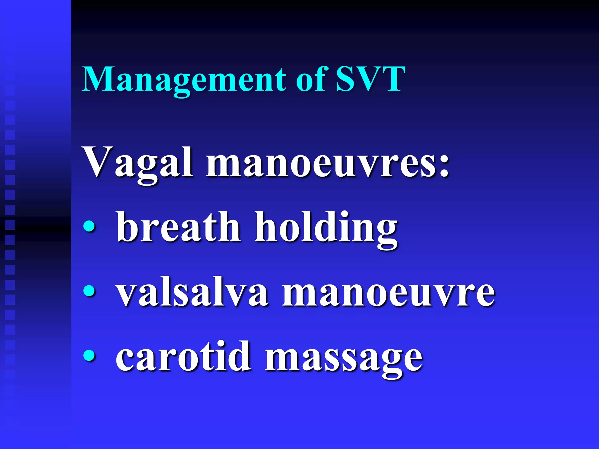
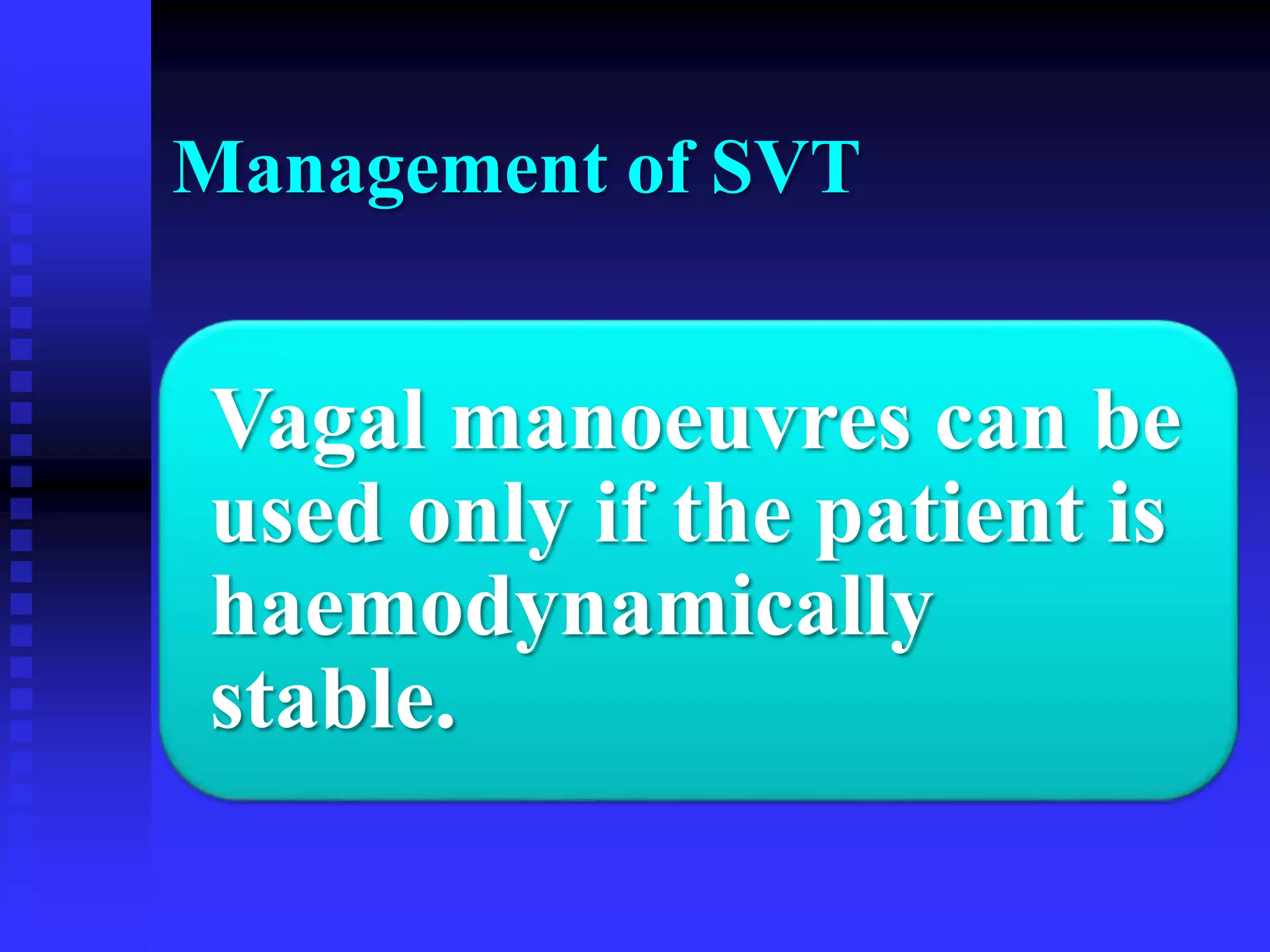


This document discusses narrow complex tachycardia, defining it as a heart rate over 100 bpm with a QRS duration under 120 ms. It describes various types of tachycardia including sinus tachycardia, supraventricular tachycardia, atrial fibrillation, atrial flutter, atrial tachycardia, multifocal atrial tachycardia, and junctional tachycardia. It provides details on management of supraventricular tachycardia including vagal maneuvers, adenosine, verapamil, beta blockers, and cardioversion. Other conditions discussed include Wolff-Parkinson-White syndrome, Long Ganong Levine syndrome, and Holiday heart syndrome


![Cardiccccac Arrhythmias [Autosaved].pptx](https://cdn.slidesharecdn.com/ss_thumbnails/cardiacarrhythmiasautosaved-241108153215-72acce97-thumbnail.jpg?width=640&height=640&fit=bounds)


