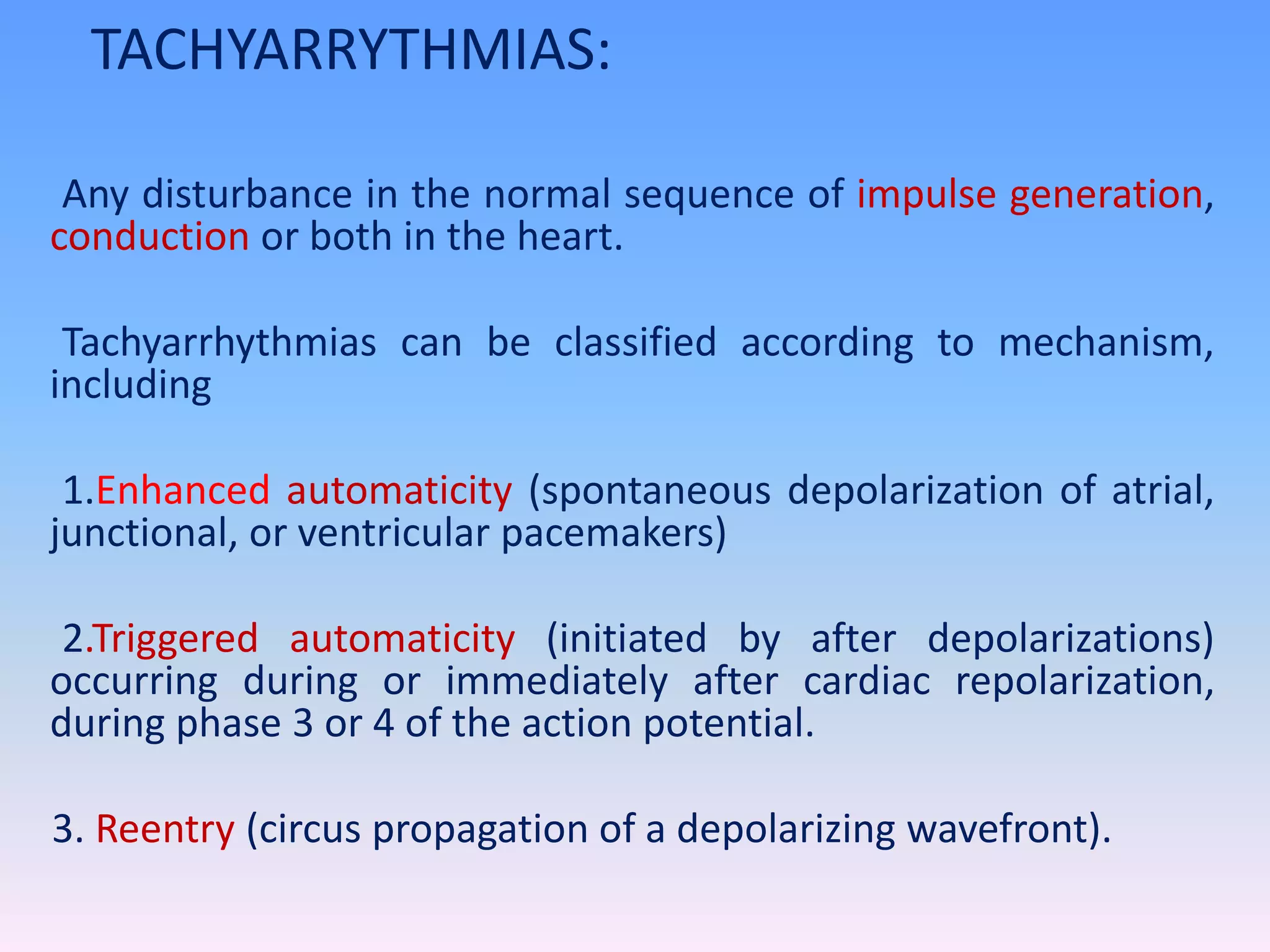
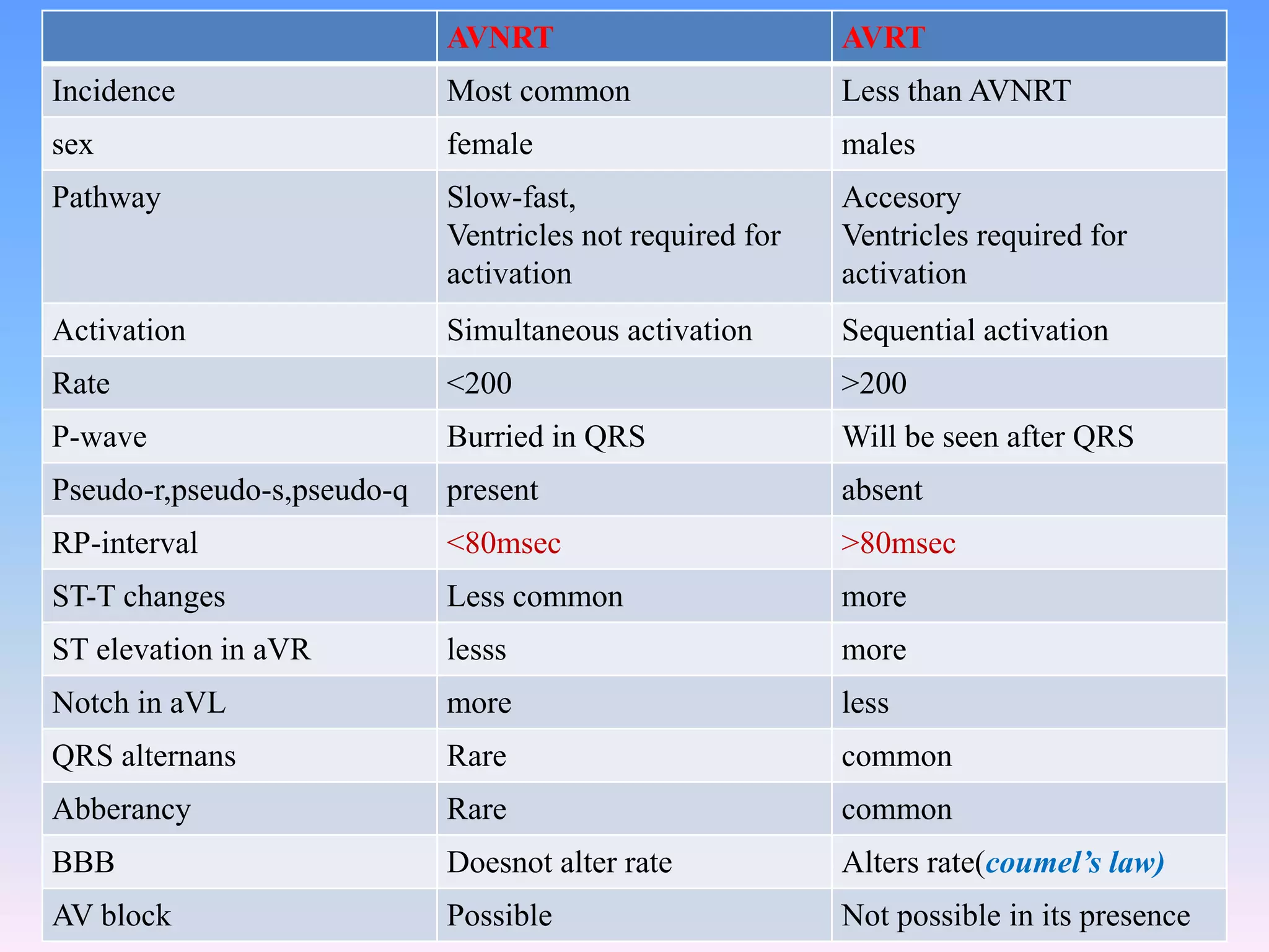
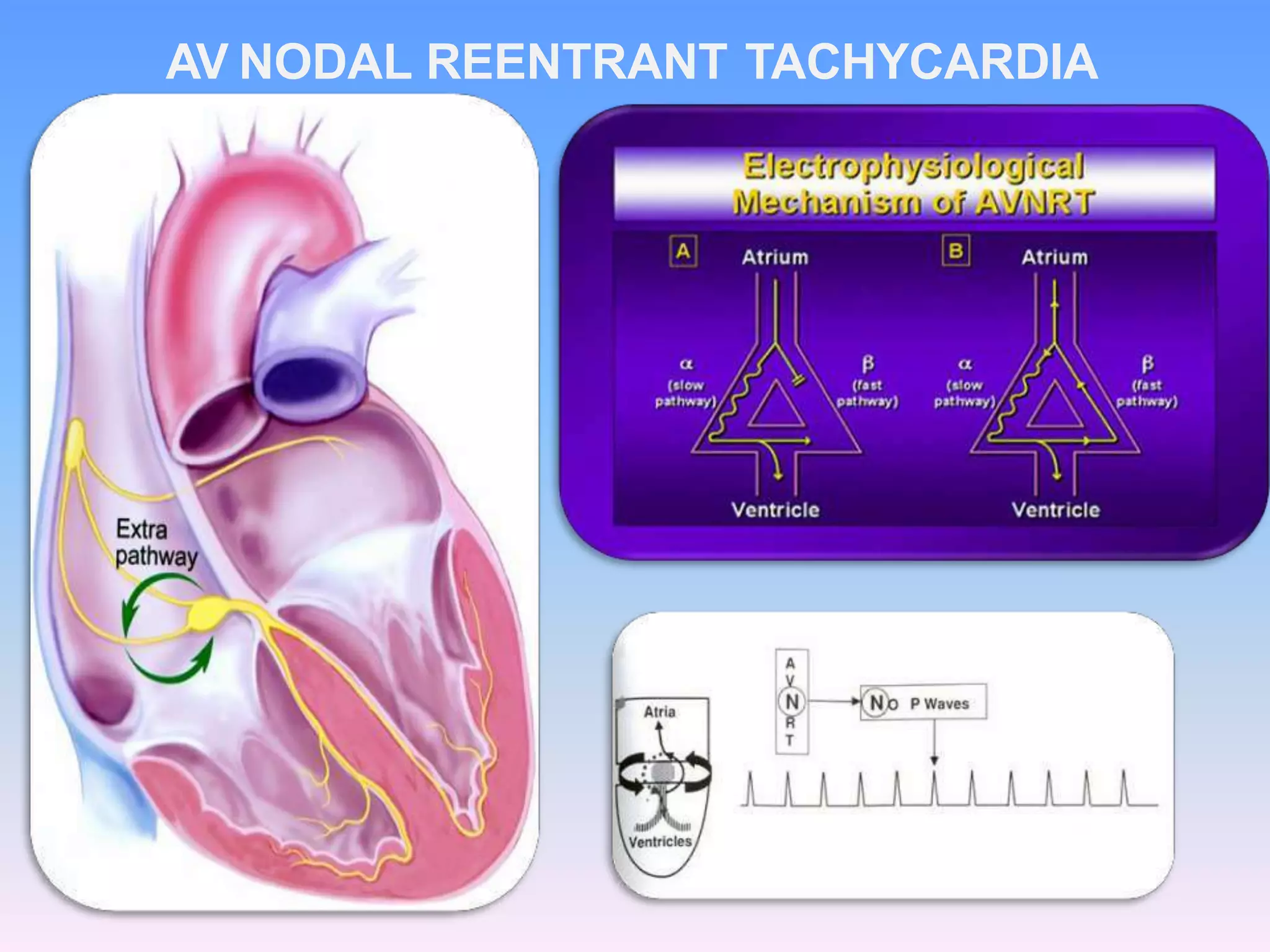
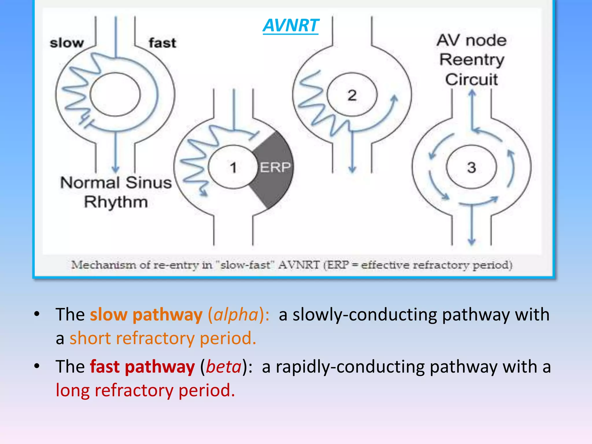
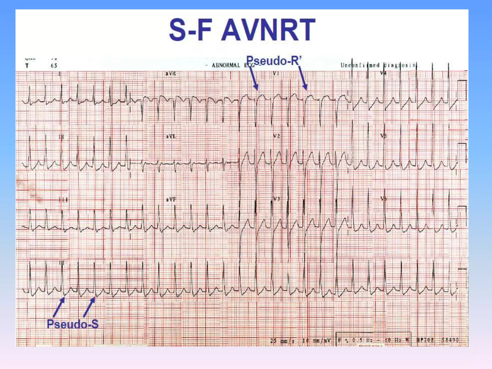
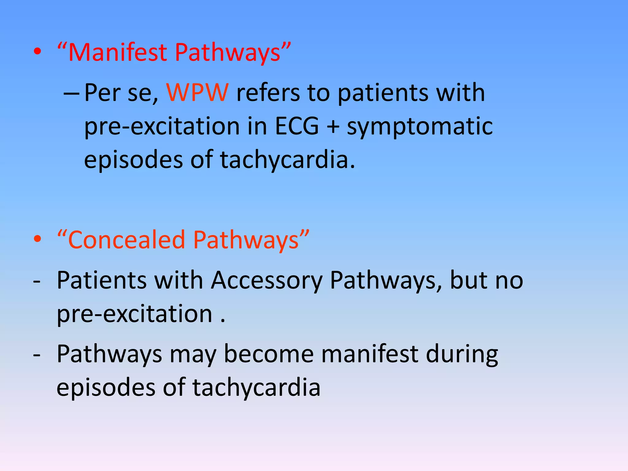
APPROACH TO A
PATIENT WITH
NARROW COMPLEX
TACHYCARDIA
Speaker-Dr Ramdhan kumar
kamat
MD(Medicine),Sr cardiology igims
4
0
3
(If )
TACHYARRYTHMIAS:
Any disturbance in the normal sequence of impulse generation,
conduction or both in the heart.
Tachyarrhythmias can be classified according to mechanism,
including
1.Enhanced automaticity (spontaneous depolarization of atrial,
junctional, or ventricular pacemakers)
2.Triggered automaticity (initiated by after depolarizations)
occurring during or immediately after cardiac repolarization,
during phase 3 or 4 of the action potential.
3. Reentry (circus propagation of a depolarizing wavefront).
Enhanced cardiac automaticity refers to the accelerated
generation of an action potential by either normal pacemaker
tissue (enhanced normal automaticity) or by abnormal tissue
within the myocardium (abnormal automaticity).
The discharge rate of normal or abnormal pacemakers may be
accelerated by drugs, various forms of cardiac disease,
reduction in extracellular potassium, or alterations of autonomic
nervous system tone.
Enhanced normal automaticity accounts for the occurrence of
sinus tachycardia, while abnormal automaticity may result in
various atrial or ventricular arrhythmias, for example, an
accelerated idioventricular rhythm or an ectopic atrial
tachycardia.
Abnormality in impulse formation:
An increase in automaticity normally causes
an increase in sinus rate and sinus tachycardia.
Abnormal automaticity is due to an increase in
the slope of phase 4 depolarization in
myocardium or reduced threshold for action
potential depolarization in myocardium other
than the sinus node. Abnormal automaticity
is thought to be responsible for most atrial
premature complexes (APC) and VPCs.
Less commonly, abnormal impulse formation is due to
the development of triggered activity.
Triggered activity is related to cellular afterdepolarization
that occur at the end of the action potential, during
phase 3, and are referred to as early afterdepolarization,
or they occur after the action potential, during phase 4,
and are referred to as late afterdepolarization.
ABNORMALITY IN IMPULSE PROPAGATION:
The most common arrhythmia mechanism is reentry.
Reentry is defined as circulation of an activation wave
around an inexcitable obstacle. Thus, the requirements
for reentry are two electrophysiologically dissimilar
pathways for impulse propagation around an inexcitable
region such electrophysiologically dissimilar pathways
for impulse propagation around an inexcitable region
such that unidirectional block occurs in one of the
pathways and a region of excitable tissue exists at the
head of the propagating wavefront .
Look for QRS duration.
QRS complex regular/irregular.
Then look for presence of p waves.
P waves morphology
P wavesand QRS relationship 1:1
AV block present.
QRS alternation
Termination initiation of tachycardia.
Effect of BBB on tachycardia cycle length.
Response to carotid sinus massage or adenosine –with
termination of arrhythmia with Pwave –AVNRT with atrial
premature beat .
Tachycardia persists with AV block –AT, AFL, SANRT
Pseudo r ‘ wave in V1 – AVNRT
SHORT RP interval – AVNRT, AVRT
Long RP interval – AT, SANRT, AVNRT atypical
Classification
• AV NODE DEPENDENT
1. AVNRT 2. AVRT ANTIDROMIC
ORTHODROMIC
AV NODE INDEPENDENT
1. ATRIAL TACHYCARDIA
2. JUNCTIONAL ECTOPIC TACHYCARDIA
3. ATRIAL FLUTTER
4. ATRIAL FIBRILLATION
s
AVNRT AVRT
Incidence Most common Less than AVNRT
sex female males
Pathway Slow-fast,
Ventricles not required for
activation
Accesory
Ventricles required for
activation
Activation Simultaneous activation Sequential activation
Rate <200 >200
P-wave Burried in QRS Will be seen after QRS
Pseudo-r,pseudo-s,pseudo-q present absent
RP-interval <80msec >80msec
ST-T changes Less common more
ST elevation in aVR lesss more
Notch in aVL more less
QRS alternans Rare common
Abberancy Rare common
BBB Doesnot alter rate Alters rate(coumel’s law)
AV block Possible Not possible in its presence
AV NODAL REENTRANT TACHYCARDIA
• The slow pathway (alpha): a slowly-conducting pathway with
a short refractory period.
• The fast pathway (beta): a rapidly-conducting pathway with a
long refractory period.
AVNRT
Presence of a narrow complex tachycardia with regular R-R
intervals and no visible p waves.
P wave are buried in the QRS complexes –simultaneous
activation of atria and ventricles – most common presentation
of AVNRT –66%.
If not synchronous –pseudo s wavein inferior leads ,pseudo r’
wavein lead V1---30%cases .
P waves are retrograde and are inverted in leads II,III,AVF
P wavemay be farther awayfrom QRS complex distorting the
ST segment ---AVNRT ,mostly AVRT.
Slow-Fast (Typical) AVNRT:
•Narrow complex tachycardia at ~ 150 bpm.
•No visible P waves.
•There are pseudo R’ waves in V1-2.
• RP <PR
What are “Pre-excitation syndromes” ?
• Term coined by Ohnell
• First described in 1930 by Louis Wolff, John Parkinson and Paul Dudley
White.
• A group of ECG and Electrophysiological abnormalities in which
– The atrial impulses are conducted partly or completely, PREMATURELY, to
the ventricles via a mechanism other than the normal AV-node
– Associated with a wide array of tachycardias with both normal QRS and
prolonged QRS durations
NARROW QRS TACHYCARDIA
• “Manifest Pathways”
–Per se, WPW refers to patients with
pre-excitation in ECG + symptomatic
episodes of tachycardia.
• “Concealed Pathways”
- Patients with Accessory Pathways, but no
pre-excitation .
- Pathways may become manifest during
episodes of tachycardia
WPW
• PR interval <120ms
• Delta wave – slurring slow rise of
initial portion of the QRS
• QRS prolongation >120ms
• ST Segment and T wave discordant
changes – i.e. in the opposite
direction to the major component
of the QRS complex
• Pseudo-infarction pattern can be
seen in up to 70% of patients – due
to negatively deflected delta waves
in the inferior / anterior leads
(“pseudo-Q waves”), or as a
prominent R wave in V1-3
(mimicking posterior infarction).
WPW in sinus rhythm
NARROW QRS TACHYCARDIA
Two types
Orthodromic
Antidromic
Antidromic is wide complex tachycardia
In NSR detected by delta wave.
Can ppt into AF and VF on use of AV nodal blockers
MEMBRANE ACTIVE ANTIARRHTYHMIC DRUGS are safe.
CONCEALED WPW syndrome – no delta wave .less risk of
AF
Typical – RP interval <PR interval
RP interval >80 millisec
Atypical –RP interval >PR interval
Concealed bypass tract – only retrograde conduction
Manifest bypass tract– both anterograde and
retrograde.
Electrical alternans –the amplitude of QRS
complexes varies by 5 mm alternatively.
Rate related BBB occuring and the rate of tachycardia
is decreasing –then the bypass tract is on the same
side of the block.
ORT - URAP
AV REENTRANT TACHYCARDIA
NARROW QRS TACHYCARDIA
Antidromic WPW
ANTIDROMIC AVRT-REGULAR BROAD COMPLEX TACHYCAR
Short PR interval
Normal QRS complex
PSVT
Lown-Ganong-Levine Syndrome
Acute
Treatment
Of
SVT
 P wave morphology changes.
 PR interval > 0.12 sec .
 Second,third degree AV block can occur.
 Tachycardia terminates with a qrs complex ..
 Right atrial origin– p wave inverted in V1.
 If biphasic in V1—initially positive then negative.
 Upright in lead AVL
 Opposite if of left atrial origin
 Superior origin –upright p waves in inferior leads
 Inferior origin –p waves are inverted in inferior leads.
At least three consequtive p waves with different morphologies
with a rate >100 bpm to be present.
Isoelectric baseline between p waves.
Also called as choatic atrial tachycardia
Mostly seen in COPD ,electrolyte abn,theophylline
Rate usually does not exceed 130-140 bpm.
Microreentrant tachycardia
Usually precipitated and terminated by premature
atrial complexes.
Atrial rate is usually 120-150 bpm.
IART -Large or small reentrant circuit.
AVblock canoccur.
Non paroxysmal – accelerated junctional rhythm
Rate < 100 bpm Usually junctional node 40-60 bpm
Paroxysmal or focal junctional tachycardia is
rare – automaticity.
110-250bpm.
P waves may be before or after QRS complex
Infrequent and nonsustained episodes –no treatment
Acute termination of SVT and establish the mechanism
of SVT in case of acute setting.
Long term goal is abolishing the arryhthmia substrate.
Precipitating factors – electrolyte
imbalance,hypoxia,ischemia,hyperthyroidism to be
sought out.
Acute
Treatment
Of
SVT
A12 lead ECG during tachycardia and NSR.
No delay in therapy if the mechanism of SVT is not
known.
Perform CAROTID SINUS MASSAGE,or give6mg
bolus adenosine.
In case of severe hemodynamic compromise a
synchronised cardioversion to be given.
Check for carotid bruit before massage.
At the level of cricoid cartilage,at the angle of mandible
the carotid sinus is situated.
Gentle pressure is applied over the carotid sinus for 5
-10 seconds.
ECG recording to be present.
In case of no response – try on the other side.
Simultaneous pressure not to be applied both sides.
Alternative manuevres are valsalva,gagreflex,ice water
pouring over the face.
If SVT is suspected to be AVnode dependent – drug
of choice is adenosine and CCBs verapamil and
diltiazem.
Useful for sustained cases of AV node independent
tachycardias.
But digoxin,BBs,CCBs better control of ventricular
response in atrial tachycardias
Class I agents to be combined withAV nodal blocking
drugs – to eliminate 1:1 conduction of atrial to
ventricles.
HEMODYNAMIC
STATUS
STABLE BP
>90/60mmHg
NarrowQRS
andregular
R-R
Vagalmaneuveres
IV adenosine
IVverapamil,diltiazem
IV sotalol
Refractory
WideQRS
complex
Vagalmanuevres
IV adenosine
procainamide
Digoxin
Verapamil
Arecontraindicated
UNSTABLE
BP< 90/60
mmHg
Direct
cardioversion
DRUG DOSE SIDE EFFECTS
AVNODAL
BLOCKERS
ADENOSINE 6-12 mgbolus Flushing,dyspnea
Chest pain
VERAPAMIL 0.15 mg/kg over2 min Hypotension bradycardia
DILTIAZEM 0.25-0.35 mg/kg -2min same
DIGOXIN 0.5-1.0 mg --- 2-10 min Digoxintoxicity
PROPANOLOL 1-3mg over I min Hypotension bradycardia
CLASSIAAD QUINIDINE 6-10MG/KGat 10mg/min hypotension
PROCAINAMIDE 10-15mg/kg at 50mg/min hypotension
DISOPYRAMIDE 1-2 mg/kg at10mg/min hypotension
PROPAFENONE 1-2mg/min at10mg/min Bradycardia,GIdisturbance
FLECAINIDE 2 mg/kg at10mg/min Bradycardia,dizziness
CLASSIII SOTALOL 1-1.5mg/kg at 10mg/min Hypotension,proarrythmic
AMIODARONE 1.5mg/kg during15min Hypotension,bradycardia
PharmacologicAgents forShort-Term tofSuprav Tachycardia(SVT)
Delacrétaz E. N Engl JMed 2006;354:1039-1051.
Refractory
cases
Narrow
QRS
complexes
IV adenosine
IV procainamide
IV amiodarone
Atrial pacing
Directcardioversion
Wide QRS
Complexes
Atrial pacing
Direct
cardioversion
 In whom recurrences areinfrequent.
 But sustained.well tolerated hemodynamically.
 Patients who have had only asingle episode of SVT..
 100-200mg of flecainide at the onset of SVTis areasonable
approach…until he reaches the hospital.
 40-160 mg verapamil –without preexcitation,
 Betablockers
 Propafenone 150-450 mg.
 80%casesinterrupted with acombination of CCBandBBin2
hrs…
Frequency and severity ofepisodes.
LVF
Costbenefits of radiofrequency ablation over the
pharmacotherapy .
Pharmacotherapy isconsidered in patients who defer
catheter ablation,whom in which ablation failed,or
carries arisk ofAVblock.
Multifocal atrialtachycardia
Trial anderror
Accessory pathway –classIa,Ic,III
AVnode blockingdrugs
Young patients –Iadrugs
ClassI agents LVD<35%notused.
Longterm
treatment
Membrane
activeAAD
Catheter
ablation
Curativesurgery
Antitachycardia
pacing
 not to be used in bronchospastic pulmonary
disease.
 Adenosine precipitates asthma
 Given rapidly in 1-2 sec.
 If given by peripheral vein uplift the arm..
 Max dose is 30 mg
 6- 12-12 mg
 Terminates AVNRT .AFL with 2:1 block
 Potentiated by dipyradimole,carbamazepine –
 Calcium channel blockers,beta blockers
,digoxin are the next drugs to be used if
not responded to adenosine
 Usually 60 % cases respond to a dose of 6
mg and 95 % cases at 12 mg.
 Type 1 a AAD, 1c,iii,AMIODARONE in
refractory cases.
 Beta blockers not to used IV in heart
failure.
Several multipolar catheters are introduced
High right atrium ,bundle of his ,RVapex,Coronary
sinus.
Radiofrequency is delivered at the site of earlier
activation
Success is defined by elimination of the tachycardia or
loss of pre excitation.
90-98% success in AV node dependent
60-80% in case of AV node independent.
Cryoablation more useful…
CatheterAblation of
CardiacArrhythmias.
Temporary role in caseof digoxintoxicity.
Permanent in caseof long termcontrol
Toterminate thetachycardia
Revert into sinusrhythm
Prevent the occurrence.
Overdrive suppression
RFinduced atrial pacing areused
No role of surgery presently in PSVTrx.
ACUTE LONGTERM
PHYSIOLOGICAL rest ,sedation valsalva
Valsalva maneuvre Carotid sinusmassage
Carotid sinusmassage
PHARAMACOLOGICAL vagomimmetic Suppresstriggering
arrhythmias
Direct effect onAVnode Change propertiesof
reentrant pathways
SlowVR ControlVR
CATHETERABLATION
SURGERY
Ablation or sectioning of
reentrant pathway
ELECTRONICDEVICES Temp.pacing
cardioversio
n
Permanent pacemaker
Antitachycardiapacing
Rxof PSVT given for patient comfort except
in IHD,MS
When the QRS complex is wide and VT is
mistaken as SVT with ABERRANT conduction
IV verapamil – not recommended decreases
BP.
If DC cardioversion to be avoided because of
possible adverse response to digitalis adm
…pacing Rt atrium and ventricle via temp
pacing.
In WPW syndrome avoid
• Rx of ectopic atrial
tachycardia – consider
digitalis toxicity,chronic lung disease,metabolic
abn,electrolyte abnormalities,acute MI ----
temporary pacing.
Unsuccessful is EC
Removal or reversal of inciting factor
Surgical excision of focus.
Rx of MAT –chronic lung
disease,metabolic,rare is digitlais toxicity ---
CCBS,BBs ..no role of cardioversion,devices
In case of WPW syndrome
symptomatic concealed or
manifested ..and evidence of
preexcitation on NSR …send
the patient for catheter
ablation…
 1. carotid sinus pressure
 2.IV adenosine.
 3.long term treatment depends upon episodes.
 4.any underlying abnormality to be checked
for.
 5.definitive etiology only knon by EP study.
 6.95% cases respond to RF ablation.
 7.much less complications with cryoablation.
 8.in case if SVT recurrs after ablation –opt
“Youonly getsomanyheart beats– you should save some for later in life” Dr.SamuelLevine
1.Coexisting Double Tachycardias
May not be identified during noninvasive testing ..needs EP
study.
Ex—typicalAVNRT andAT.
Concentric –eccentric –concentric.
AVNRT –both APC,VPC
AT onlyAPC
2.Pseudo AF- infrequent presentation of PSVT.
Occurs during onset and termination of tahcycardia.
Multiple accessory AV pathways.
In young who haveAF without other risk factors.
5%of AVNRT.
Group beating is seen
 CARDIOLOGYthird edition –Michael.H.Crawford
 HURST’STHEHEART– 12 th edition.
 BRAUNWALD’SHEARTDISEASE–ATEXTBOOKOFCARDIOVASCULAR
MEDICINE– 7 th ED
 HARRISON’SPRINICPLESOFINTERNALMEDICINE-17 th ED
 SUPRAVENTRICULARTACHYCARDIA–NEJM 2006
 CARDIOVASCULARMEDICINE– SVT– JERONIMOFERRE’
 BASICANDBEDSIDEELECTROCARDIOGRAPHY–ROMULO.F.BALTAZAR
 SCHAMROTH–ELECTROCARDIOGRAPHY
 www.medscape.com
 www.ecglibrary.com
 www.googleimages.com
 www.acc.org.
 www.clinicaltrials.gov
 www.nejm.org
approach to narrow comlex tachycardia

approach to narrow comlex tachycardia

  • 1.
    APPROACH TO A PATIENTWITH NARROW COMPLEX TACHYCARDIA Speaker-Dr Ramdhan kumar kamat MD(Medicine),Sr cardiology igims
  • 2.
  • 5.
    TACHYARRYTHMIAS: Any disturbance inthe normal sequence of impulse generation, conduction or both in the heart. Tachyarrhythmias can be classified according to mechanism, including 1.Enhanced automaticity (spontaneous depolarization of atrial, junctional, or ventricular pacemakers) 2.Triggered automaticity (initiated by after depolarizations) occurring during or immediately after cardiac repolarization, during phase 3 or 4 of the action potential. 3. Reentry (circus propagation of a depolarizing wavefront).
  • 12.
    Enhanced cardiac automaticityrefers to the accelerated generation of an action potential by either normal pacemaker tissue (enhanced normal automaticity) or by abnormal tissue within the myocardium (abnormal automaticity). The discharge rate of normal or abnormal pacemakers may be accelerated by drugs, various forms of cardiac disease, reduction in extracellular potassium, or alterations of autonomic nervous system tone. Enhanced normal automaticity accounts for the occurrence of sinus tachycardia, while abnormal automaticity may result in various atrial or ventricular arrhythmias, for example, an accelerated idioventricular rhythm or an ectopic atrial tachycardia. Abnormality in impulse formation:
  • 13.
    An increase inautomaticity normally causes an increase in sinus rate and sinus tachycardia. Abnormal automaticity is due to an increase in the slope of phase 4 depolarization in myocardium or reduced threshold for action potential depolarization in myocardium other than the sinus node. Abnormal automaticity is thought to be responsible for most atrial premature complexes (APC) and VPCs.
  • 14.
    Less commonly, abnormalimpulse formation is due to the development of triggered activity. Triggered activity is related to cellular afterdepolarization that occur at the end of the action potential, during phase 3, and are referred to as early afterdepolarization, or they occur after the action potential, during phase 4, and are referred to as late afterdepolarization.
  • 15.
    ABNORMALITY IN IMPULSEPROPAGATION: The most common arrhythmia mechanism is reentry. Reentry is defined as circulation of an activation wave around an inexcitable obstacle. Thus, the requirements for reentry are two electrophysiologically dissimilar pathways for impulse propagation around an inexcitable region such electrophysiologically dissimilar pathways for impulse propagation around an inexcitable region such that unidirectional block occurs in one of the pathways and a region of excitable tissue exists at the head of the propagating wavefront .
  • 17.
    Look for QRSduration. QRS complex regular/irregular. Then look for presence of p waves. P waves morphology P wavesand QRS relationship 1:1 AV block present. QRS alternation Termination initiation of tachycardia. Effect of BBB on tachycardia cycle length.
  • 20.
    Response to carotidsinus massage or adenosine –with termination of arrhythmia with Pwave –AVNRT with atrial premature beat . Tachycardia persists with AV block –AT, AFL, SANRT Pseudo r ‘ wave in V1 – AVNRT SHORT RP interval – AVNRT, AVRT Long RP interval – AT, SANRT, AVNRT atypical
  • 25.
    Classification • AV NODEDEPENDENT 1. AVNRT 2. AVRT ANTIDROMIC ORTHODROMIC AV NODE INDEPENDENT 1. ATRIAL TACHYCARDIA 2. JUNCTIONAL ECTOPIC TACHYCARDIA 3. ATRIAL FLUTTER 4. ATRIAL FIBRILLATION
  • 28.
    s AVNRT AVRT Incidence Mostcommon Less than AVNRT sex female males Pathway Slow-fast, Ventricles not required for activation Accesory Ventricles required for activation Activation Simultaneous activation Sequential activation Rate <200 >200 P-wave Burried in QRS Will be seen after QRS Pseudo-r,pseudo-s,pseudo-q present absent RP-interval <80msec >80msec ST-T changes Less common more ST elevation in aVR lesss more Notch in aVL more less QRS alternans Rare common Abberancy Rare common BBB Doesnot alter rate Alters rate(coumel’s law) AV block Possible Not possible in its presence
  • 29.
    AV NODAL REENTRANTTACHYCARDIA
  • 30.
    • The slowpathway (alpha): a slowly-conducting pathway with a short refractory period. • The fast pathway (beta): a rapidly-conducting pathway with a long refractory period. AVNRT
  • 32.
    Presence of anarrow complex tachycardia with regular R-R intervals and no visible p waves. P wave are buried in the QRS complexes –simultaneous activation of atria and ventricles – most common presentation of AVNRT –66%. If not synchronous –pseudo s wavein inferior leads ,pseudo r’ wavein lead V1---30%cases . P waves are retrograde and are inverted in leads II,III,AVF P wavemay be farther awayfrom QRS complex distorting the ST segment ---AVNRT ,mostly AVRT.
  • 35.
    Slow-Fast (Typical) AVNRT: •Narrowcomplex tachycardia at ~ 150 bpm. •No visible P waves. •There are pseudo R’ waves in V1-2. • RP <PR
  • 37.
    What are “Pre-excitationsyndromes” ? • Term coined by Ohnell • First described in 1930 by Louis Wolff, John Parkinson and Paul Dudley White. • A group of ECG and Electrophysiological abnormalities in which – The atrial impulses are conducted partly or completely, PREMATURELY, to the ventricles via a mechanism other than the normal AV-node – Associated with a wide array of tachycardias with both normal QRS and prolonged QRS durations
  • 38.
  • 39.
    • “Manifest Pathways” –Perse, WPW refers to patients with pre-excitation in ECG + symptomatic episodes of tachycardia. • “Concealed Pathways” - Patients with Accessory Pathways, but no pre-excitation . - Pathways may become manifest during episodes of tachycardia
  • 40.
    WPW • PR interval<120ms • Delta wave – slurring slow rise of initial portion of the QRS • QRS prolongation >120ms • ST Segment and T wave discordant changes – i.e. in the opposite direction to the major component of the QRS complex • Pseudo-infarction pattern can be seen in up to 70% of patients – due to negatively deflected delta waves in the inferior / anterior leads (“pseudo-Q waves”), or as a prominent R wave in V1-3 (mimicking posterior infarction). WPW in sinus rhythm
  • 41.
  • 42.
    Two types Orthodromic Antidromic Antidromic iswide complex tachycardia In NSR detected by delta wave. Can ppt into AF and VF on use of AV nodal blockers MEMBRANE ACTIVE ANTIARRHTYHMIC DRUGS are safe. CONCEALED WPW syndrome – no delta wave .less risk of AF
  • 43.
    Typical – RPinterval <PR interval RP interval >80 millisec Atypical –RP interval >PR interval Concealed bypass tract – only retrograde conduction Manifest bypass tract– both anterograde and retrograde. Electrical alternans –the amplitude of QRS complexes varies by 5 mm alternatively. Rate related BBB occuring and the rate of tachycardia is decreasing –then the bypass tract is on the same side of the block.
  • 44.
  • 45.
  • 50.
  • 53.
  • 55.
  • 58.
    Short PR interval NormalQRS complex PSVT
  • 59.
  • 60.
  • 62.
     P wavemorphology changes.  PR interval > 0.12 sec .  Second,third degree AV block can occur.  Tachycardia terminates with a qrs complex ..  Right atrial origin– p wave inverted in V1.  If biphasic in V1—initially positive then negative.  Upright in lead AVL  Opposite if of left atrial origin  Superior origin –upright p waves in inferior leads  Inferior origin –p waves are inverted in inferior leads.
  • 65.
    At least threeconsequtive p waves with different morphologies with a rate >100 bpm to be present. Isoelectric baseline between p waves. Also called as choatic atrial tachycardia Mostly seen in COPD ,electrolyte abn,theophylline Rate usually does not exceed 130-140 bpm.
  • 68.
    Microreentrant tachycardia Usually precipitatedand terminated by premature atrial complexes. Atrial rate is usually 120-150 bpm. IART -Large or small reentrant circuit. AVblock canoccur.
  • 69.
    Non paroxysmal –accelerated junctional rhythm Rate < 100 bpm Usually junctional node 40-60 bpm Paroxysmal or focal junctional tachycardia is rare – automaticity. 110-250bpm. P waves may be before or after QRS complex Infrequent and nonsustained episodes –no treatment Acute termination of SVT and establish the mechanism of SVT in case of acute setting. Long term goal is abolishing the arryhthmia substrate. Precipitating factors – electrolyte imbalance,hypoxia,ischemia,hyperthyroidism to be sought out.
  • 71.
  • 73.
    A12 lead ECGduring tachycardia and NSR. No delay in therapy if the mechanism of SVT is not known. Perform CAROTID SINUS MASSAGE,or give6mg bolus adenosine. In case of severe hemodynamic compromise a synchronised cardioversion to be given.
  • 74.
    Check for carotidbruit before massage. At the level of cricoid cartilage,at the angle of mandible the carotid sinus is situated. Gentle pressure is applied over the carotid sinus for 5 -10 seconds. ECG recording to be present. In case of no response – try on the other side. Simultaneous pressure not to be applied both sides. Alternative manuevres are valsalva,gagreflex,ice water pouring over the face.
  • 75.
    If SVT issuspected to be AVnode dependent – drug of choice is adenosine and CCBs verapamil and diltiazem. Useful for sustained cases of AV node independent tachycardias. But digoxin,BBs,CCBs better control of ventricular response in atrial tachycardias Class I agents to be combined withAV nodal blocking drugs – to eliminate 1:1 conduction of atrial to ventricles.
  • 76.
    HEMODYNAMIC STATUS STABLE BP >90/60mmHg NarrowQRS andregular R-R Vagalmaneuveres IV adenosine IVverapamil,diltiazem IVsotalol Refractory WideQRS complex Vagalmanuevres IV adenosine procainamide Digoxin Verapamil Arecontraindicated UNSTABLE BP< 90/60 mmHg Direct cardioversion
  • 77.
    DRUG DOSE SIDEEFFECTS AVNODAL BLOCKERS ADENOSINE 6-12 mgbolus Flushing,dyspnea Chest pain VERAPAMIL 0.15 mg/kg over2 min Hypotension bradycardia DILTIAZEM 0.25-0.35 mg/kg -2min same DIGOXIN 0.5-1.0 mg --- 2-10 min Digoxintoxicity PROPANOLOL 1-3mg over I min Hypotension bradycardia CLASSIAAD QUINIDINE 6-10MG/KGat 10mg/min hypotension PROCAINAMIDE 10-15mg/kg at 50mg/min hypotension DISOPYRAMIDE 1-2 mg/kg at10mg/min hypotension PROPAFENONE 1-2mg/min at10mg/min Bradycardia,GIdisturbance FLECAINIDE 2 mg/kg at10mg/min Bradycardia,dizziness CLASSIII SOTALOL 1-1.5mg/kg at 10mg/min Hypotension,proarrythmic AMIODARONE 1.5mg/kg during15min Hypotension,bradycardia
  • 78.
    PharmacologicAgents forShort-Term tofSupravTachycardia(SVT) Delacrétaz E. N Engl JMed 2006;354:1039-1051.
  • 82.
    Refractory cases Narrow QRS complexes IV adenosine IV procainamide IVamiodarone Atrial pacing Directcardioversion Wide QRS Complexes Atrial pacing Direct cardioversion
  • 84.
     In whomrecurrences areinfrequent.  But sustained.well tolerated hemodynamically.  Patients who have had only asingle episode of SVT..  100-200mg of flecainide at the onset of SVTis areasonable approach…until he reaches the hospital.  40-160 mg verapamil –without preexcitation,  Betablockers  Propafenone 150-450 mg.  80%casesinterrupted with acombination of CCBandBBin2 hrs…
  • 85.
    Frequency and severityofepisodes. LVF Costbenefits of radiofrequency ablation over the pharmacotherapy . Pharmacotherapy isconsidered in patients who defer catheter ablation,whom in which ablation failed,or carries arisk ofAVblock. Multifocal atrialtachycardia Trial anderror Accessory pathway –classIa,Ic,III AVnode blockingdrugs Young patients –Iadrugs ClassI agents LVD<35%notused. Longterm treatment Membrane activeAAD Catheter ablation Curativesurgery Antitachycardia pacing
  • 86.
     not tobe used in bronchospastic pulmonary disease.  Adenosine precipitates asthma  Given rapidly in 1-2 sec.  If given by peripheral vein uplift the arm..  Max dose is 30 mg  6- 12-12 mg  Terminates AVNRT .AFL with 2:1 block  Potentiated by dipyradimole,carbamazepine –
  • 87.
     Calcium channelblockers,beta blockers ,digoxin are the next drugs to be used if not responded to adenosine  Usually 60 % cases respond to a dose of 6 mg and 95 % cases at 12 mg.  Type 1 a AAD, 1c,iii,AMIODARONE in refractory cases.  Beta blockers not to used IV in heart failure.
  • 88.
    Several multipolar cathetersare introduced High right atrium ,bundle of his ,RVapex,Coronary sinus. Radiofrequency is delivered at the site of earlier activation Success is defined by elimination of the tachycardia or loss of pre excitation. 90-98% success in AV node dependent 60-80% in case of AV node independent. Cryoablation more useful…
  • 89.
  • 90.
    Temporary role incaseof digoxintoxicity. Permanent in caseof long termcontrol Toterminate thetachycardia Revert into sinusrhythm Prevent the occurrence. Overdrive suppression RFinduced atrial pacing areused No role of surgery presently in PSVTrx.
  • 91.
    ACUTE LONGTERM PHYSIOLOGICAL rest,sedation valsalva Valsalva maneuvre Carotid sinusmassage Carotid sinusmassage PHARAMACOLOGICAL vagomimmetic Suppresstriggering arrhythmias Direct effect onAVnode Change propertiesof reentrant pathways SlowVR ControlVR CATHETERABLATION SURGERY Ablation or sectioning of reentrant pathway ELECTRONICDEVICES Temp.pacing cardioversio n Permanent pacemaker Antitachycardiapacing
  • 92.
    Rxof PSVT givenfor patient comfort except in IHD,MS When the QRS complex is wide and VT is mistaken as SVT with ABERRANT conduction IV verapamil – not recommended decreases BP. If DC cardioversion to be avoided because of possible adverse response to digitalis adm …pacing Rt atrium and ventricle via temp pacing. In WPW syndrome avoid
  • 93.
    • Rx ofectopic atrial tachycardia – consider digitalis toxicity,chronic lung disease,metabolic abn,electrolyte abnormalities,acute MI ---- temporary pacing. Unsuccessful is EC Removal or reversal of inciting factor Surgical excision of focus. Rx of MAT –chronic lung disease,metabolic,rare is digitlais toxicity --- CCBS,BBs ..no role of cardioversion,devices
  • 94.
    In case ofWPW syndrome symptomatic concealed or manifested ..and evidence of preexcitation on NSR …send the patient for catheter ablation…
  • 95.
     1. carotidsinus pressure  2.IV adenosine.  3.long term treatment depends upon episodes.  4.any underlying abnormality to be checked for.  5.definitive etiology only knon by EP study.  6.95% cases respond to RF ablation.  7.much less complications with cryoablation.  8.in case if SVT recurrs after ablation –opt
  • 97.
    “Youonly getsomanyheart beats–you should save some for later in life” Dr.SamuelLevine
  • 116.
    1.Coexisting Double Tachycardias Maynot be identified during noninvasive testing ..needs EP study. Ex—typicalAVNRT andAT. Concentric –eccentric –concentric. AVNRT –both APC,VPC AT onlyAPC 2.Pseudo AF- infrequent presentation of PSVT. Occurs during onset and termination of tahcycardia. Multiple accessory AV pathways. In young who haveAF without other risk factors. 5%of AVNRT. Group beating is seen
  • 117.
     CARDIOLOGYthird edition–Michael.H.Crawford  HURST’STHEHEART– 12 th edition.  BRAUNWALD’SHEARTDISEASE–ATEXTBOOKOFCARDIOVASCULAR MEDICINE– 7 th ED  HARRISON’SPRINICPLESOFINTERNALMEDICINE-17 th ED  SUPRAVENTRICULARTACHYCARDIA–NEJM 2006  CARDIOVASCULARMEDICINE– SVT– JERONIMOFERRE’  BASICANDBEDSIDEELECTROCARDIOGRAPHY–ROMULO.F.BALTAZAR  SCHAMROTH–ELECTROCARDIOGRAPHY  www.medscape.com  www.ecglibrary.com  www.googleimages.com  www.acc.org.  www.clinicaltrials.gov  www.nejm.org

Editor's Notes

  • #31 ATRIAL TACHYCARDIA – FOCAL, MACRO RE ENTRANT, SINOATRIAL REENTRY ATRIAL FLUTTER- RT- CLOCKWISE , COUNTER CLOCKWISE LT- MITRAL RE ENTRY, SCAR MEDIATED,PULMONARY VEIN ALMOST ALL IRREGULAR TACHYS ARE AV NOT INDEPENDENT