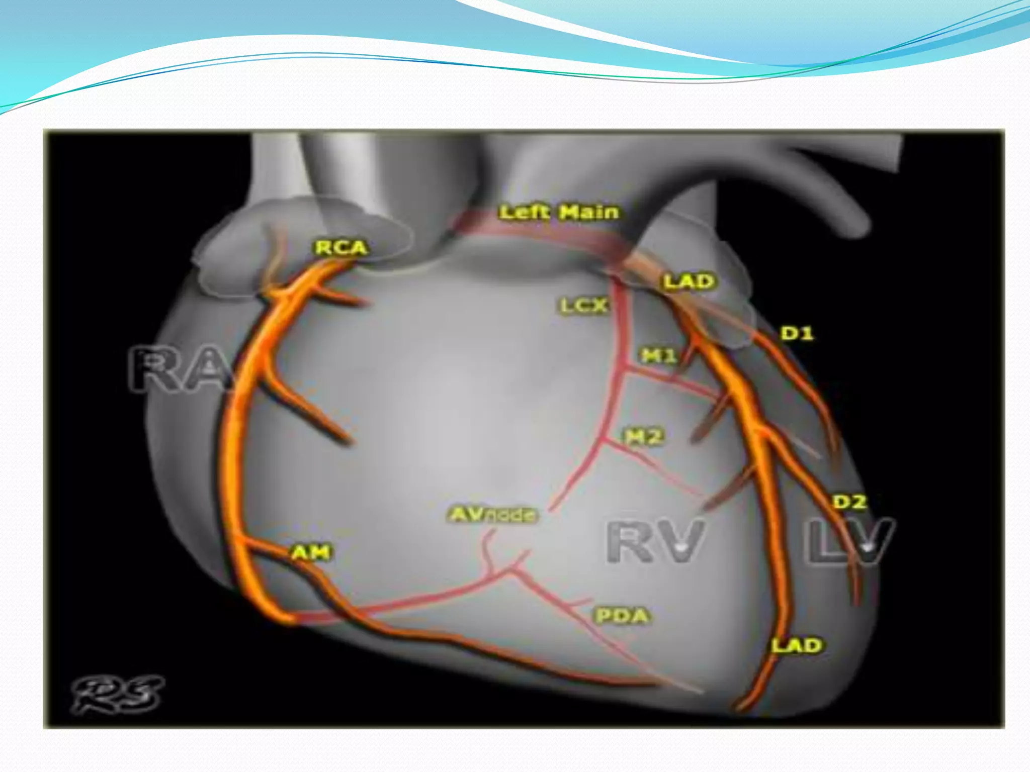
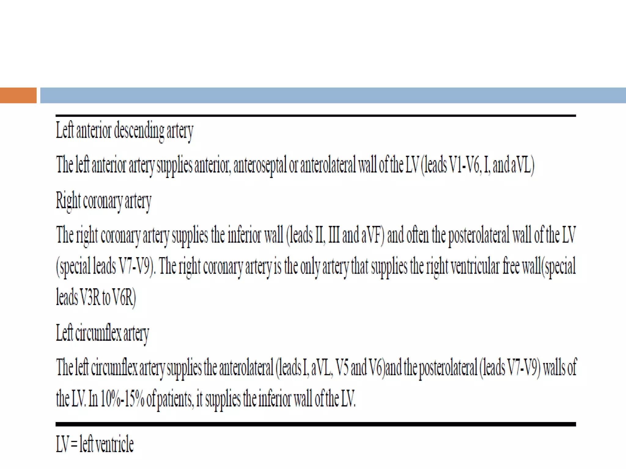
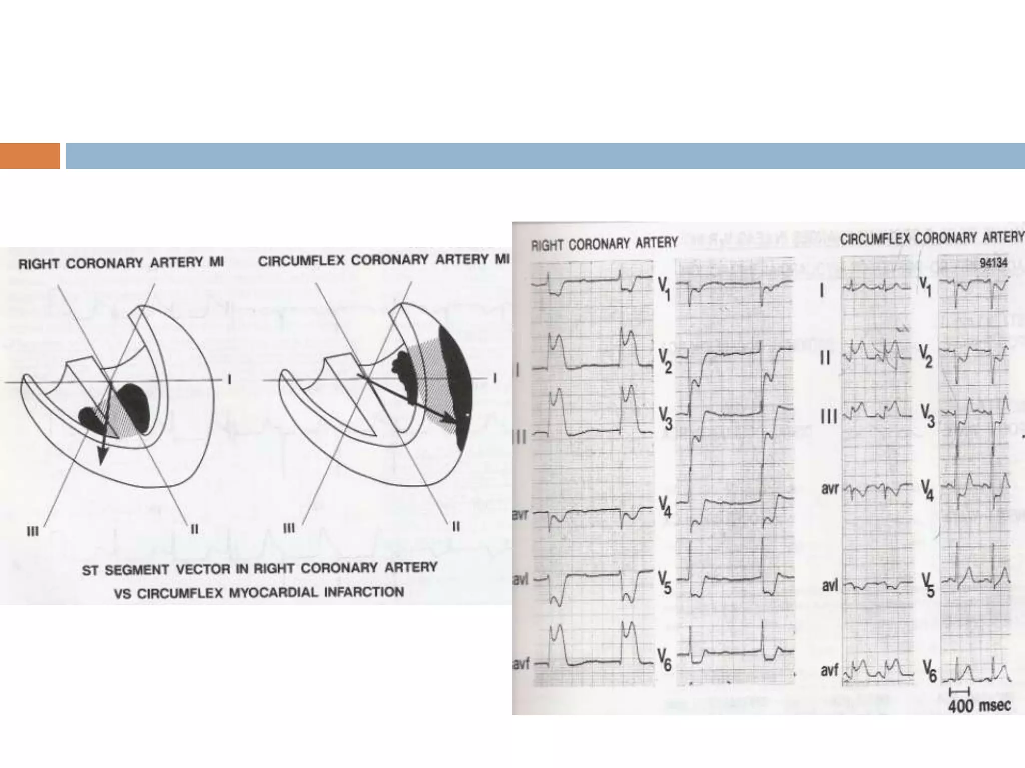
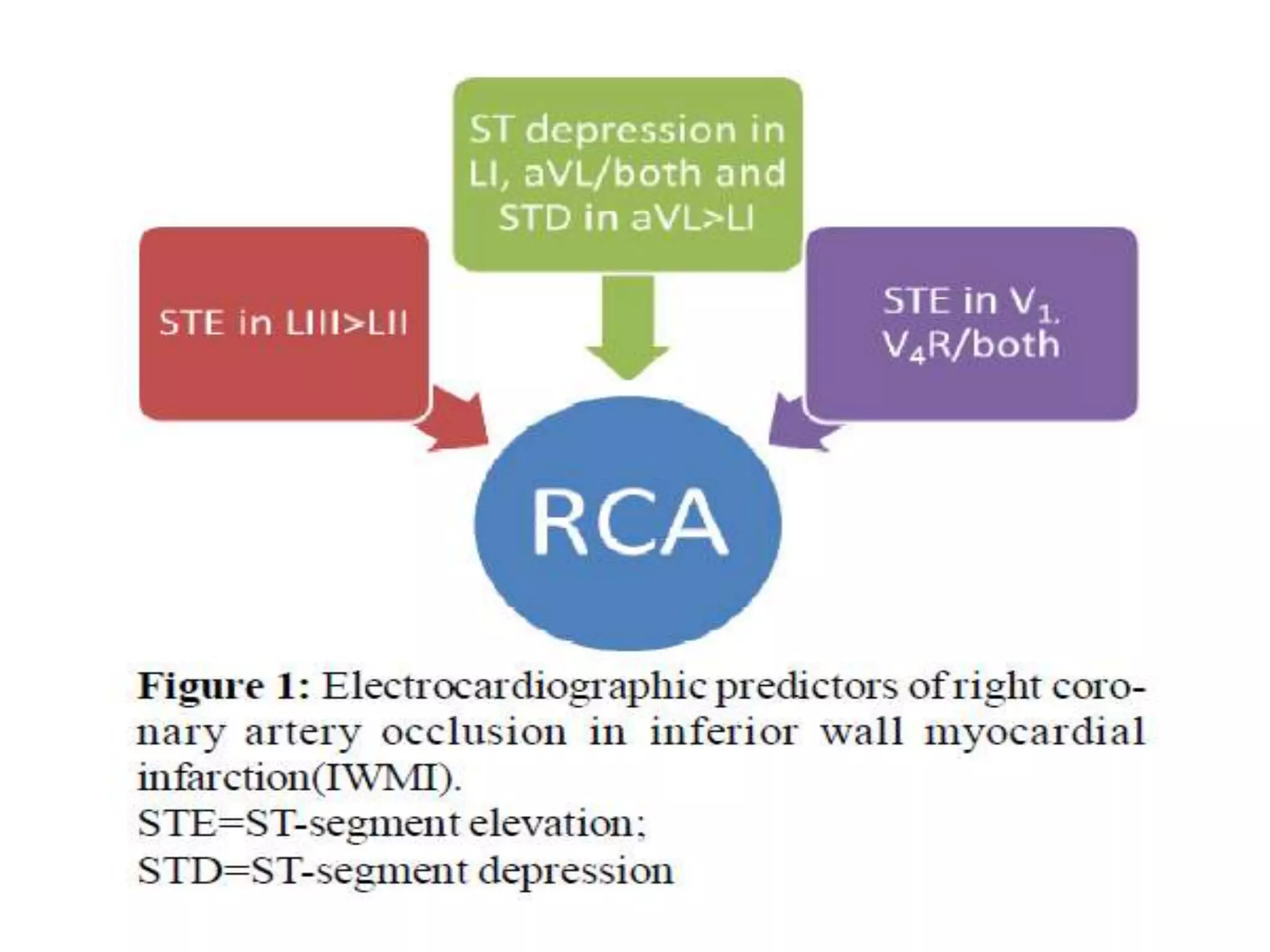
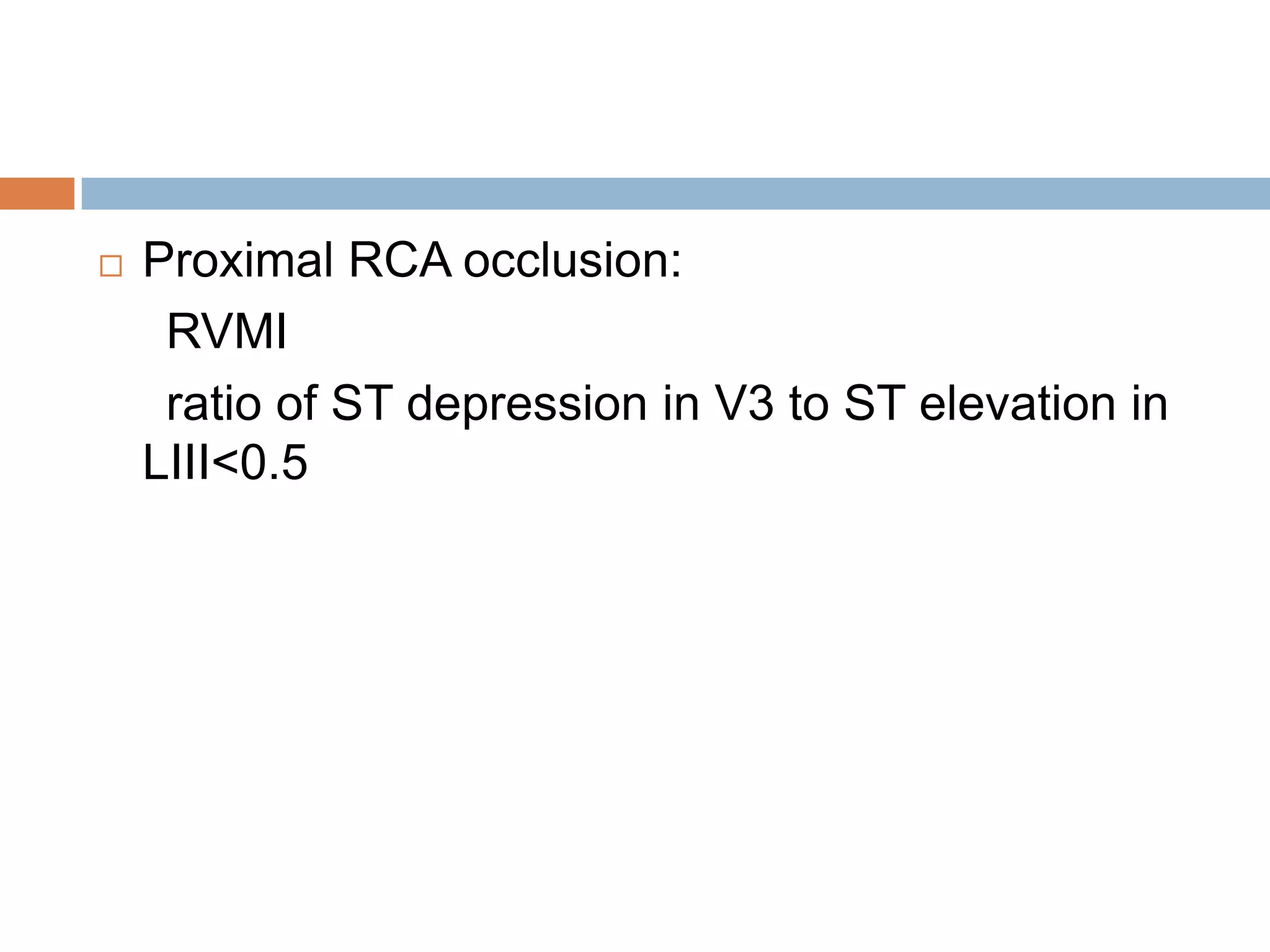
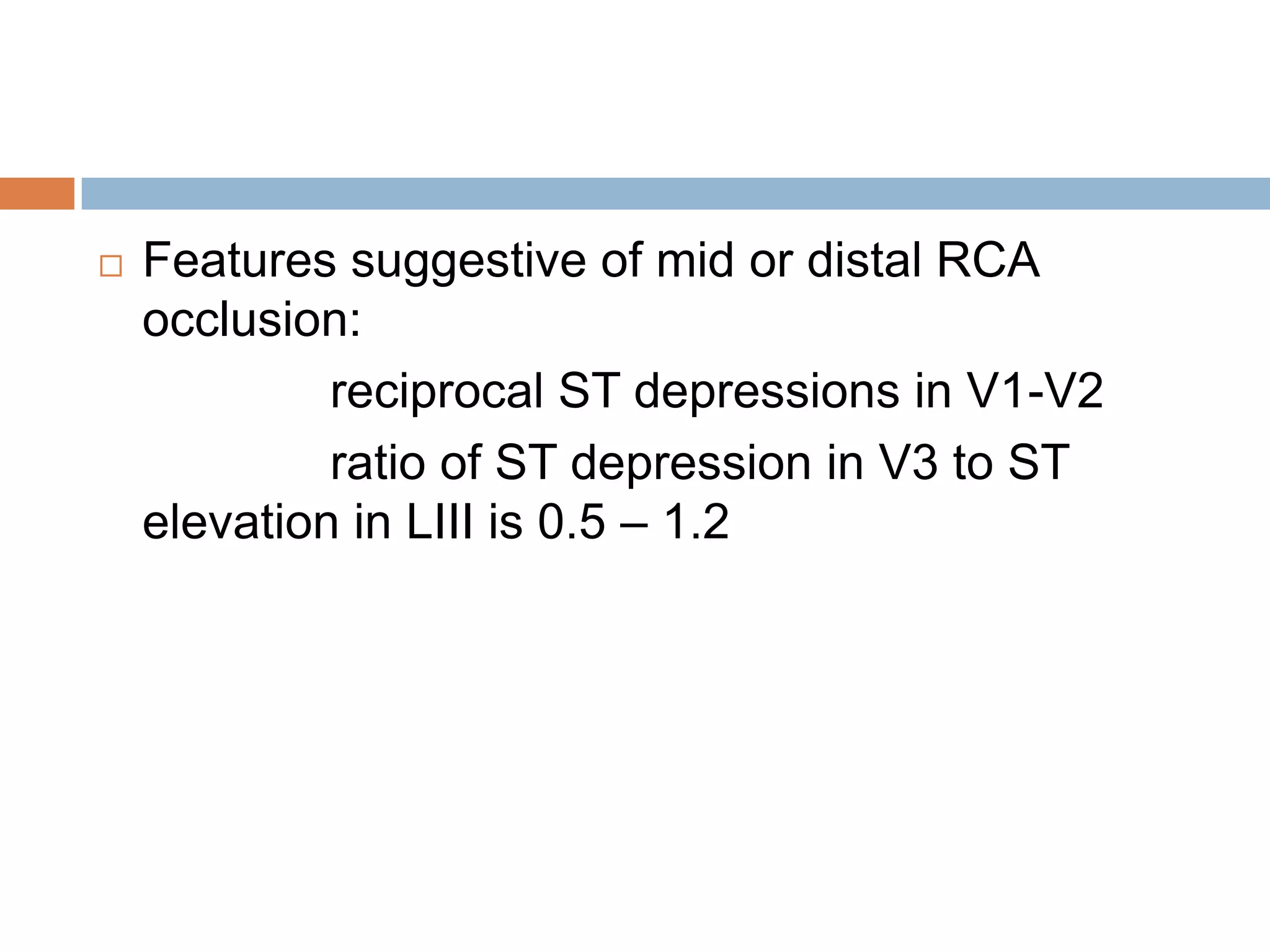
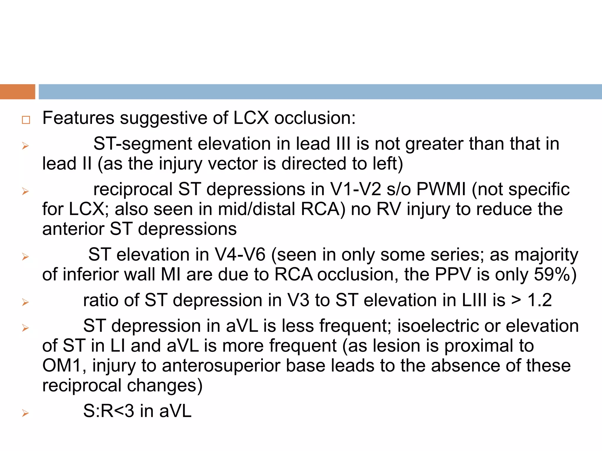
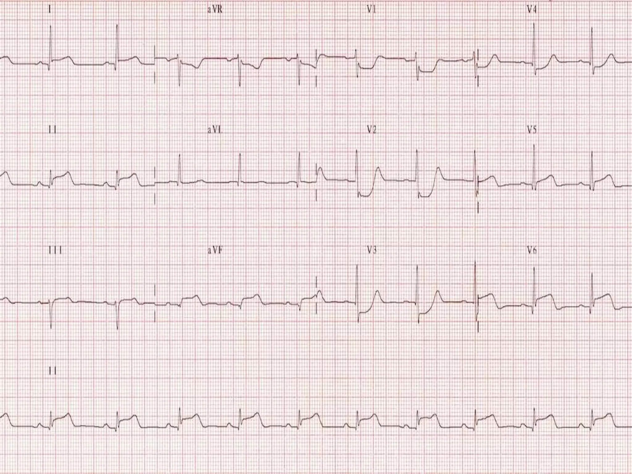
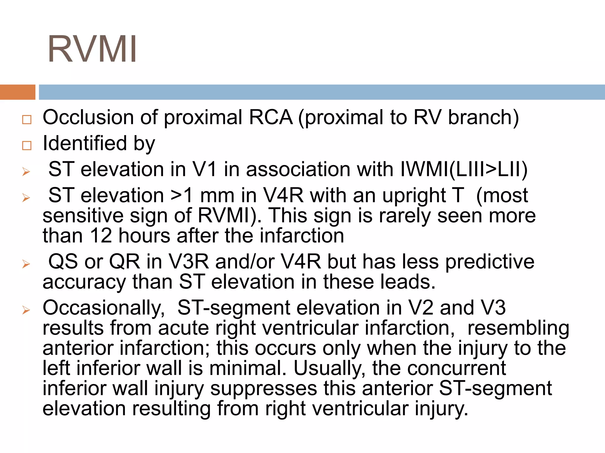
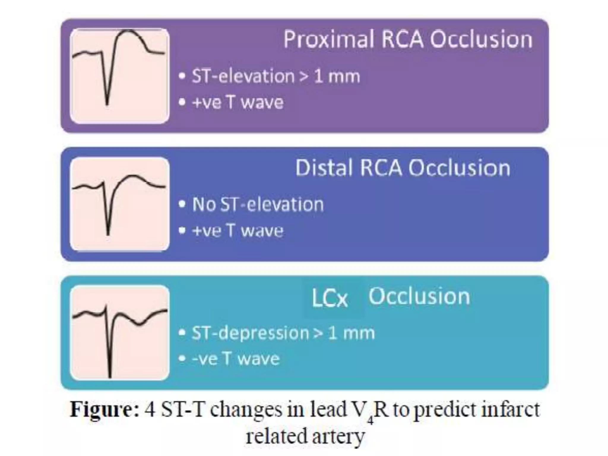
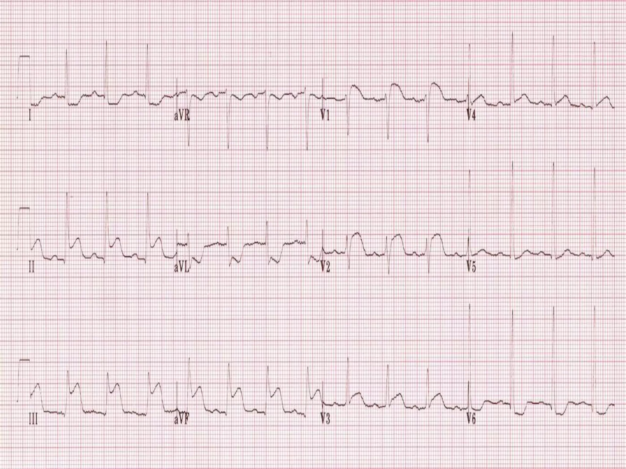
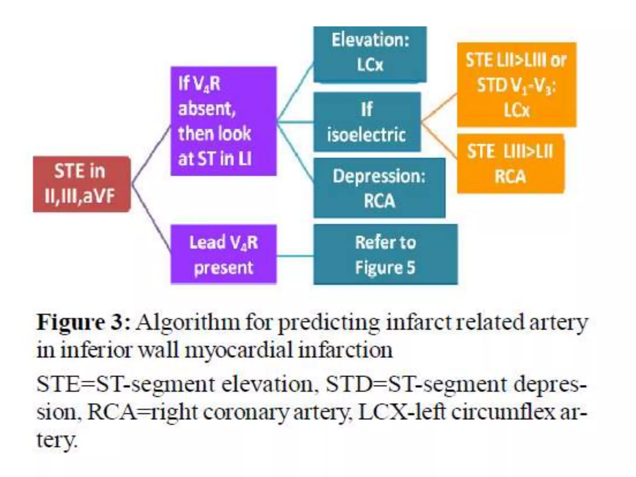
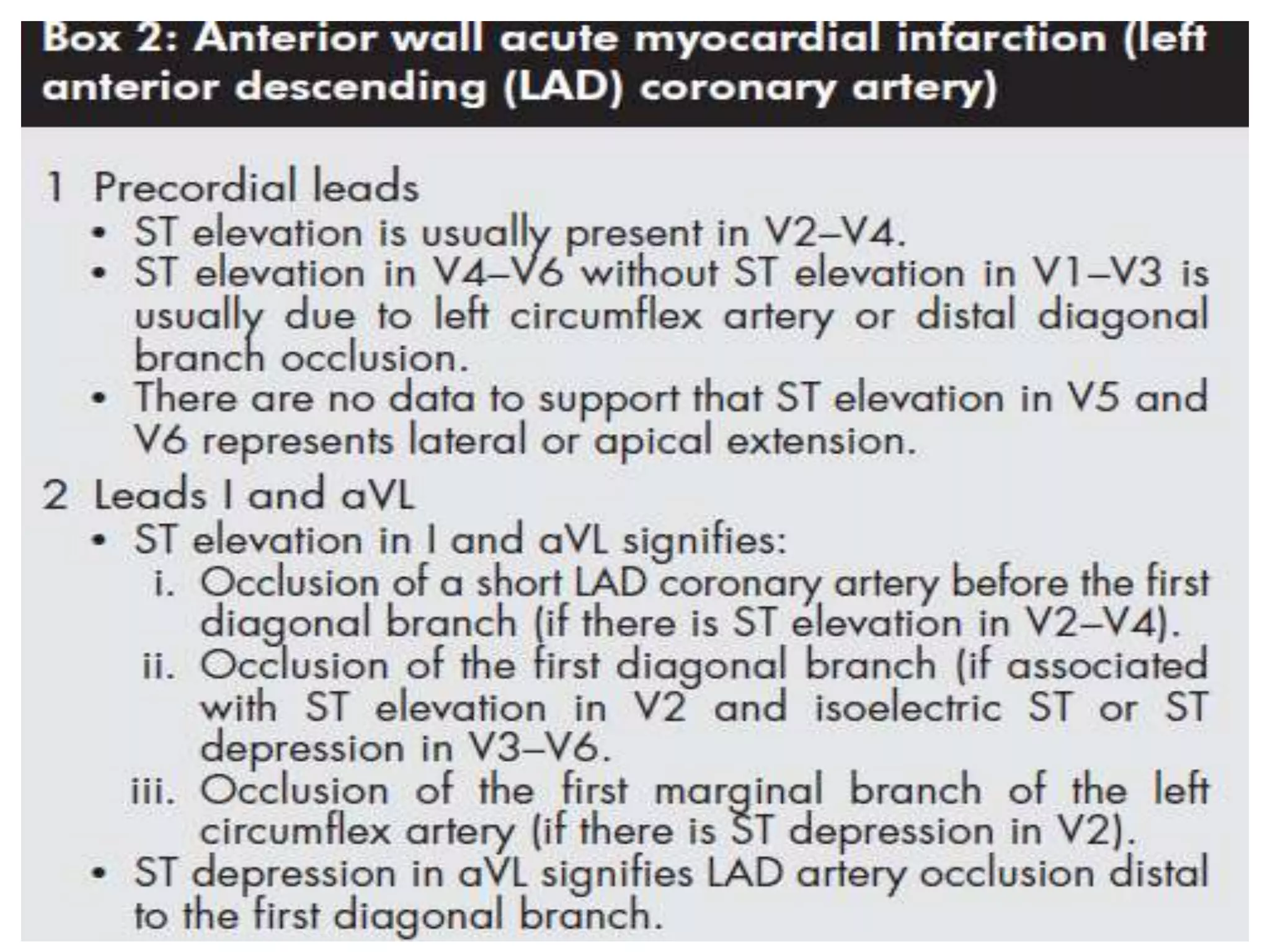
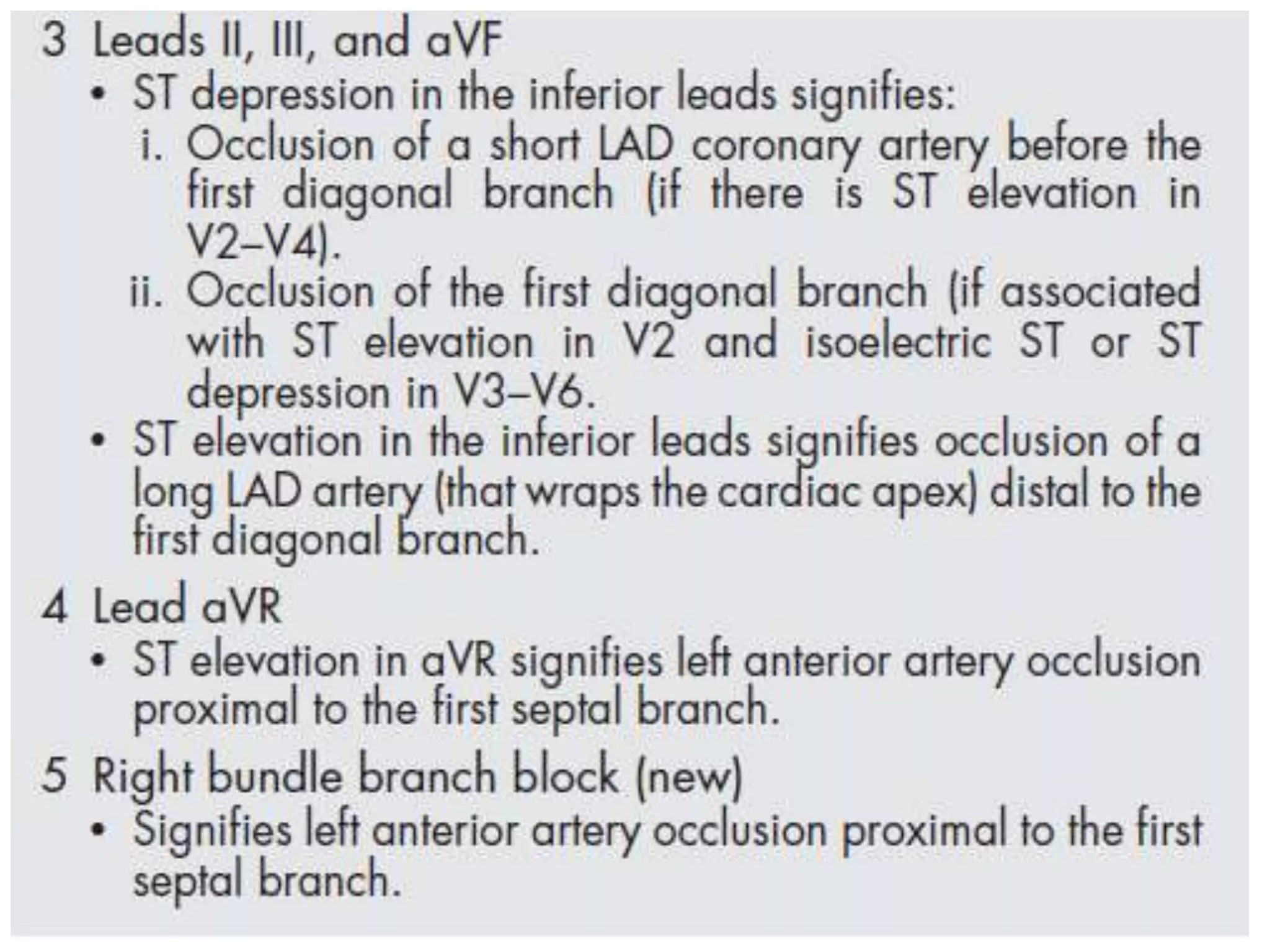
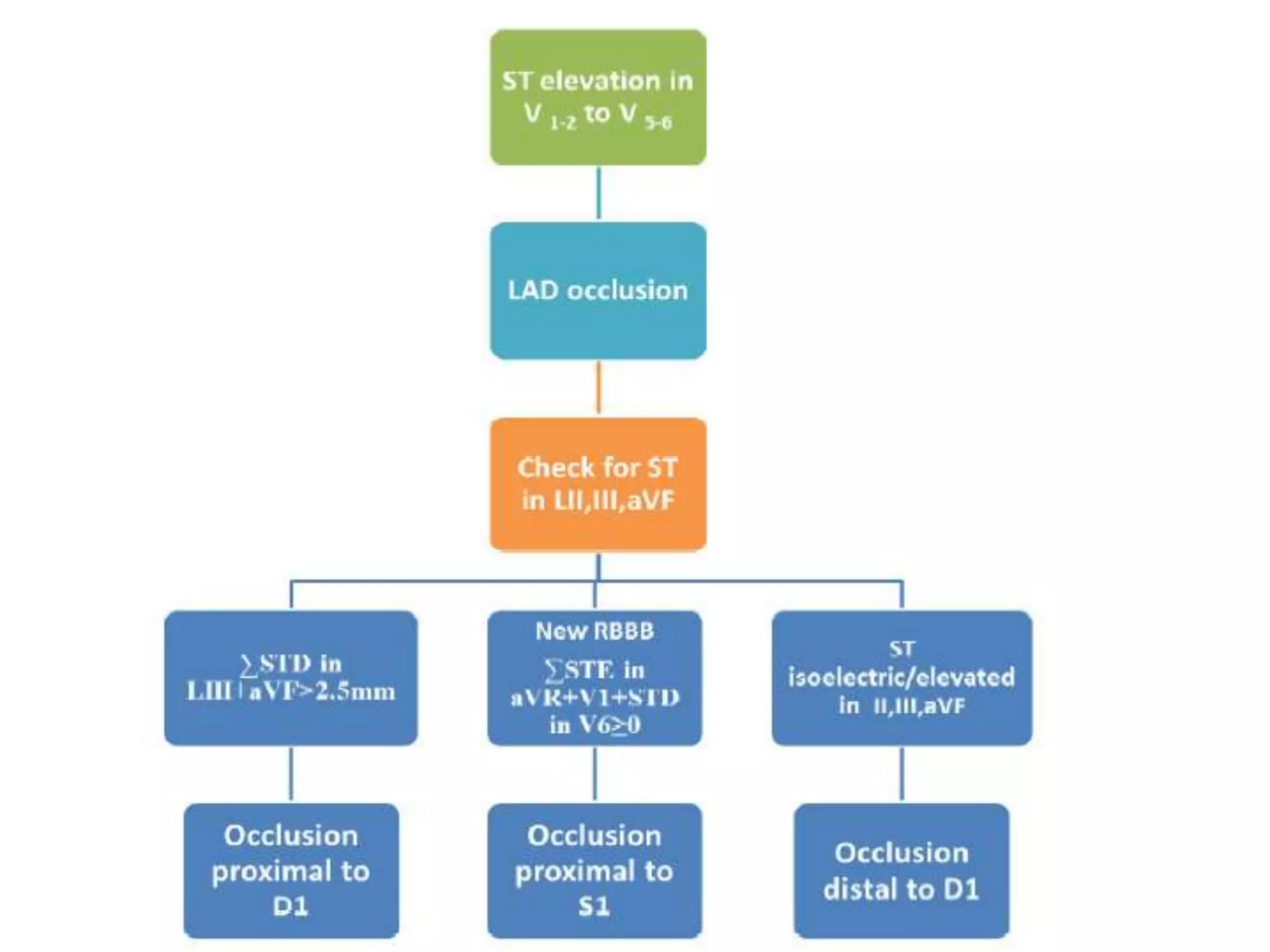
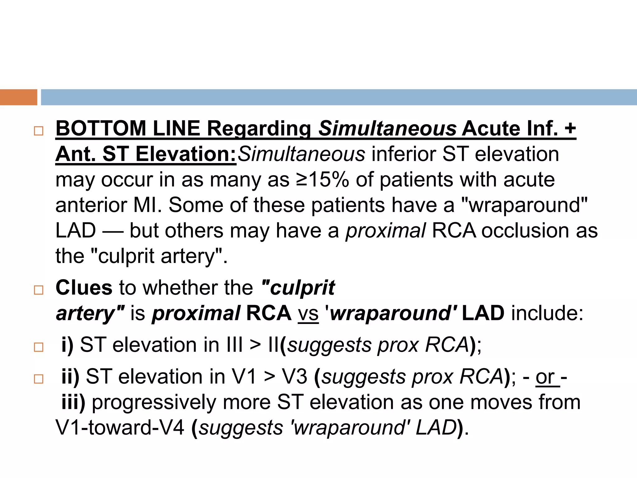
The document discusses coronary artery anatomy and ECG localization of the culprit vessel during acute myocardial infarction. It describes the typical blood supply and branches of the right coronary artery, left main coronary artery, left anterior descending artery, and left circumflex artery. It then covers how the ECG can be used to localize whether the right coronary artery, left circumflex artery, or other vessels are responsible for an acute MI based on the leads showing ST elevation and depression. Factors such as the ratio of ST changes in different leads help indicate whether proximal or distal vessels are involved.
2. Blood supply of the heart
The two coronary arteries, the right coronary artery
(RCA) and left coronary artery (LCA), originate from
their respective sinuses of Valsalva—the RCA from the
right sinus of Valsalva and the LCA from the left sinus of
Valsalva.
The right sinus of Valsalva is located anteriorly and the
left sinus of Valsalva posteriorly and to the left. The third
sinus of Valsalva, located posteriorly and to the right,
does not give rise to a coronary artery, and is referred to
as ―noncoronary cusp/sinus.‖
There may be variations in the number, shape, and
location of coronary ostia or origins of the coronary
arteries, most of which are of no clinical significance.
3.
4. RCA
The RCA arises from the right sinus of Valsalva,
inferior to the origin of the LCA.
It courses anteriorly and inferiorly under the right
atrial appendage along the right atrioventricular
(AV) groove, toward the acute margin of the heart,
where it turns posteriorly and inferiorly toward the
crux of the heart and divides into the posterior
descending coronary artery (PDA) and the
posterolateral ventricular branch (PLB)
The RCA supplies the right atrium, right ventricle,
posterior third of the interventricular septum, and
5. The conus branch arises as the first branch of the
RCA
The conus branch courses anteriorly and to the
right and supplies the pulmonary outflow tract
The sinoatrial nodal artery is the second branch
that arises from the proximal RCA in
65.4%, immediately distal to the RCA origin. In
16.6% of cases, the sinoatrial nodal artery arises
from the LCX
This artery courses toward the superior vena cava
near the cranial aspect of the interatrial septum.
6. The next branches of the RCA are the
marginal branches that supply the right
ventricular myocardium.
The acute marginal artery comes off the
acute margin of the heart and courses
anteriorly and to the right, anterior to the right
ventricle.
The acute marginal branches supply the free
wall of the right ventricular myocardium.
7. In 10% to 20% of patients, an acute marginal
branch runs on the diaphragmatic surface of the
heart to supply the distal posterior
interventricular septum.
After the RCA gives off the acute marginal
branches, it continues in the right AV groove
toward the diaphragmatic aspect of the heart. At
the crux of the heart, the RCA makes a U-turn
and branches into the PDA and PLB
8. The PDA is of variable size and runs along the
diaphragmatic surface in the posterior
interventricular groove toward the inferior
septum. Short septal branches arising
perpendicularly from the PDA supply the
posterior third of the septum and can connect
with the septal branches from the LAD to form a
collateral circulation.
The PLB runs in the posterior left AV groove and
gives off multiple branches that supply the
posterior and inferior wall of the left ventricle.
9. Within 1 to 2 cm of the crux, the PLB runs on
the diaphragmatic surface of the left ventricle
parallel to the PDA to supply the posterolateral
diaphragmatic surface of the left ventricle.
Here the RCA can serve as a collateral for an
occluded LCX.
Also close to the crux of the heart, just distal to
the PDA origin, the RCA gives rise to a small
AV nodal artery that supplies the AV node of
the conduction system . The AV nodal artery
arises from the LCX in a left dominant system.
10. LMCA
LMCA arises from the left sinus of Valsalva . It
courses to the left, beneath the left atrial
appendage and posterior to the right ventricular
outflow tract, before branching into the LAD and
the LCX
A normal variation in the anatomy is a true
trifurcation of the LM, when the middle branch
between the LAD and LCX is called the ramus
intermedius (RI)
11. LAD
The LAD runs anteriorly and inferiorly in the
anterior interventricular groove to the apex of
the heart, and supplies the anterior and
anterolateral wall of the left ventricular
myocardium and the anterior two thirds of the
interventricular septum.
In approximately 82% of cases, the LAD curves
around the cardiac apex to supply part of the
inferior wall of the left ventricle.
12. In 7%, it may not reach the apex of the
heart, and in about 11% of cases, the LAD
terminates in the distal anterior
interventricular groove or even more
proximally.
In such cases, the distal territory may be
supplied by an unusually long diagonal
branch or by RCA branches that traverse the
posterior interventricular groove or the
inferior surface of the heart, a normal variant.
This is one of the potential collateral routes if
either the RCA or the LAD is occluded.
13. The LAD gives off septal perforators and diagonal
branches.
The diagonal branches course along and supply
the anterior and anterolateral wall of the left
ventricular myocardium.
They can vary in size and number, and are
sequentially numbered as they arise from the LAD).
The septal perforator branches arise at right angles
from the LAD and supply the anterior two thirds of
the interventricular septum.
14. They are numbered sequentially as they
arise from the LAD and are of smaller caliber
then diagonal branches and vary in number
(one to five) and distribution.
The first septal branch is more constant in
position than the first diagonal. It may branch
early with both branches running parallel
within the septum. Occasionally, a septal
branch runs parallel to the LAD within the
myocardium of the septum.
15.
16. LCX
The LCX runs posteriorly and to the left in the left AV
groove . It gives rise to obtuse marginal branches that are
also numbered sequentially as they arise from the LCX
(OM1, OM2, and OM3)
The LCx artery is the dominant vessel in 15% of
patients, supplying the left PDA from the distal continuation
of the LCx.
In the remaining patients, the distal LCx varies in size and
length, depending on the number of posterolateral
branches supplied by the distal RCA.
The LCX and its branches supply the lateral and
posterolateral wall of the left ventricle. Additional branches
of the LCX are small atrial branches that supply the lateral
17. The RI is the most common variation of LCA
anatomy, occurring when the LM trifurcates; the
branch between the LAD and the LCX is the RI.
The RI can supply the myocardial territory of the
diagonal branch or the obtuse marginal branch
depending on whether it supplies the anterior wall
or the lateral wall of the left ventricular
myocardium.
When large, the RI perfuses a significant portion
of the myocardial territory of the diagonal branch
and OM1
18. DOMINANCE OF CORONARY CIRCULATION
The artery that supplies the inferior portion of the posterior
interventricular septum is considered to be the dominant artery.
In 80% to 85% of cases, the RCA is dominant; when at the crux
of the heart, it gives rise to the PDA and PLB .
In a left dominant system, the LCX continues in the posterior left
AV groove and gives rise to the PDA and PLB; this is seen in 7%
to 8% of the population.
In the remaining 7% to 8%, there is a codominant system or
balanced circulation in which the RCA gives rise to the PDA and
terminates in the posterior interventricular groove; the LCX may
also give rise to a PDA with two PDAs running parallel in the
interventricular septum, or the LCX may give rise to all
posterolateral branches
The nondominant artery is usually smaller in size and terminates
early in its respective AV groove.
19. SA Nodal Artery — is supplied by
the RCA in ~60% of patients. In the remaining
~40% — the SAN arises from either the LCx or
from both the RCA and LCx.
AV Nodal Artery — is supplied by
the PDA branch from the RCA in ~80-90% of
patients. In the remainder — the AVN arises from
the PDA off the LCx in a left-dominant circulation
.
Bundle of HIS — receives a dual blood supply
(from the AVN and LAD). Within the septum — the
HIS divides into right and left bundle branches.
20. Right Bundle Branch — is a relatively thin conduction
fascicle; it
is primarily supplied by septal perforatorsfrom
the LAD (it may also receive collaterals from the RCA
or LCx).
The common Left Bundle Branch divides after a short
distance into the LAH (Left Anterior Hemifascicle)
and LPH (Left Posterior Hemifascicle).
LAH — is supplied by septal perforators from
the LAD. LAHB (Left Anterior HemiBlock) is commonly seen
with acute anterior MI (since the LAH is very susceptible to
ischemia with anterior MI).
LPH — is much thicker and more diffuse than the LAH. The
LPH has a dual blood supply (from RCA andLCA). LPHB
(Left Posterior HemiBlock) is rare compared to
21.
22. ECG localisation
The electrocardiogram (ECG) is a key investigation in
diagnosing acute ST-segment elevation myocardial
infarction (STEMI).
During acute transmural ischaemia, one of the important
determinants of the site of coronary artery occlusion is the
direction of the vector of ST-segment deviation.
The injury vector is always oriented toward the injured
area.
The lead facing the injury vector head shows ST-segment
elevation and the lead facing the vector tail (opposite
leads) shows ST segment depression.
23. Ischaemia at a distance Vs reciprocal
changes
Patients with ST elevation in one territory often have ST
depression in other territories.
The additional ST deviation may represent acute
ischaemia due to coronary artery disease in non infarct
related arteries (ischaemia at a distance) or may
represent pure "mirror image" reciprocal changes.
Most of the common patterns of remote ST depression
probably represent reciprocal changes and not ―ischaemia
at a distance‖.
24. AWMI
In anterior myocardial infarction, ST depression
in the inferior leads is reciprocal to involvement
of the basal anterolateral region, supplied by the
first diagonal branch and represented by ST
elevation in leads I and aVL.
25. IWMI
In patients with IWMI, ST depression in aVL is a pure
reciprocal change and is found in almost all patients, and
ST depression in V1–V3 probably do not represent
―ischaemia at a distance‖, but rather reciprocal changes
due to more posterior, inferoseptal, apical, or lateral left
ventricular involvement.
In contrast, among patients with IWMI, ST depression in
V4–V6 is associated with concomitant LAD stenosis or
three vessel disease.
Thus, presence of an atypical pattern of ST
depression, and especially ST depression in leads V4–V6
in IWMI may signify ―ischaemia at a distance‖.
26. In some circumstances, both types of ST depression may
be present.
In acute myocardial infarction due to occlusion of the D1,
in addition to ST elevation in leads I, aVL and V2, there is
usually reciprocal ST depression in the inferior leads. The
reciprocal ST depression is associated with negative T
wave.
In contrast, in this type of myocardial infarction if there is
ST depression in leads V3–V5, it signifies subendocardial
involvement. This type of ST depression is associated with
tall peaked T waves.
27. IWMI
The leads showing the greatest magnitude of ST
elevation are, in descending order: leads III, aVF, and II.
Caused by occlusion of RCA (80%) or LCX
In addition to ST elevation in the inferior leads
, reciprocal ST depression in lead aVL is seen in almost
all patients with acute inferior myocardial infarction.
28. ECG confirmation of the infarct related artery
during acute inferior myocardial infarction may
be particularly valuable when coronary
angiography indicates lesions in both the right
and left circumflex coronary arteries
29.
30. Features favouring RCA as the culprit lesion:
ST elevation in LIII > LII (as injury vector is directed to
right in RCA occlusion)
ST depression in aVL > ST depression in L1 with ST
depression > 1 mm in LI and aVL
RVMI suggested by ST elevation in V3R,V4R (as RV
branch arises from proximal RCA, right ventricular injury
cancels reciprocal ST depression in V1,V2 in acute IWMI)
ratio of ST depression in V3 to ST elevation in LIII<0.5
decrease in R wave amplitude and an increase in S
wave amplitude with a S:R ratio of >3 in aVL
31.
32.
33.
34.
35. Proximal RCA occlusion:
RVMI
ratio of ST depression in V3 to ST elevation in
LIII<0.5
36. Features suggestive of mid or distal RCA
occlusion:
reciprocal ST depressions in V1-V2
ratio of ST depression in V3 to ST
elevation in LIII is 0.5 – 1.2
37. Features suggestive of LCX occlusion:
ST-segment elevation in lead III is not greater than that in
lead II (as the injury vector is directed to left)
reciprocal ST depressions in V1-V2 s/o PWMI (not specific
for LCX; also seen in mid/distal RCA) no RV injury to reduce the
anterior ST depressions
ST elevation in V4-V6 (seen in only some series; as majority
of inferior wall MI are due to RCA occlusion, the PPV is only 59%)
ratio of ST depression in V3 to ST elevation in LIII is > 1.2
ST depression in aVL is less frequent; isoelectric or elevation
of ST in LI and aVL is more frequent (as lesion is proximal to
OM1, injury to anterosuperior base leads to the absence of these
reciprocal changes)
S:R<3 in aVL
38.
39. RVMI
Occlusion of proximal RCA (proximal to RV branch)
Identified by
ST elevation in V1 in association with IWMI(LIII>LII)
ST elevation >1 mm in V4R with an upright T (most
sensitive sign of RVMI). This sign is rarely seen more
than 12 hours after the infarction
QS or QR in V3R and/or V4R but has less predictive
accuracy than ST elevation in these leads.
Occasionally, ST-segment elevation in V2 and V3
results from acute right ventricular infarction, resembling
anterior infarction; this occurs only when the injury to the
left inferior wall is minimal. Usually, the concurrent
inferior wall injury suppresses this anterior ST-segment
elevation resulting from right ventricular injury.
40.
41.
42. LATERAL APICAL MYOCARDIAL ZONE
In patients with acute inferior myocardial infarction, ST
elevation in leads V5 and V6 is thought to indicate
extension of the infarct to the lateral aspect of the
cardiac apex; however, there is as yet no direct
evidence for this.
The cause of such an extension may be occlusion of
either the left circumflex or a right coronary artery with
a posterior descending or posterolateral branch that
extends to the lateral apical zone.
Tsuka and coworkers found that ST elevation in lead
V6 during inferior acute myocardial infarction was
associated with a larger infarct size, a greater
frequency of major cardiac arrhythmias, and a higher
incidence of pericarditis during the patient’s hospital
43. PWMI
The standard 12-lead ECG is a relatively insensitive tool
for detecting PWMI
Usually caused by LCx occlusion but may also be seen in
dominant RCA occlusion.
ST-segment elevation in the posterior chest leads V7
through V9 > 0.5 mm in a case of IWMI
ST segment depression in leads V1 and V2 (reciprocal
changes) in a case of IWMI suggests concomitant
posterior wall MI
Abnormal R in V1 (0.04 in duration and/or R/S ratio > 1
in the absence of preexcitation or RVH), with inferior
or lateral Q waves, isolated - occlusion of a dominant
LCx without collateral circulation
44. Isolated ST elevation in leads V7–V9 without ST
elevation in the inferior leads occurs in only 4%
of patients with acute myocardial infarction and
is usually due to left circumflex coronary artery
occlusion
45. In inferior myocardial infarction due to proximal
right coronary artery occlusion with concomitant
right ventricular infarction, posterior wall injury
may be masked because the two opposed
electrical vectors may cancel each other (that is,
ST elevation in leads V1–V3 with right
ventricular infarction and reciprocal ST
depression in these same leads with concurrent
posterior infarction).
46.
47. AWMI
The amount of LV myocardium at risk of infarction in a case of
AWMI depends largely on the site of occlusion in the course of
LAD. Therefore knowing the site of LAD occlusion with the help
of ECG criteria in the emergency room is of immense help.
LAD occlusion may lead to a very extensive AWMI, or only
septal, apical-anterior or mid-anterior MI depending on the
location of occlusion.
Proximal LAD occlusion has been documented as an
independent predictor of worse outcome related to increased
mortality and recurrent MI and distal LAD occlusion is
considered to have a favourable outcome.
Thus, an early identification of proximal LAD occlusion has
crucial value not only from an academic standpoint, but also
from a therapeutic point of view.
48. AWMI
Precordial lead (V1-V6) ST-segment elevation in patients
with symptoms suggestive of ACS indicates STEMI due to
LAD occlusion.
ST segment changes in other precordial and frontal leads
depends on the presence of ischaemia in three vectorially
opposite areas, namely
(i) basal septal area perfused by proximal septal branch;
(ii) basolateral area perfused by 1st diagonal, and
(iii) inferoapical area, when distal LAD wraps around apex
49. During acute AWMI, the maximal ST-segment elevation is
best recorded in V2 or V3 (V1 – V4)
In descending order: V2, V3, V4, V5, aVL, V1, and V6
V2 is the most sensitive lead to record ST-segment
elevation (sensitivity 99%) and to identify the culprit lesion
at the LAD.
Lead V1 captures electrical phenomena from the right
paraseptal area, which has dual blood supply by the septal
branches of the LAD and by a conal branch of the RCA. So
patients with AWMI usually have no ST elevation in V1.
50. Rarely, ST elevation in V1–V4 signifies proximal RCA
occlusion with concomitant right ventricular infarction
RVMI that produces ST elevation in leads V1–V4 can be
distinguished from anterior MI by
ST elevation in lead V1 greater than in lead V2,
ST elevation in the right precordial leads V3R and V4R,
ST depression in lead V6, and
ST elevation in the inferior leads II, III, and aVF.
The magnitude of ST elevation in lead V1 correlates better
with the magnitude of ST elevation in lead V3R than with
lead V2, suggesting that ST elevation in lead V1 reflects the
right ventricle more than the left ventricle.
51. In the case of RVMI, the ST segment is
directed ant.ly and more than +90° to the right
(producing downward displacement of ST
segment in LI) while in the case of AS LVMI
the vector is also anterior but located from -30°
to -90° to the left in frontal plane ( producing
an elevation of ST segment in LI)
52. Anterosuperior myocardial zone:
The high anterolateral wall at the base of the LV is supplied by
D1 of LAD , OM1 of LCX , occ. Ramus intermedius
The lead that directly faces this zone is aVL.
ST elevation in L1 and aVL(part.) in AWMI indicates LAD
occlusion proximal to D1 – very specific but low sensitivity.This
is acc. by ST depression in inferior leads(reciprocal changes)
Patients with a long LAD artery that wraps around the cardiac
apex have concomitant injury to the inferiorapical and
anterosuperior walls of the left ventricle. When this happens, no
ST elevation may be seen in either anterosuperior leads (that
is, I, aVL) or inferior leads (that is, II, III, aVF) because the
opposing forces cancel each other
54. ST depression in aVL in AWMI indicates LAD occlusion
distal to D1
ST elevation in L1,aVL and V2 with iso electric or ST
depression in V3 and V4 indicates isolated D1 occlusion
In contrast, a LAD artery occlusion proximal to the first
diagonal branch results in ST elevation extending
beyond lead V2–V3 and occasionally, to V4–V6
ST elevation in L1,aVL with reciprocal ST depression in
V2 indicates LCX occlusion(as it supplies more
posteriorly)
55. ST depression in the ―inferior‖ leads II, III, and aVF
during acute AWMI indicates injury to the high
anterolateral wall and does not signify inferior wall
ischaemia.
Such reciprocal ST depression in the inferior leads
indicates LAD occlusion proximal to the first D1branch.
However, in patients with a long LAD artery that wraps
around the cardiac apex, proximal LAD artery occlusion
may not produce reciprocal ST depression in the inferior
leads because of extension of the infarction to the
inferoapical wall.
56. Several ECG criteria have been reported to indicate a LAD
artery occlusion proximal to the first septal perforator branch:
(1) ST elevation in lead aVR
(2) right bundle branch block
(3) ST depression in lead V5 and
(4) ST elevation in lead V1 >2.5mm
Birnbaum et al found no association between ST elevation in
lead V1 and LAD artery occlusion proximal to the first septal
branch in one series.
Criteria reported to indicate a LAD artery occlusion distal to
the first septal perforator branch include abnormal Q waves in
leads V4–V6
57.
58. Lateral and apical myocardial zones:
Anteroseptal pattern with ST elavation confined to V1-V3 indicates LAD
occlusion
In contrast, isolated ST elevation in leads V4–V6, without ST elevation
in leads V1–V3 is usually due to an occlusion of the left circumflex
artery or distal diagonal branch rather than the main LAD artery.
It is plausible that in patients with ―extensive anterior‖ myocardial
infarction (ST elevation in leads V1–V6), the injury extends to the distal
anterolateral wall and cardiac apex due to a long LAD artery and/or
prominent diagonal branches,whereas patients with an ―anteroseptal‖
pattern (ST elevation confined to leads V1–V3) have a short LAD artery
or large obtuse marginal branches or ramus intermediate branch that
supply these anterolateral and apical zones.
However, there have been no investigations to determine whether there
are differences in coronary anatomy between patients with an
―anteroseptal‖ versus an ―extensive anterior‖ myocardial infarction ECG
pattern
59. Inferior myocardial zone:
During acute anterior myocardial infarction,
injury may extend to the inferior wall, as evidenced
by ST elevation in leads II, III, and aVF, if the LAD
artery wraps around the cardiac apex.
However, anterior myocardial infarction that is
caused by a LAD artery occlusion proximal to the
first diagonal branch does not manifest such an
―anterior and inferior‖ injury pattern because of
cancellation of opposing vectors
60. Indicators of proximal LAD occlusion on surface ECG
are
ST-segment elevation in V1-V3 and aVL and aVR
and
ST-segment depression of >1 mm in lead aVF
ST-segment depression in V5
disappearance of preexistent septal Q waves in
lateral leads
(direction of ST-segment vector is upward, toward
leads V1, aVL, and aVR, and away from the inferior
leads)
New QRBBB in V1 is a specific but insensitive
61. Occlusion of the LAD beyond the origin of the first
diagonal branch:
ST-segment elevation in leads V1, V2, and V3
without significant inferior ST-segment depression
LAD occlusion distal to the origin of the first
diagonal branch, in a vessel that wraps around the
apex to supply the inferoapical region of the left
ventricle:
ST-segment elevation in leads V1, V2, and V3 with
concomitant elevation in the inferior leads
62.
63.
64.
65.
66. BOTTOM LINE Regarding Simultaneous Acute Inf. +
Ant. ST Elevation:Simultaneous inferior ST elevation
may occur in as many as ≥15% of patients with acute
anterior MI. Some of these patients have a "wraparound"
LAD — but others may have a proximal RCA occlusion as
the "culprit artery".
Clues to whether the "culprit
artery" is proximal RCA vs 'wraparound' LAD include:
i) ST elevation in III > II(suggests prox RCA);
ii) ST elevation in V1 > V3 (suggests prox RCA); - or -
iii) progressively more ST elevation as one moves from
V1-toward-V4 (suggests 'wraparound' LAD).
67. LMCA stenosis
Typical ECG findings in severe LMCA stenosis or
occlusion include ST-segment elevation in lead aVR
more than V1 with either widespread ST-segment
depression or anterior ST elevation.
Yamaji et al described an aVR ST-segment of >0.05 mV
elevation present in 88% of the LMCA obstruction group
compared with 46% in the left anterior descending
artery.
68.
69. Grading of ischaemia
Shortly after occlusion of a coronary artery, serial
ECG changes are detected by leads facing the
ischaemic zone
Grade I ischaemia: the T waves become tall,
symmetrical, and peaked
Grade II ischaemia: there is ST elevation , without
distortion of the terminal portion of the QRS
Grade III ischaemia: changes in the terminal
portion of the QRS complex appear. These changes
include an increase in the amplitude of the R waves
and disappearance of the S waves.
70.
71. Differentiation between viable and necrotic
myocardium at the ischemic area at risk
Q waves were traditionally considered as a sign of myocardial
necrosis
It has been suggested that Q waves that appear within six
hours from onset of symptoms
do not signify irreversible damage,
do not preclude myocardial salvage by thrombolytic
therapy
transient and disappear later.
Several authors have found early Q waves to be associated
with larger ischaemic zone and ultimate infarct size.
90 minutes after thrombolytic therapy, TIMI flow grade III is
achieved less often in patients with than without abnormal Q
waves on presentation
72. Early inversion of the T waves, along with ST elevation
resolution, is a sign of reperfusion
Wong et al reported that 90 minutes after thrombolytic
therapy, TIMI flow grade III was seen less often in
patients presented with ST elevation and negative T
waves than in those with positive T waves
Therefore, ST elevation with negative T waves,
especially if it occurs in patients presenting more than
two hours of onset of symptoms, might be a sign of a
more advanced stage of myocardial infarction with
lesser chance of achieving successful reperfusion and
higher mortality.
It might be a sign that irreversible damage has already
73. PRESENCE OF OLD
MYOCARDIAL INFARCTION
Presence of abnormal Q waves in leads without ST
elevation is suggestive of old myocardial infarction.
However, pathological Q waves in leads with ST
elevation do not necessarily mean old myocardial
infarction or completion of the present acute myocardial
infarction.
75. Acute INFERIOR Infarction:
Sinus Bradycardia and 1st, 2nd, or 3rd degree AV block may all
be seen with acute inferior MI. When they occur early (within the
first ~6 hours) — increased vagal tone is the most common
mechanism. As a result —Atropine (sometimes in low dose) tends
to be very effective if treatment is needed.
Complete (3°) AV block with acute inferior MI is generally at the
level of the AV Node. As a result
the QRS isusually narrow and the escape rate acceptable
(between 40-60/min) — such that the patient may not be
symptomatic even when AV block is complete.
Edema of the AV node (rather than increased vagal tone) — is
the mechanism of AV block that develops later (after 12-24
hours). Atropine is much less likely to work in such cases —
although AV block usually still resolves on its own over days-to-
weeks as edema subsides (permanent pacing is usually not
needed)
76. Acute ANTERIOR Infarction
Vagal tone is not implicated (Atropine is therefore unlikely to work).
Sinus Tachycardia — is typically seen with acute anterior MI (due to enhanced sympathetic
tone – and/orassociated heart failure). This may respond to cautious use of IV beta-blockers.
Conduction system damage is due to septal necrosis. Risk of complications is greatest
with LMain orproximal LAD occlusion (prior to S-1 takeoff ).
1st Degree AV Block — may also be seen with anterior MI (not due to AV nodal ischemia — but
rather from HIS involvement).
2nd Degree AV Block with anterior MI — is typically Mobitz II. The QRS is wide because the
level of block isbelow the AV Node. Atropine is ineffective — and pacing is essential (since
complete AV block or ventricular standstill may abruptly occur).
New RBBB with anterior MI is a sign of severe conduction system damage. There may
be bifascicular block (usually RBBB/LAHB — but occasionally with extensive necrosis
RBBB/LPHB).
PACING (both temporary and permanent) — is much more likely to be needed
with anterior MI. Additivedefects (ie, RBBB plus 1st degree; RBBB/LAHB) will increase risk of
complete AV block.
Development of severe conduction disturbance with acute anterior MI is a poor prognostic
sign (indicative of extensive myocardial necrosis = high risk of cardiogenic shock).