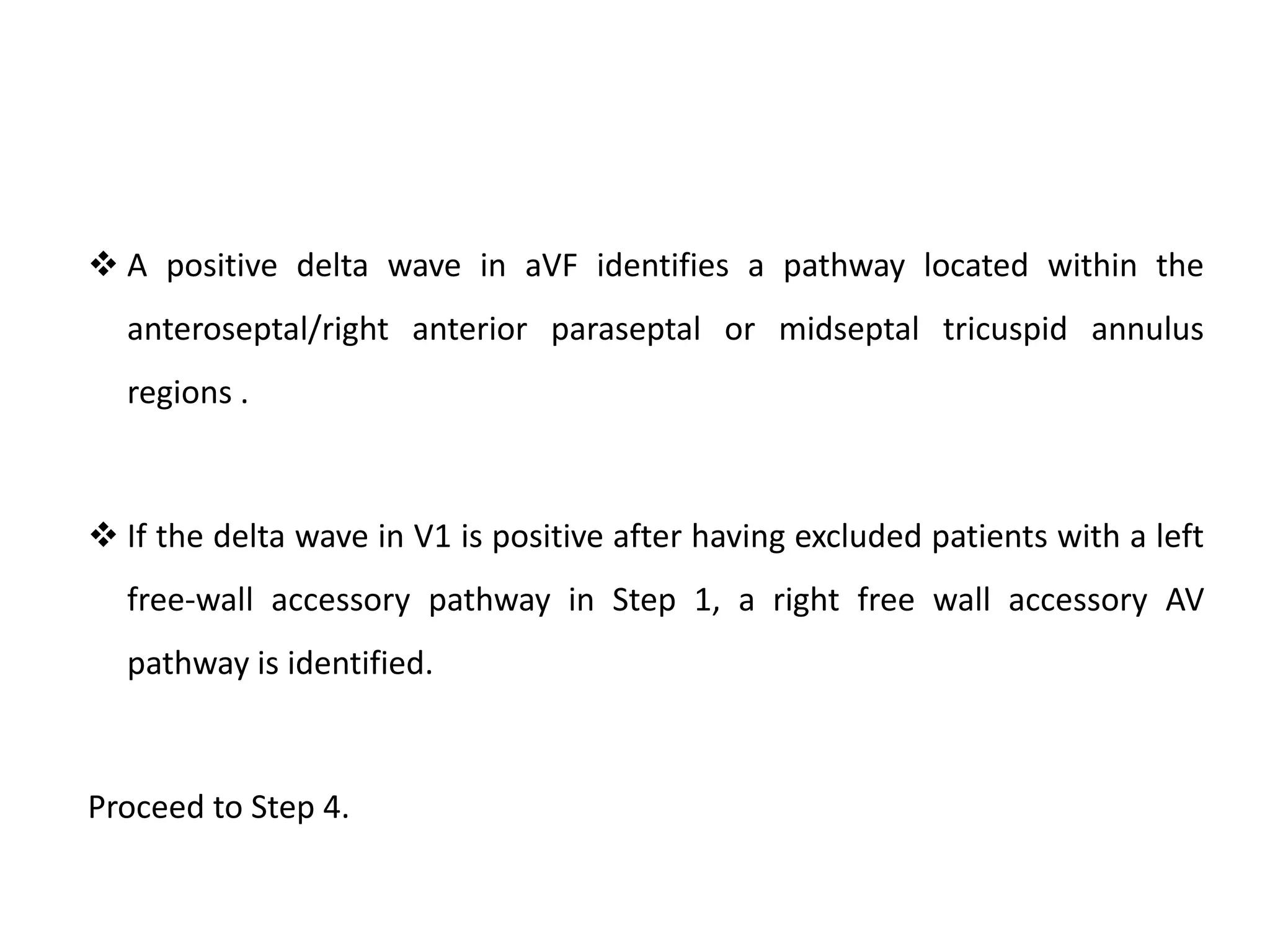
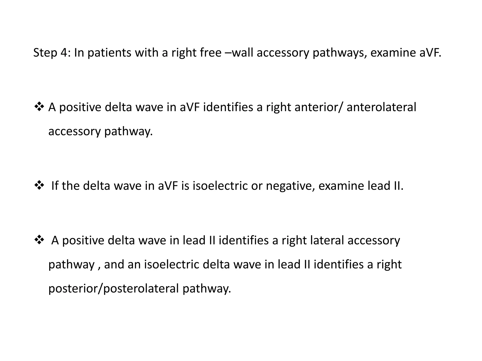
Downloaded 858 times
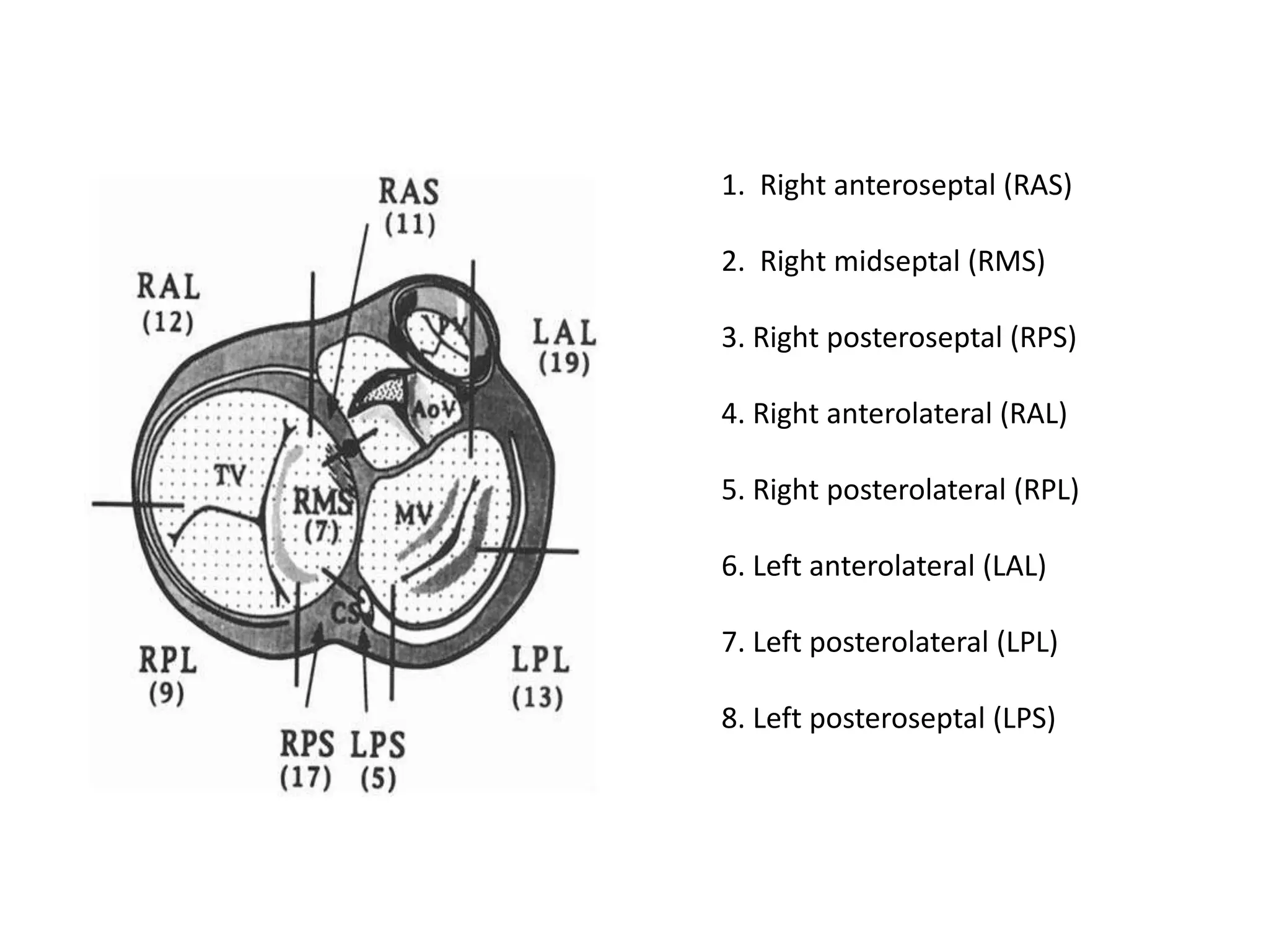
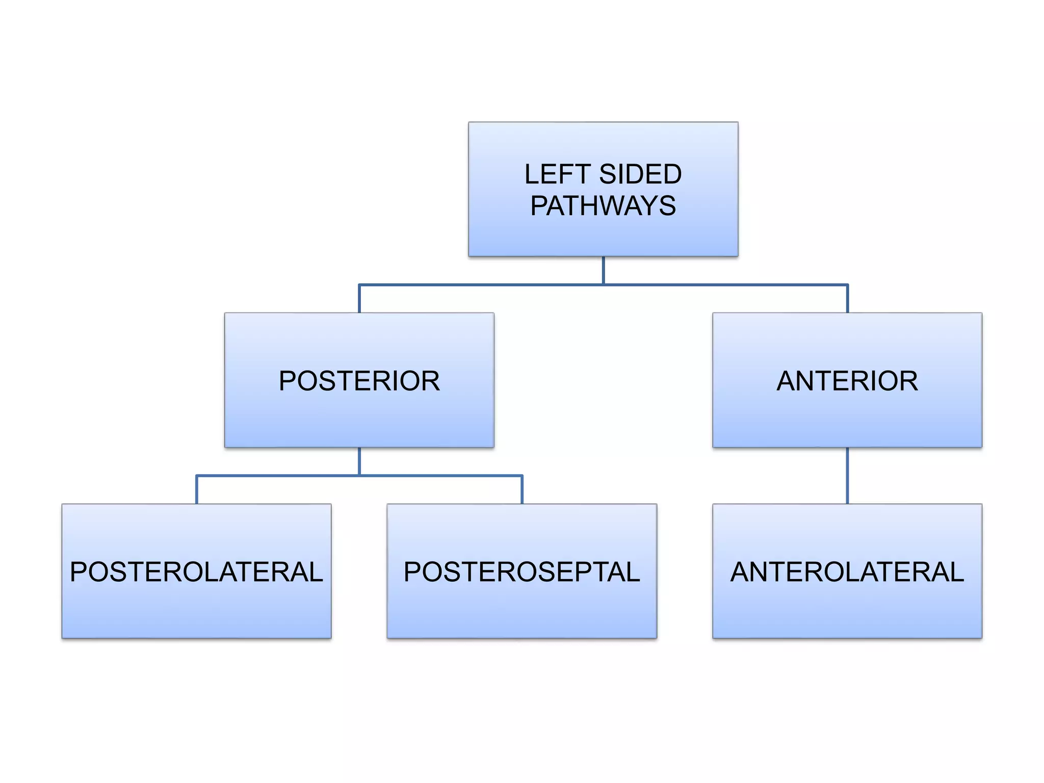
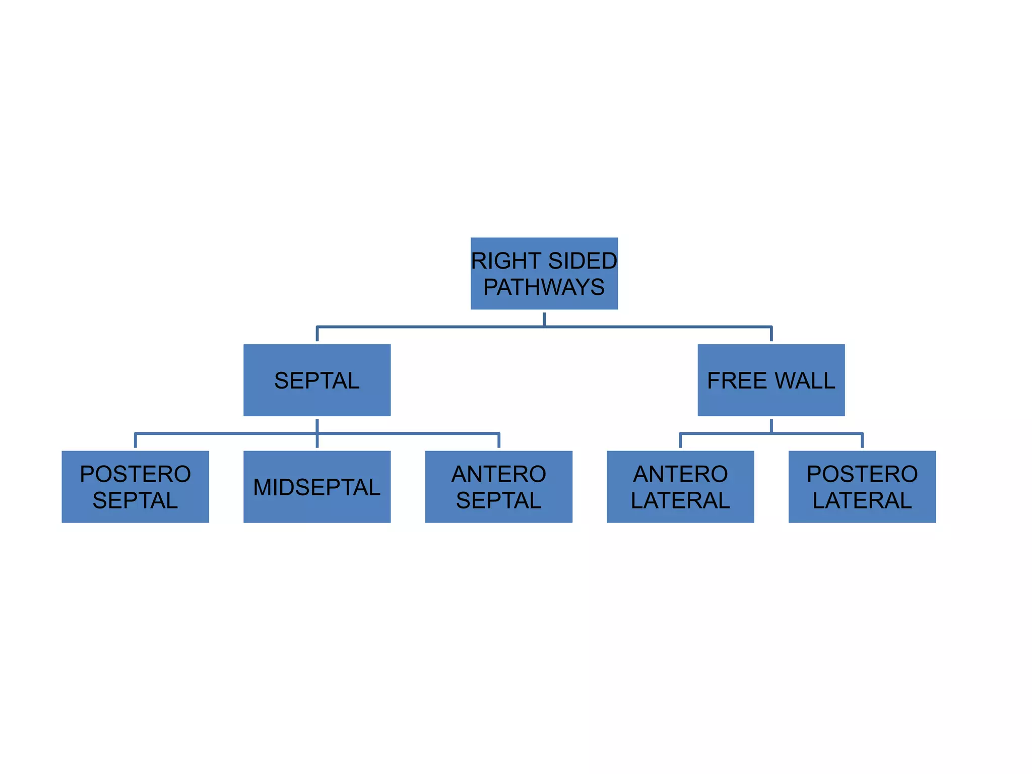
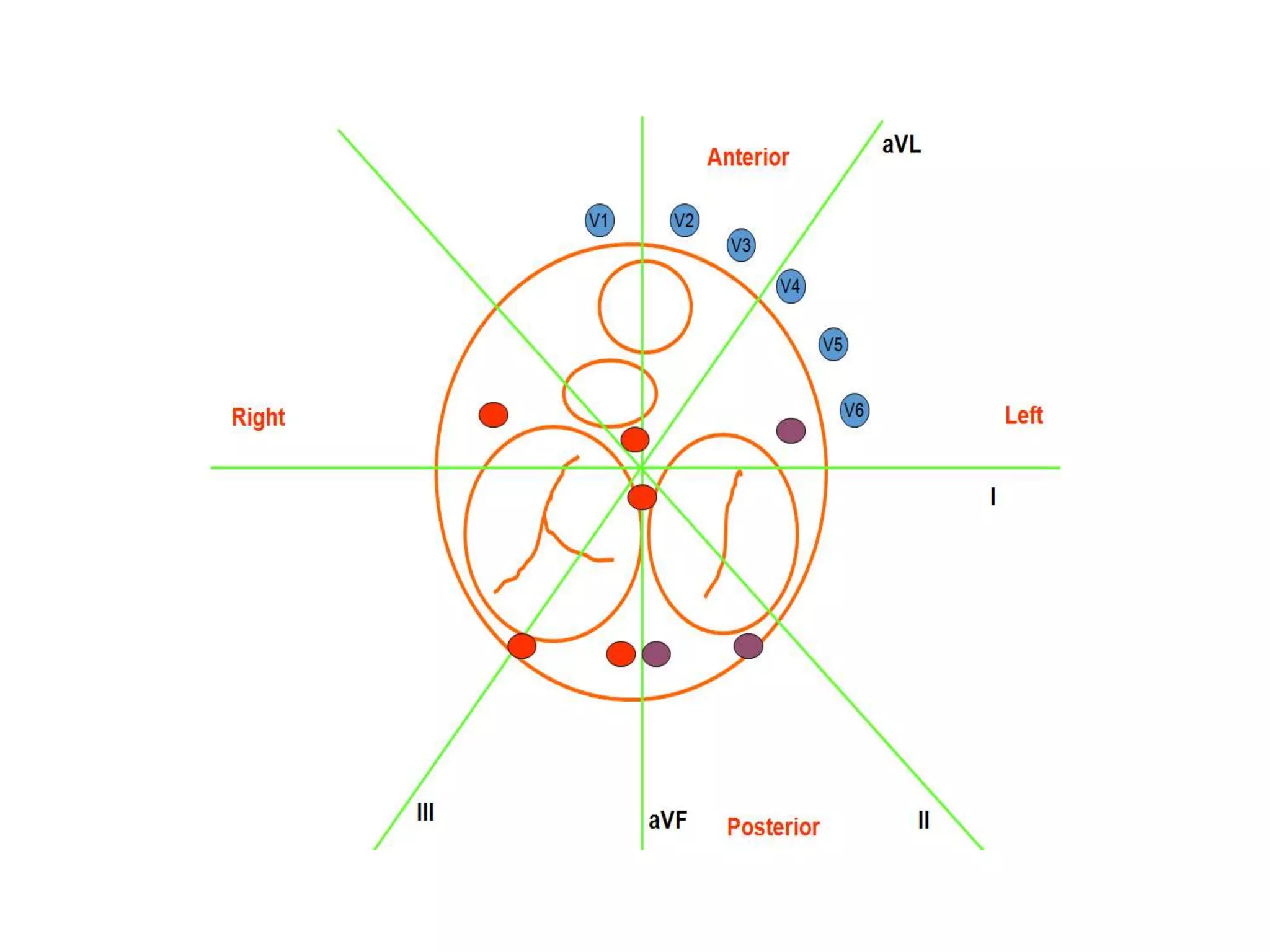
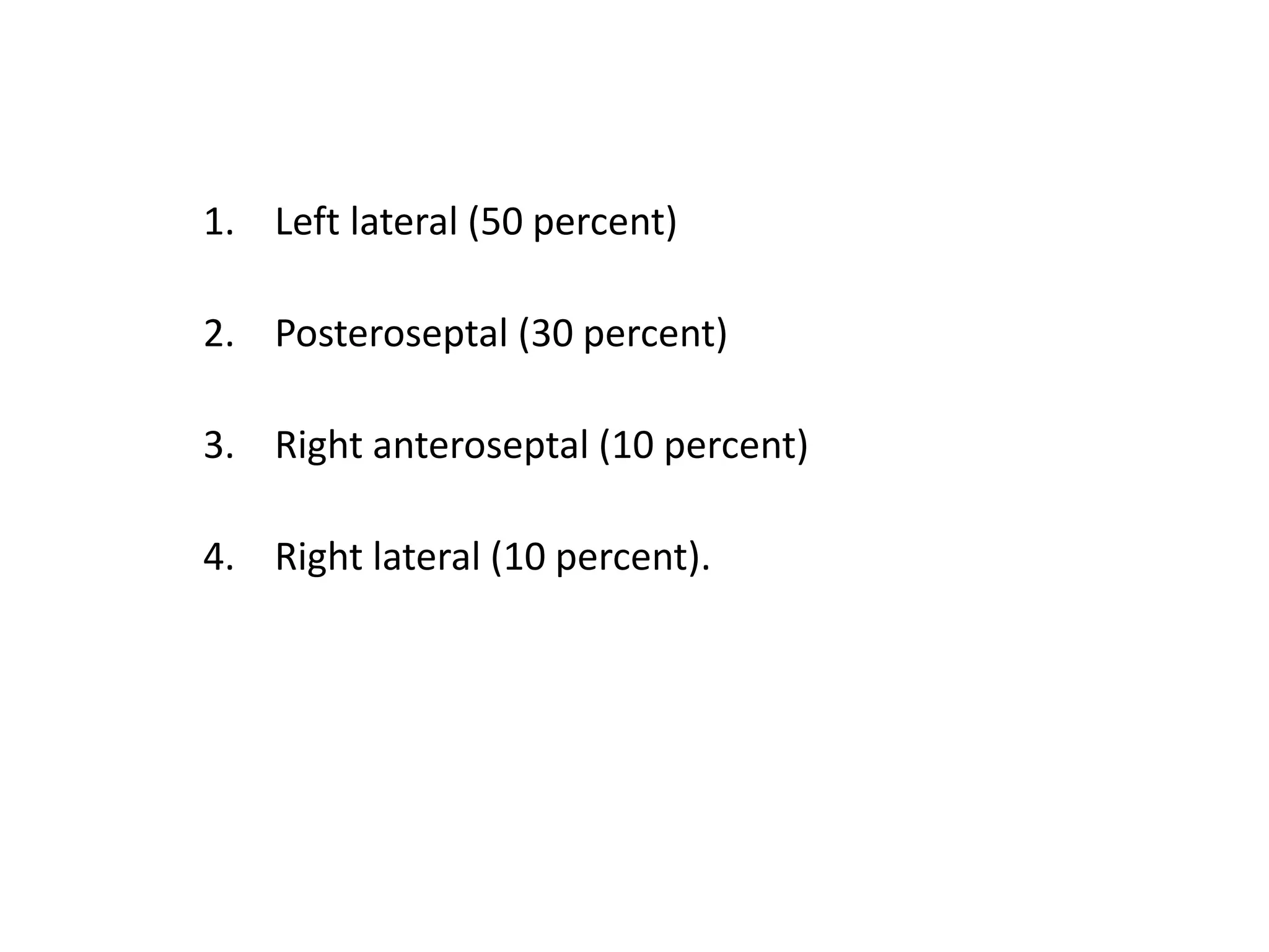
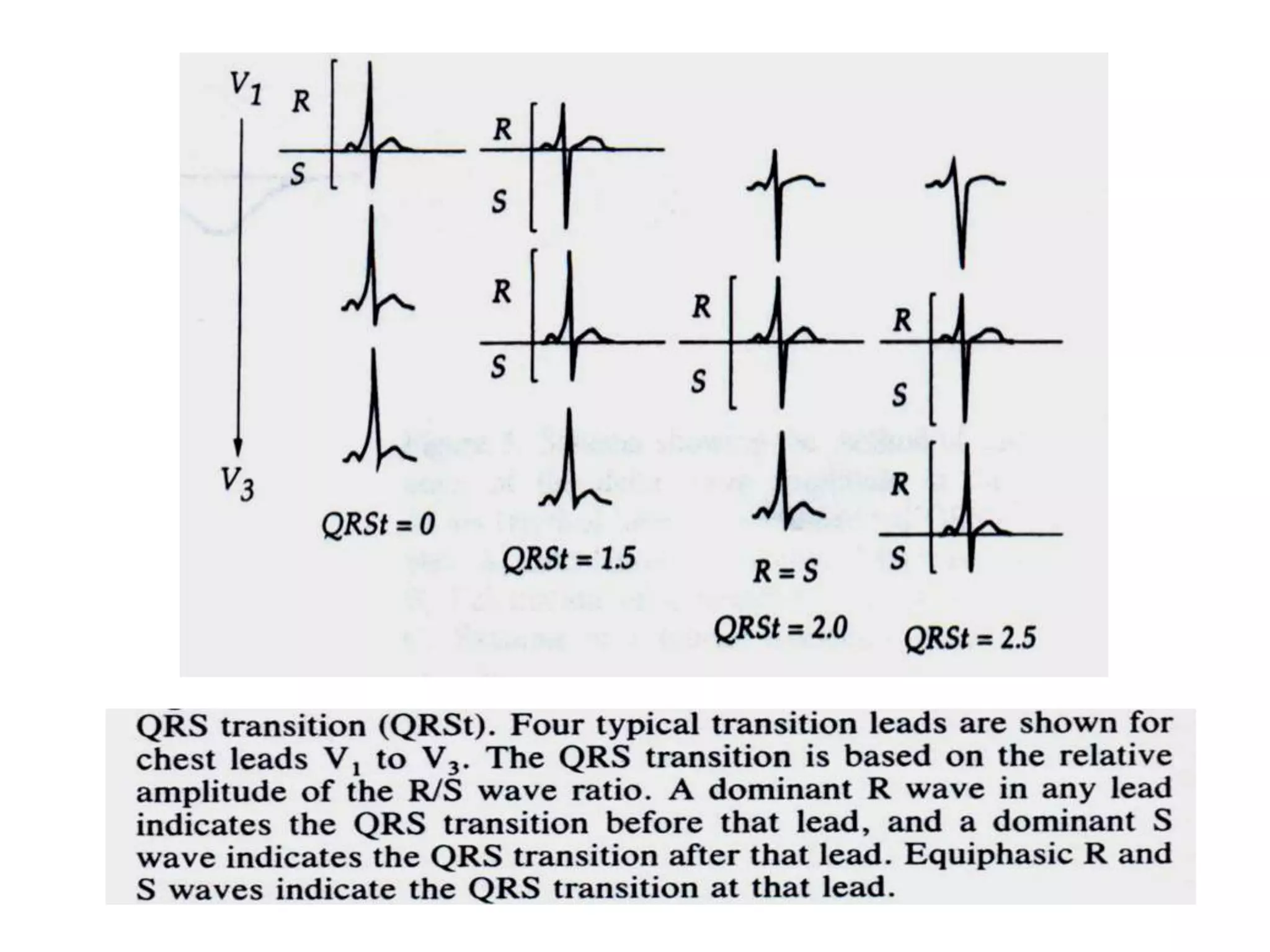
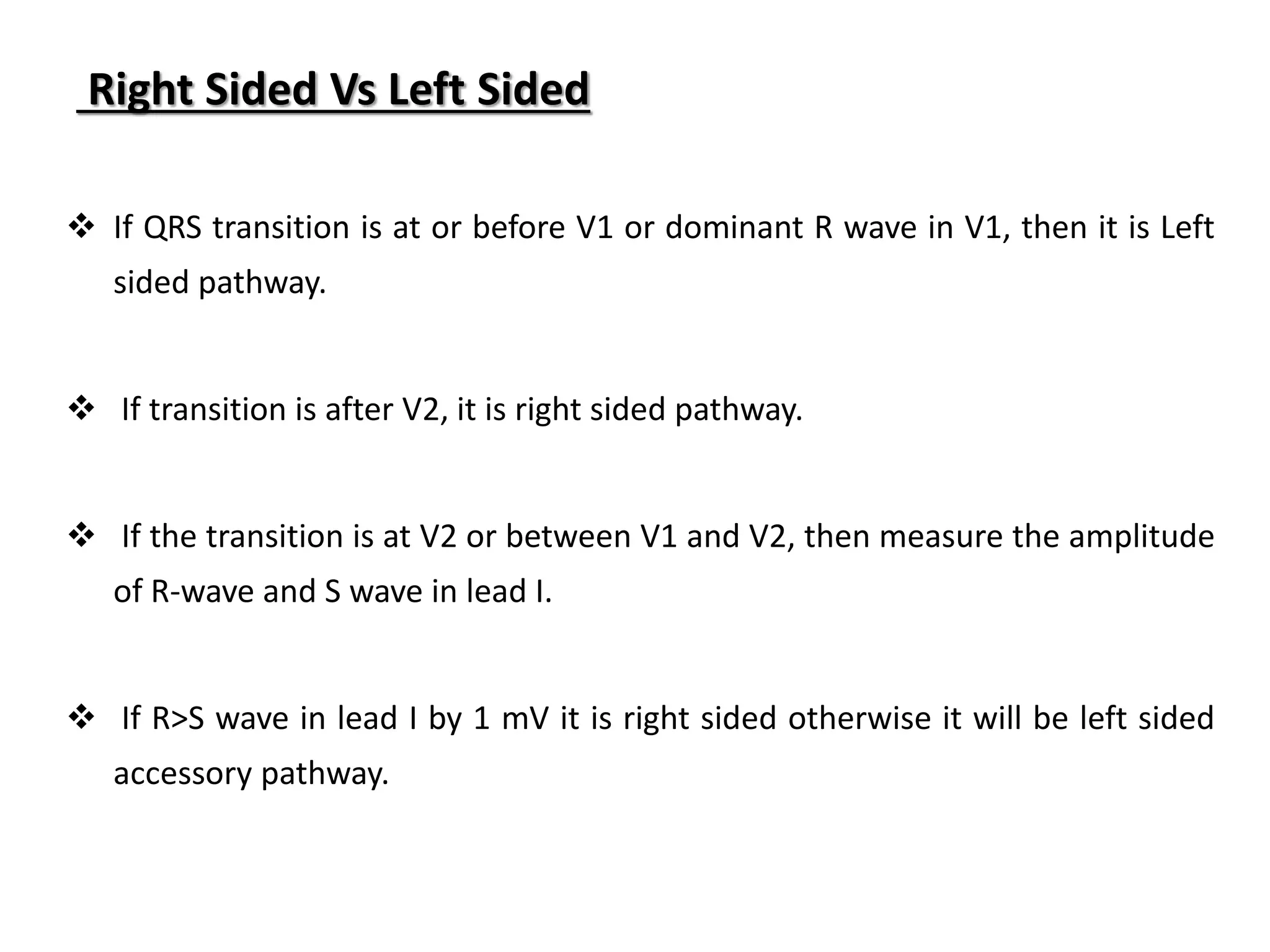
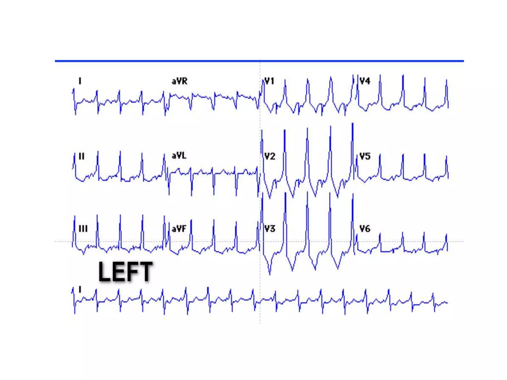
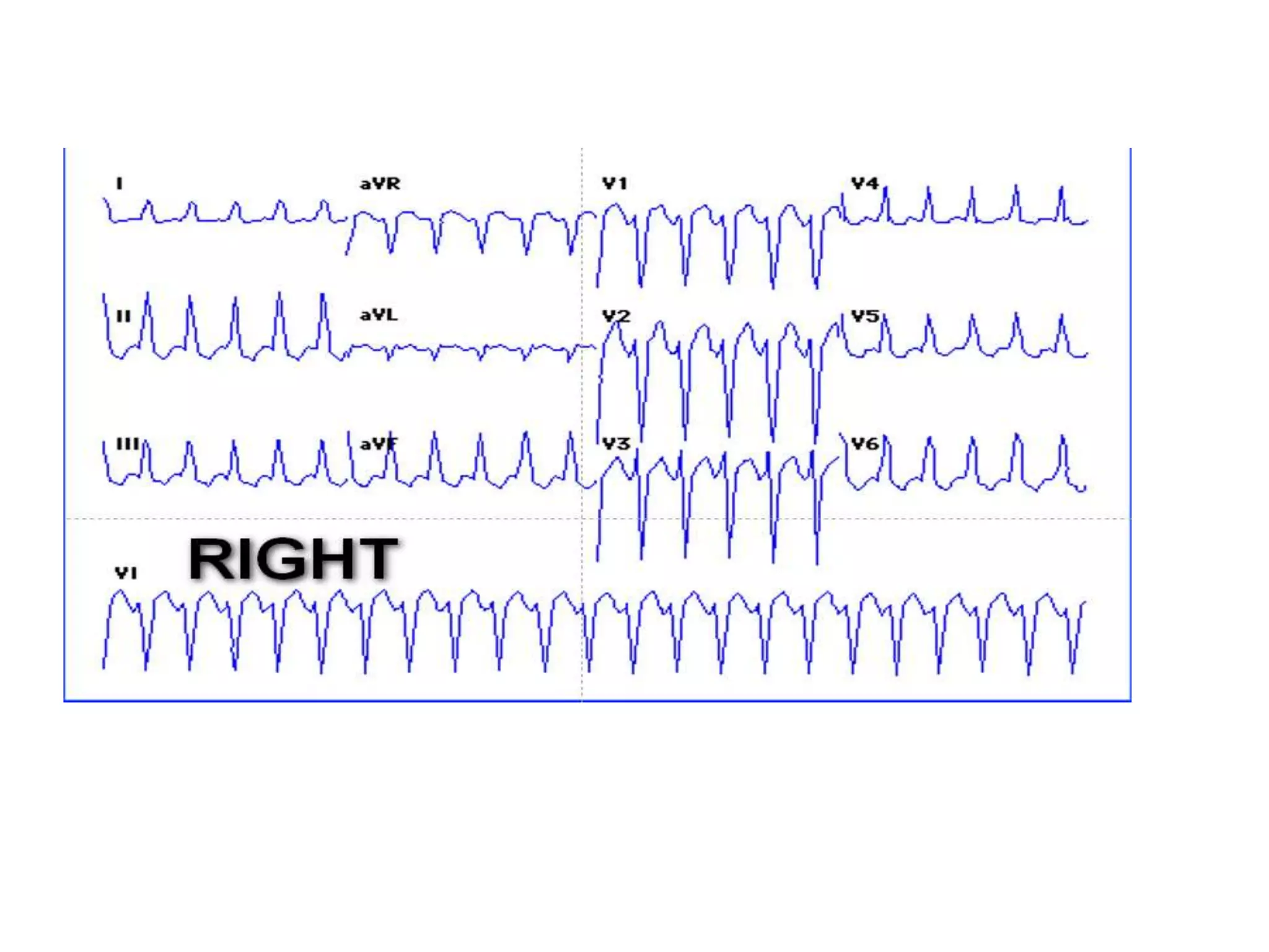
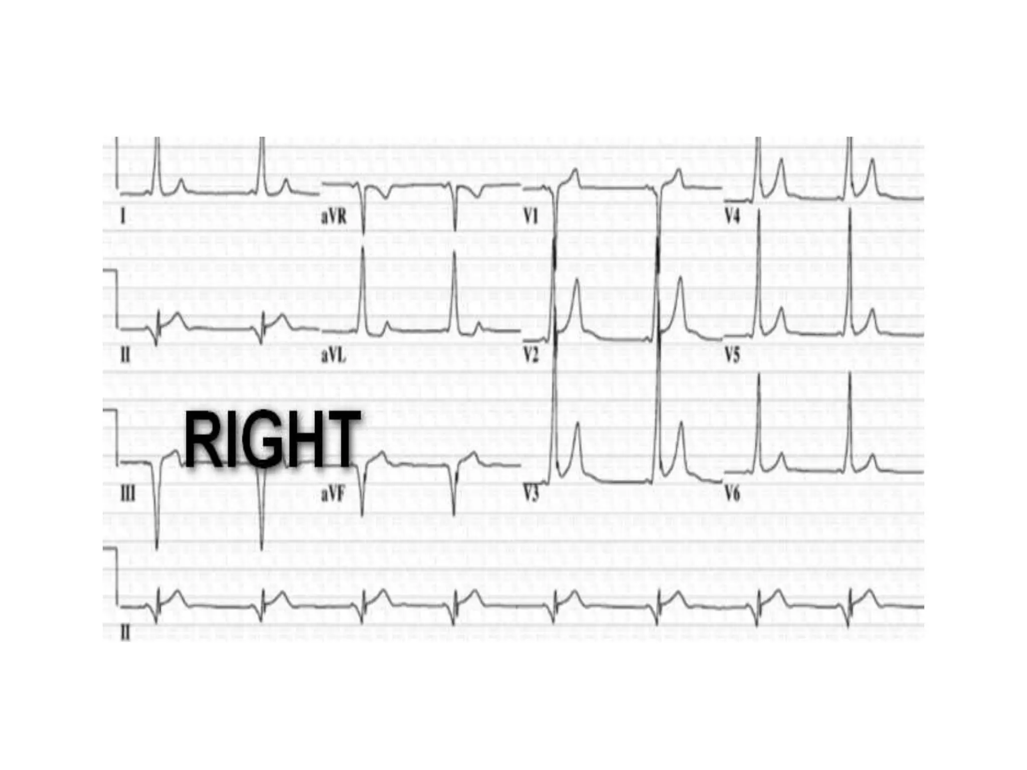
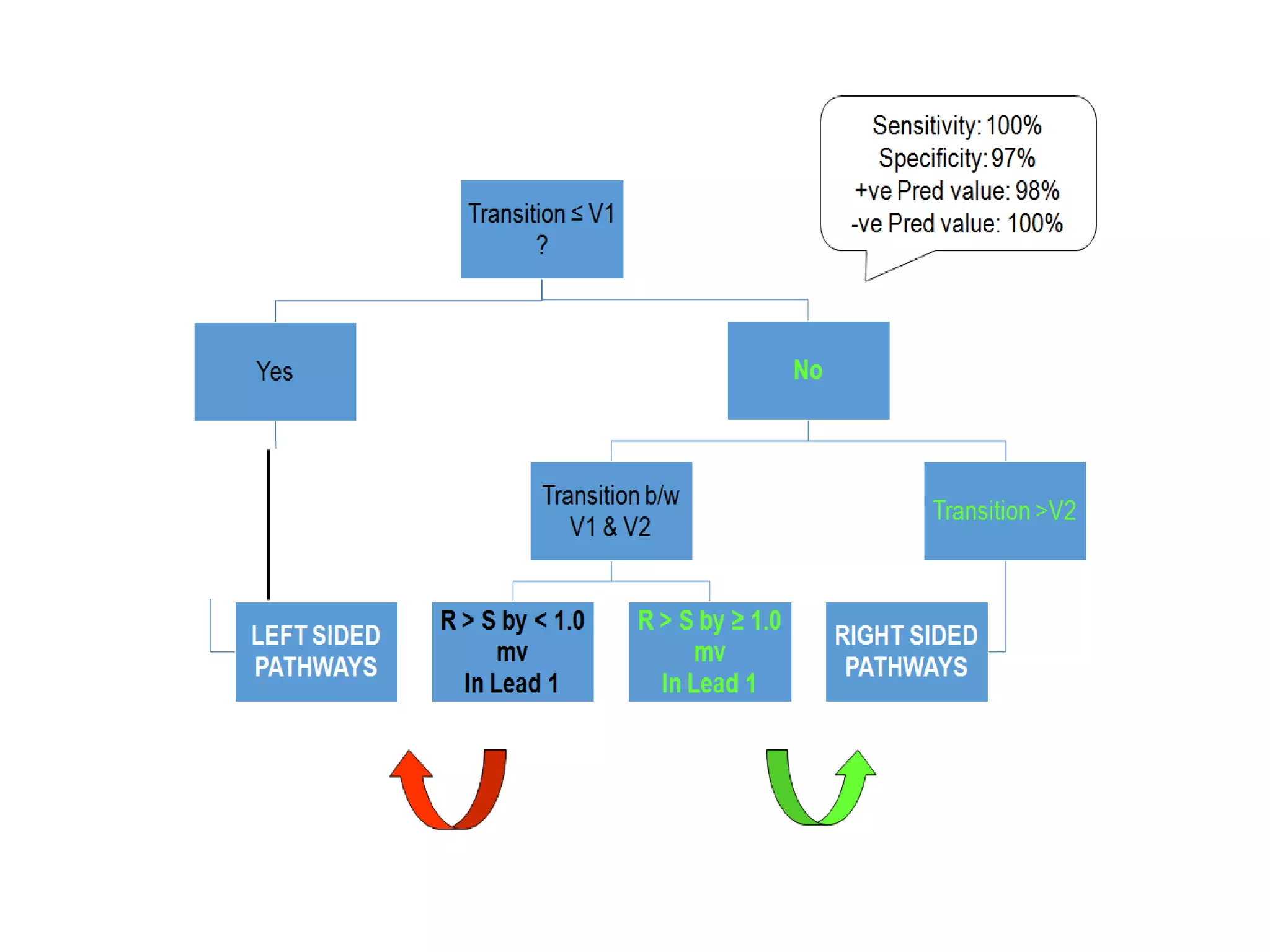
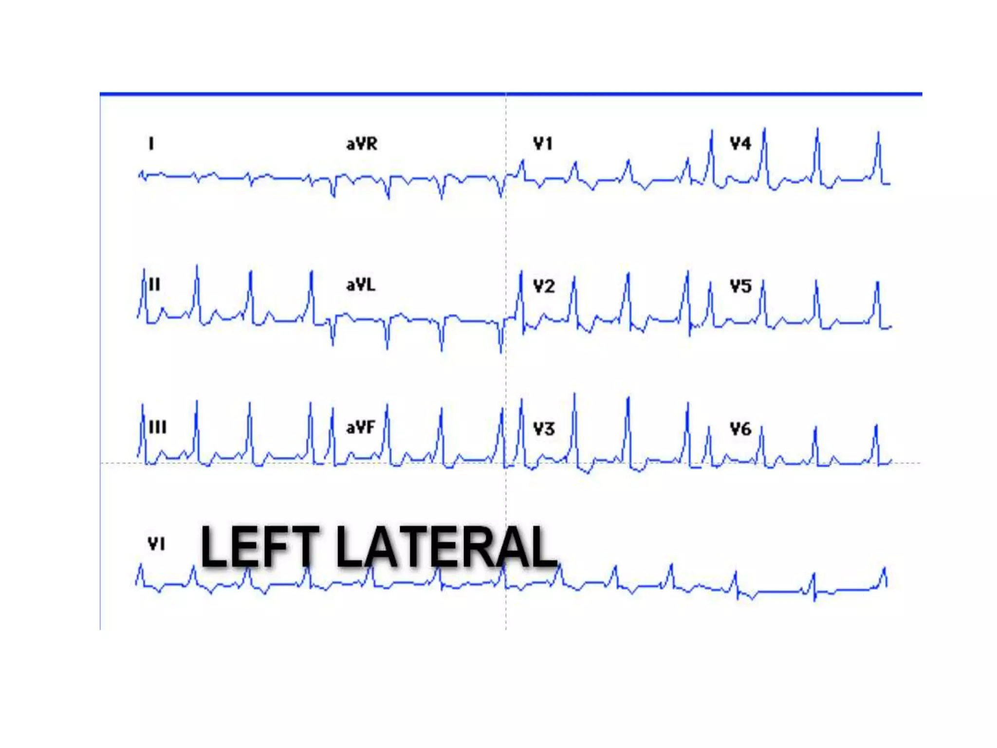
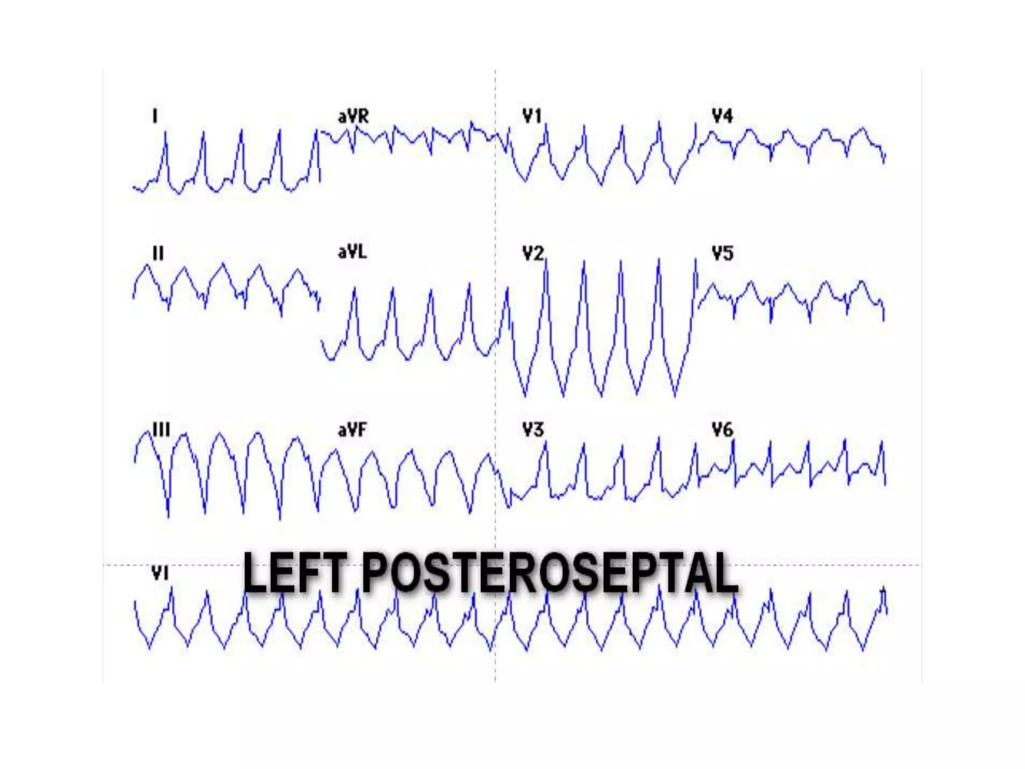
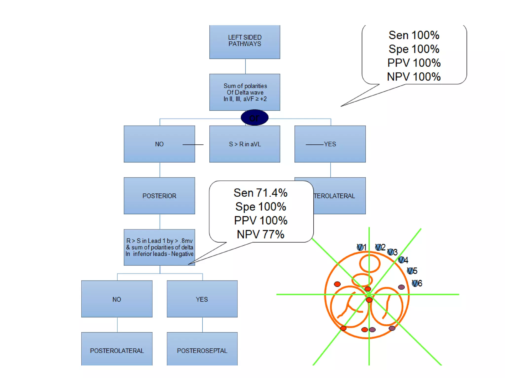
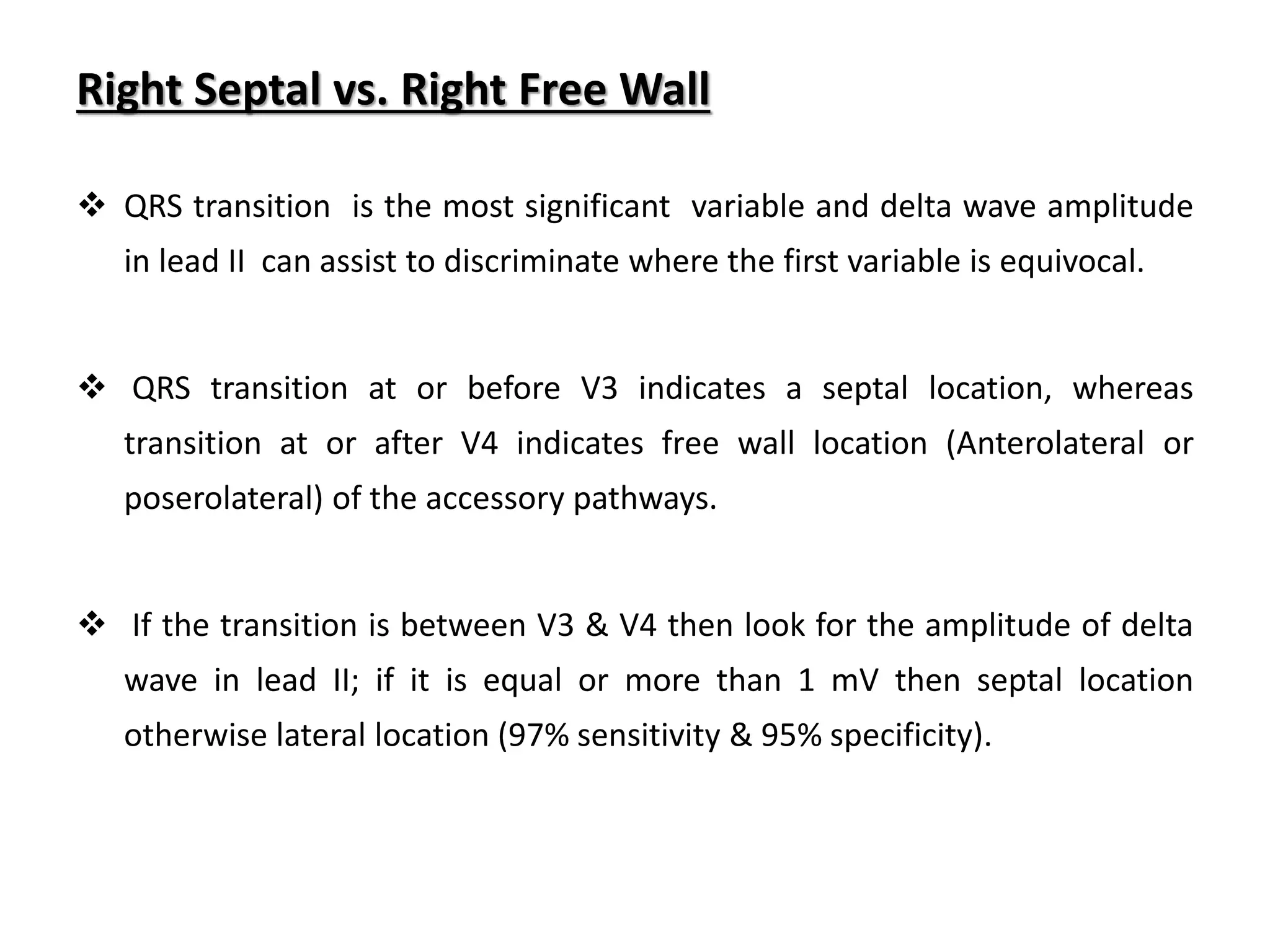
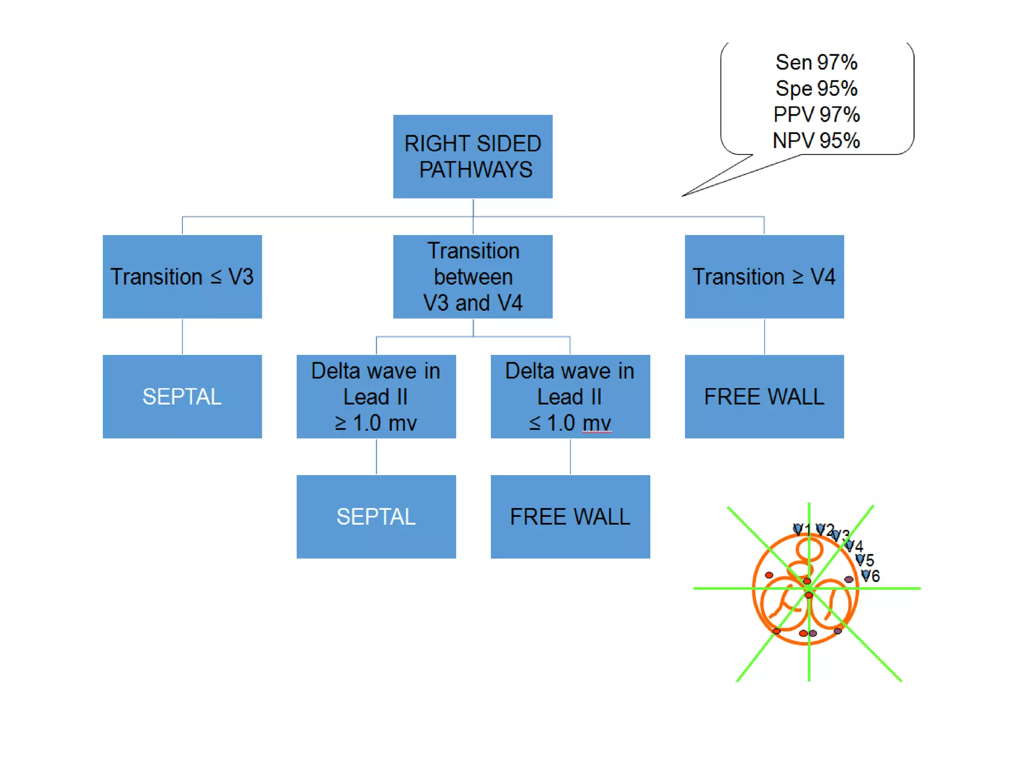
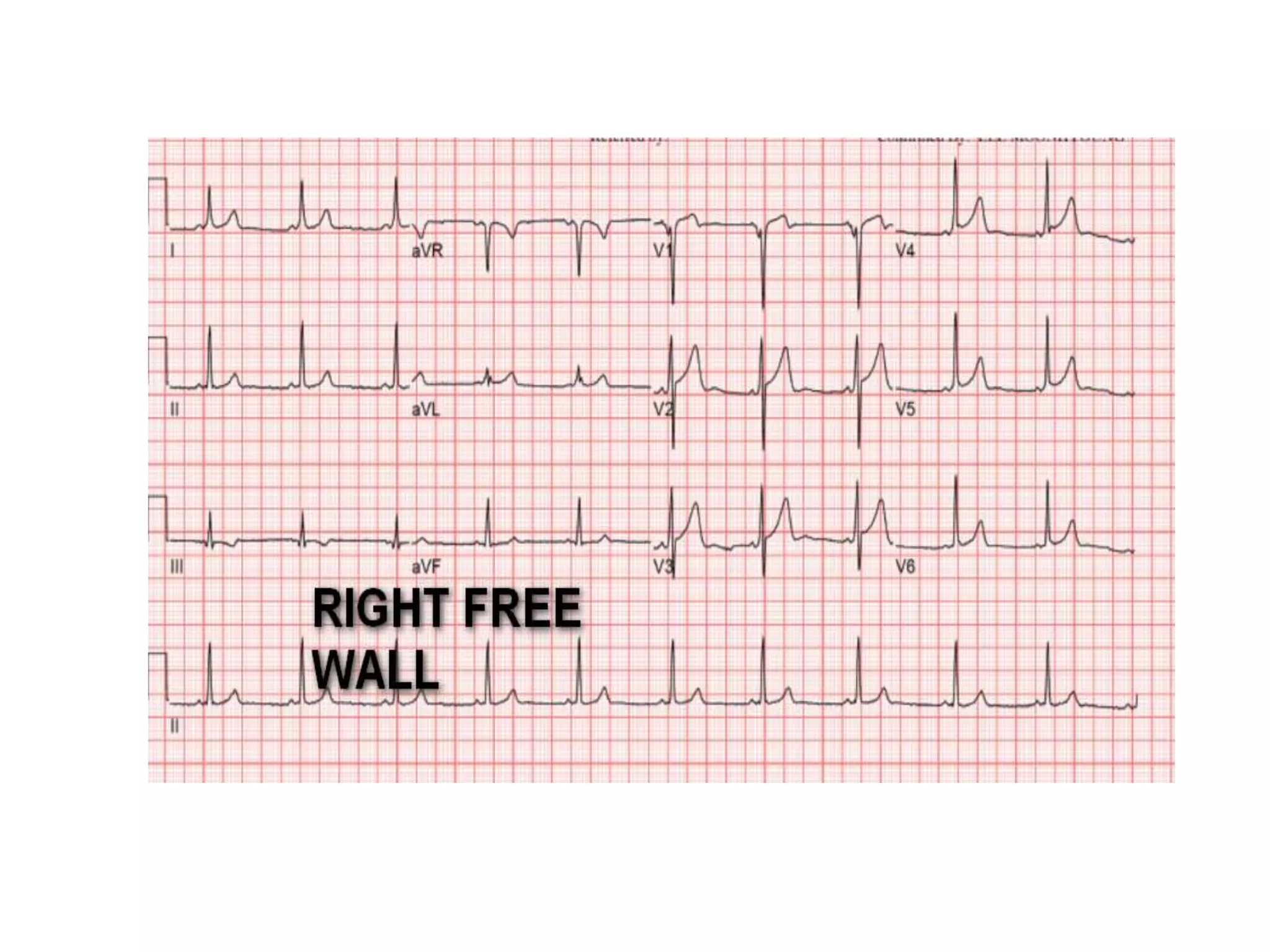
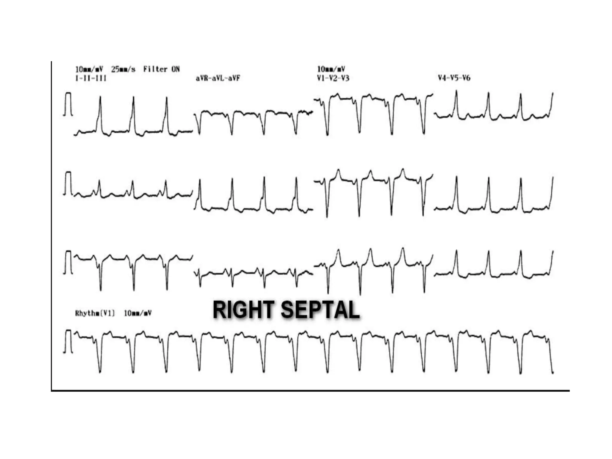

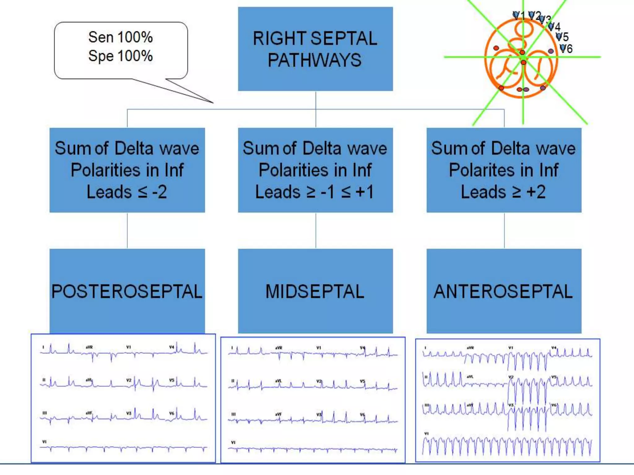
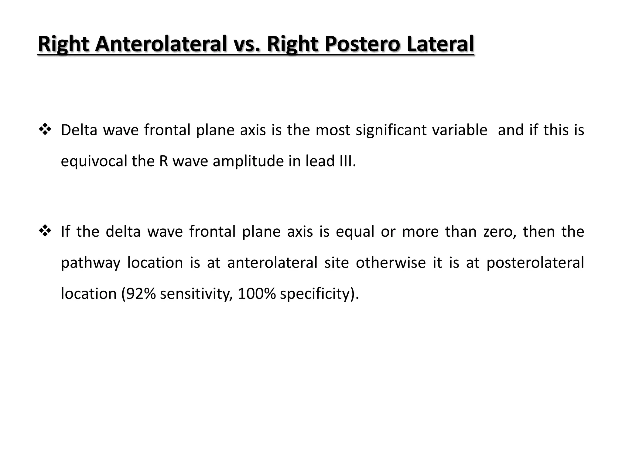
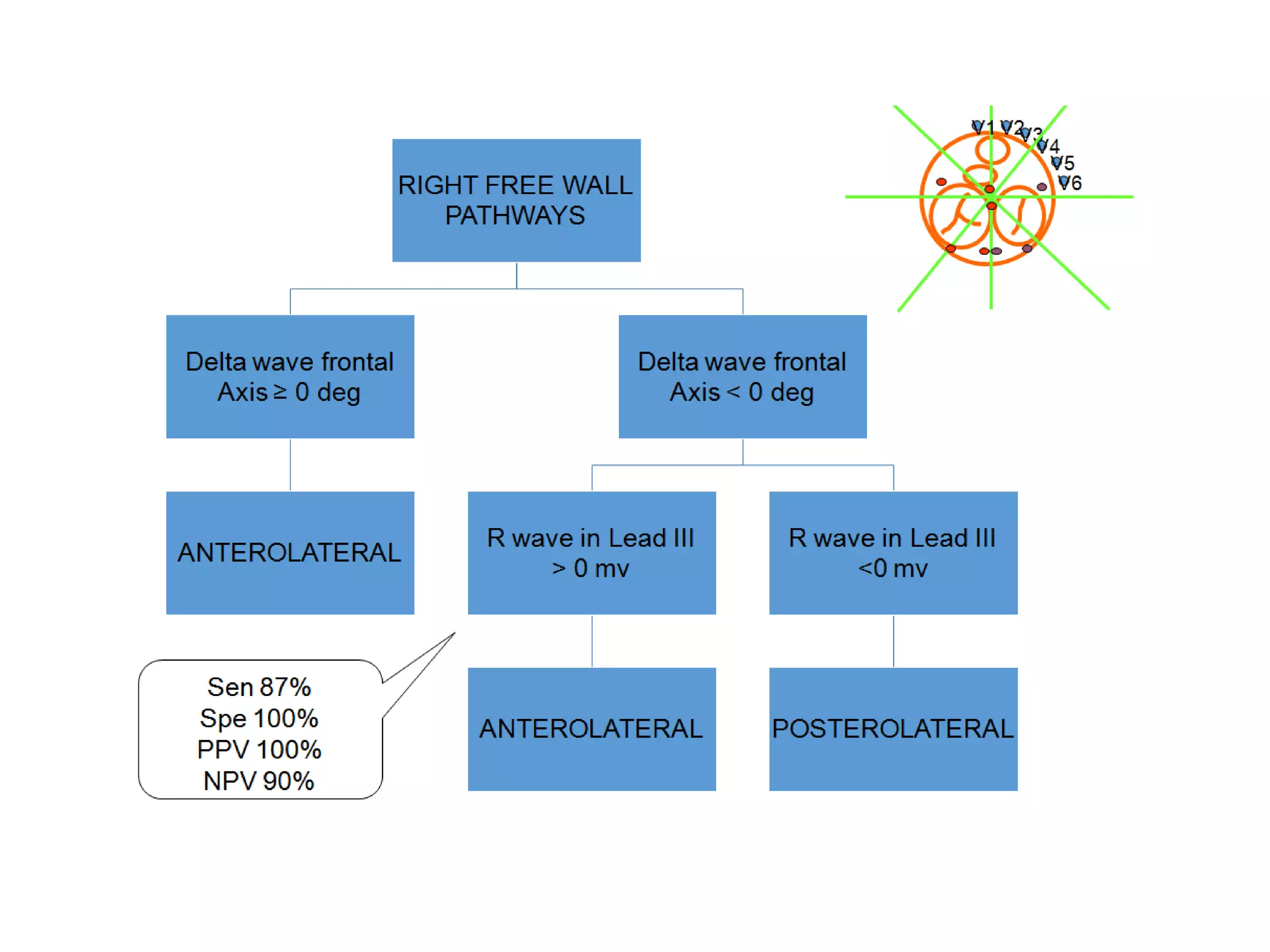
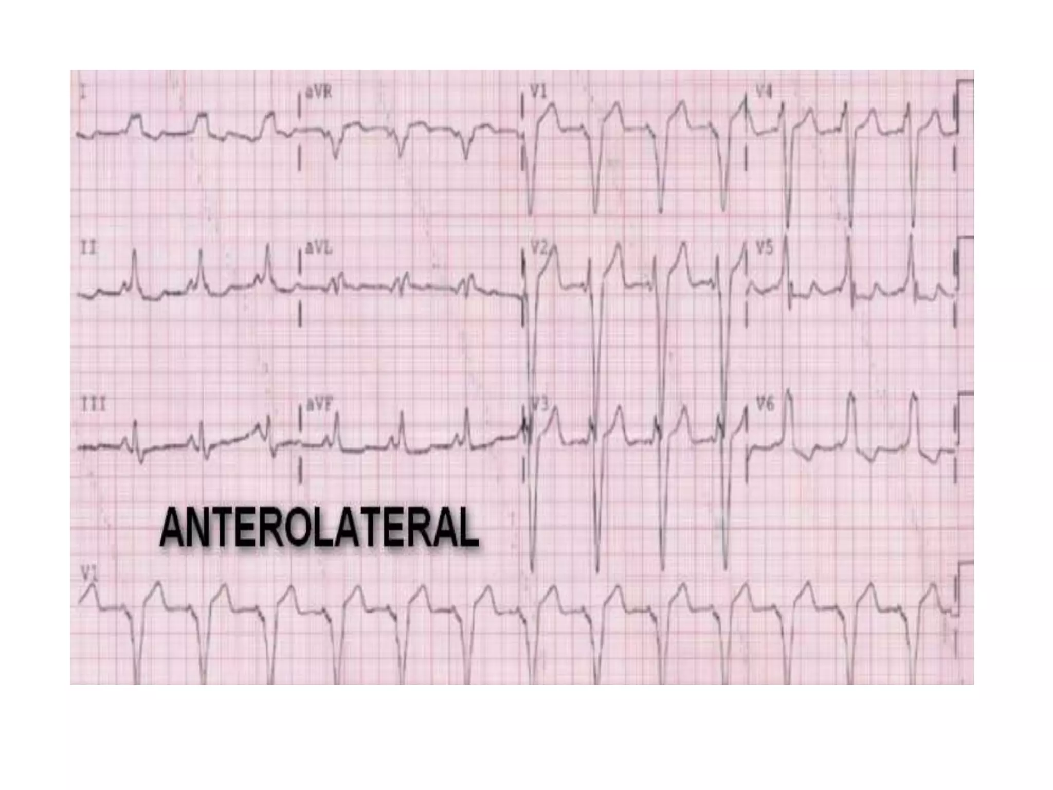
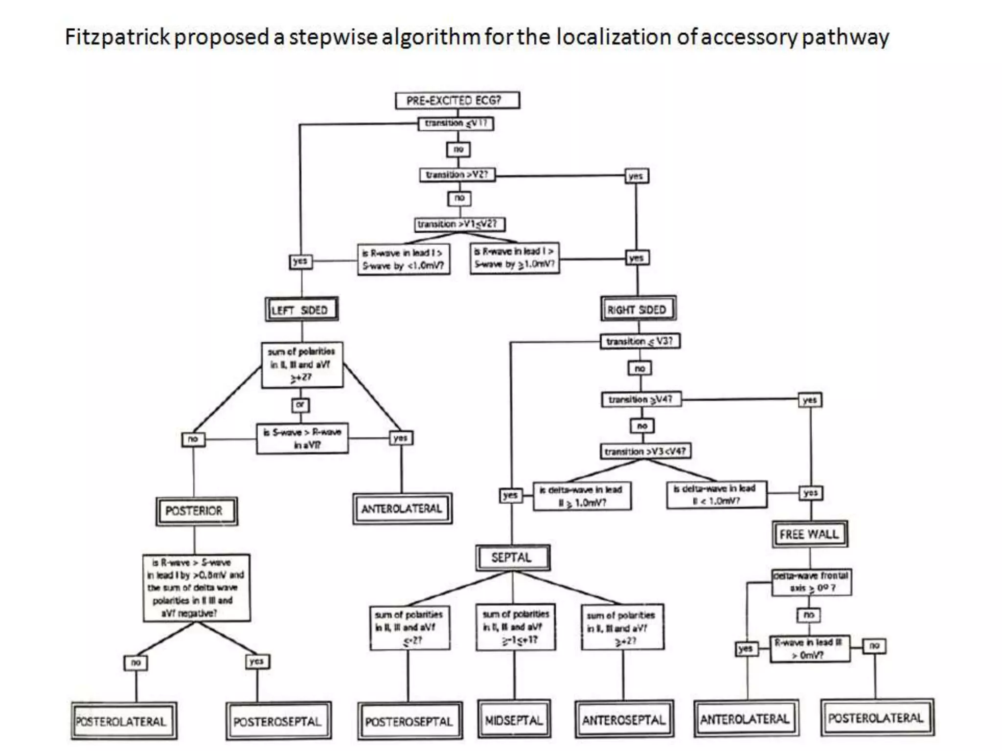
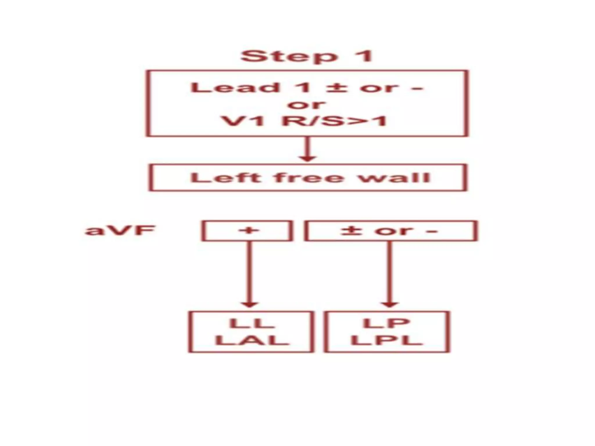

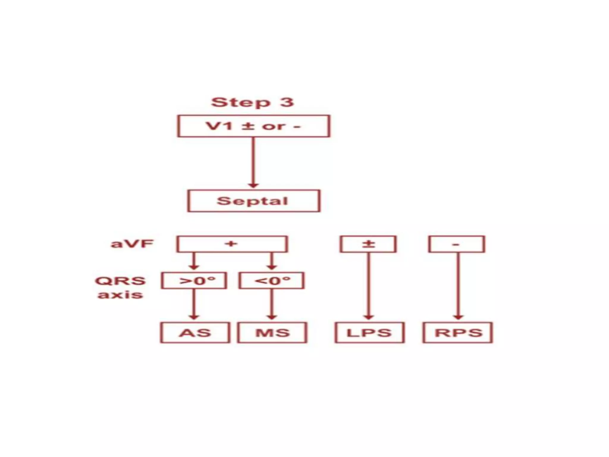

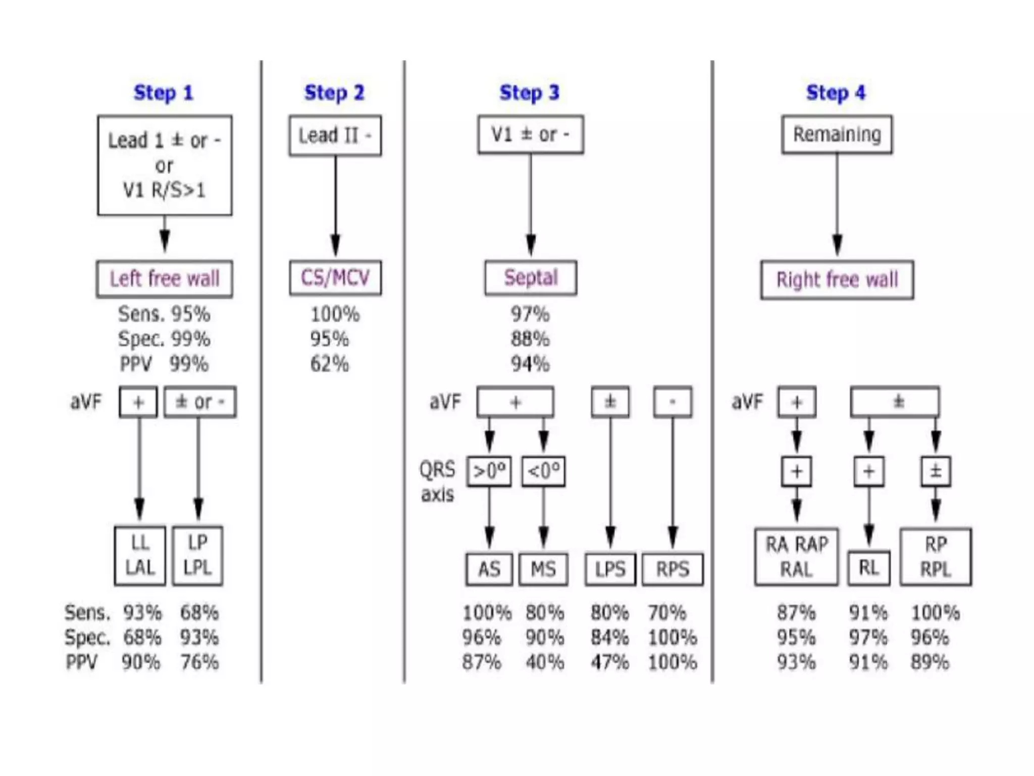


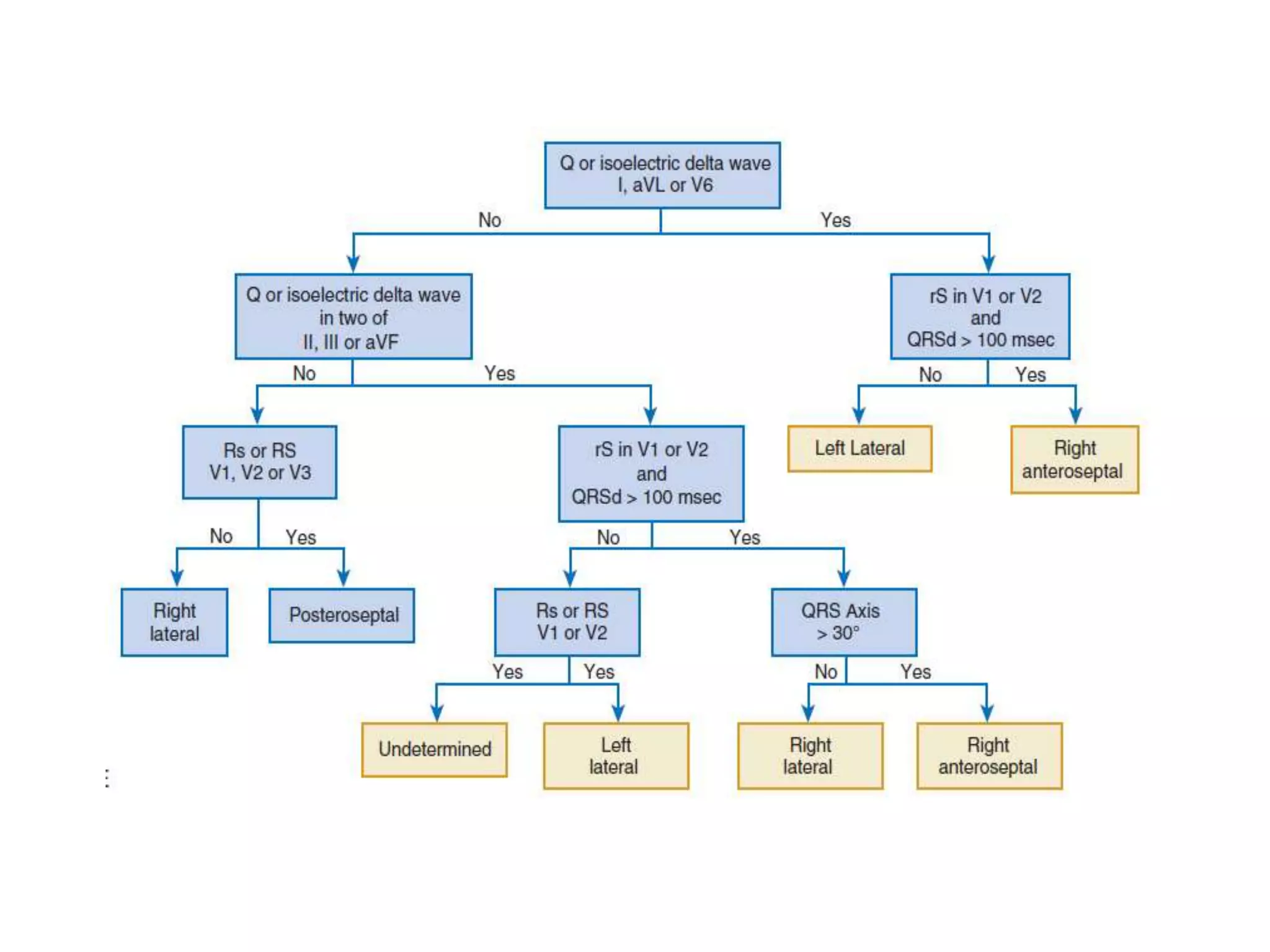
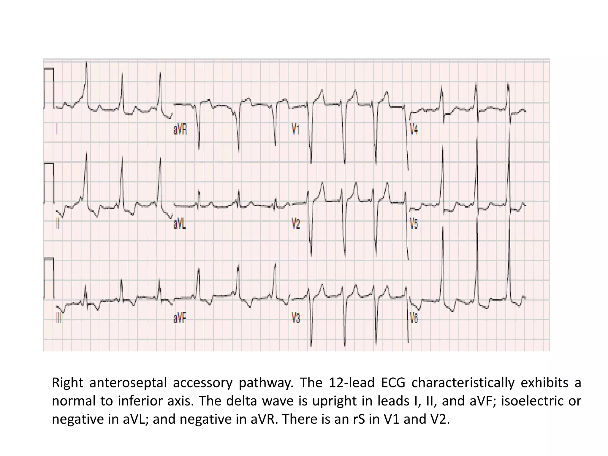
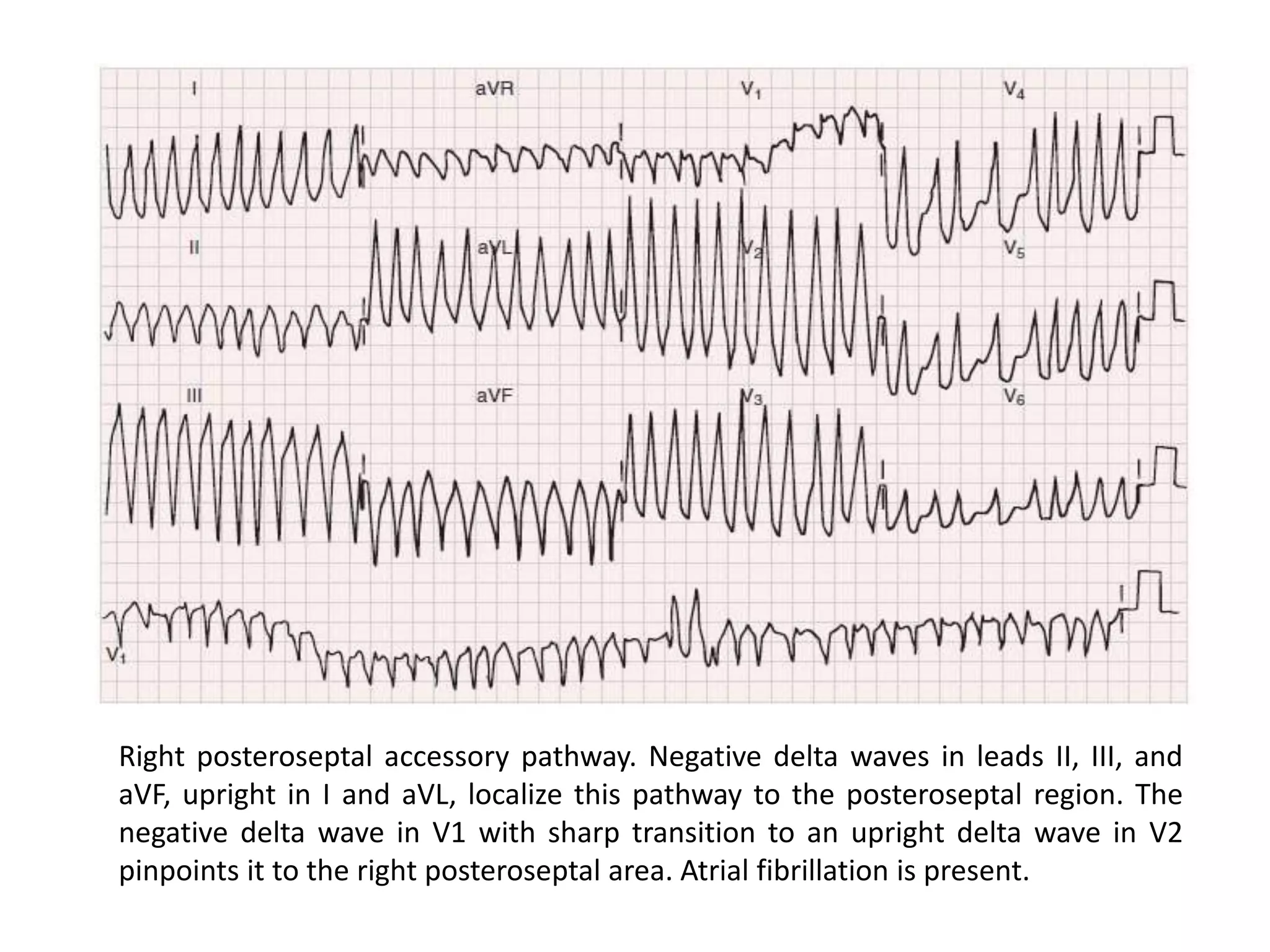
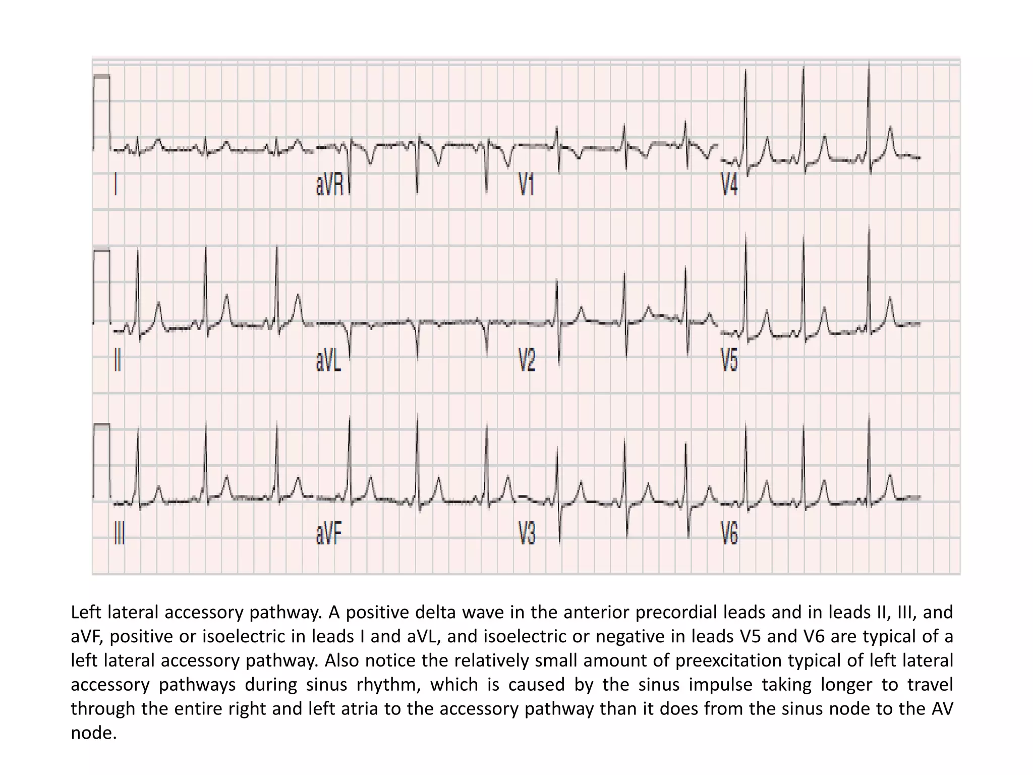
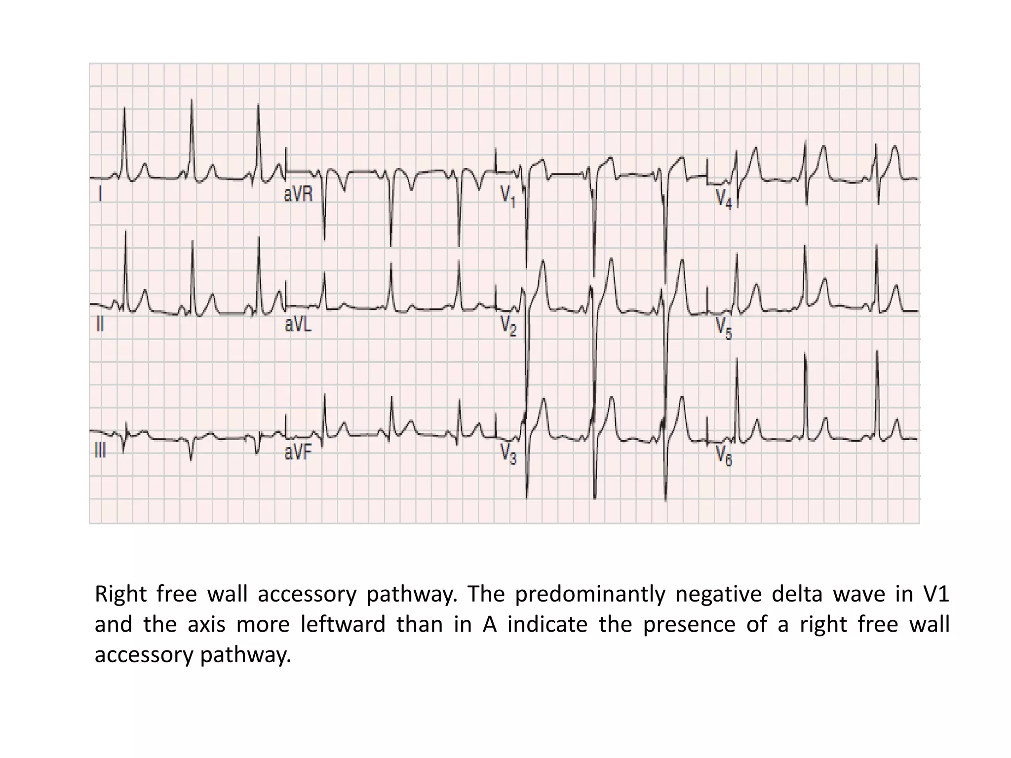

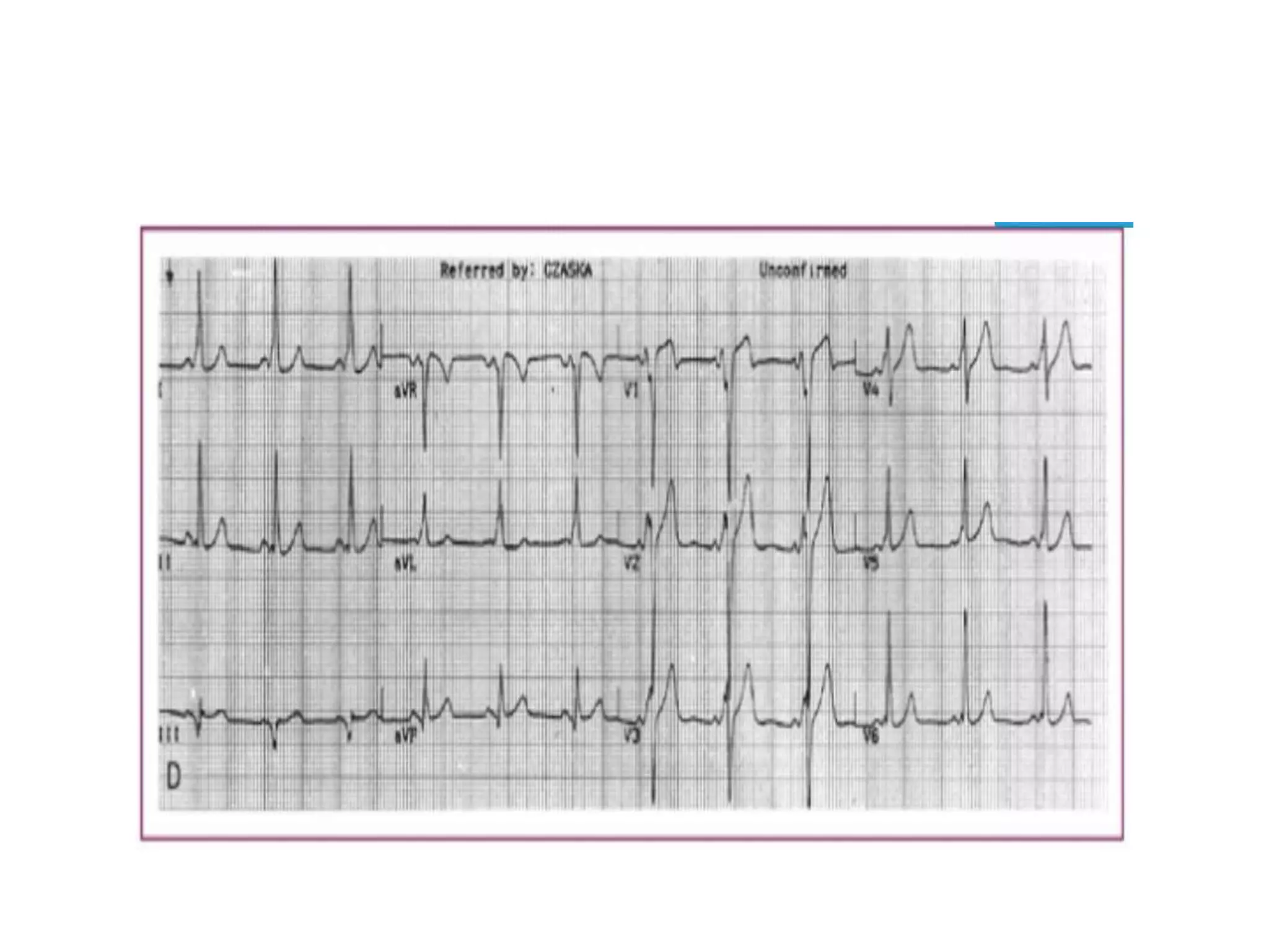
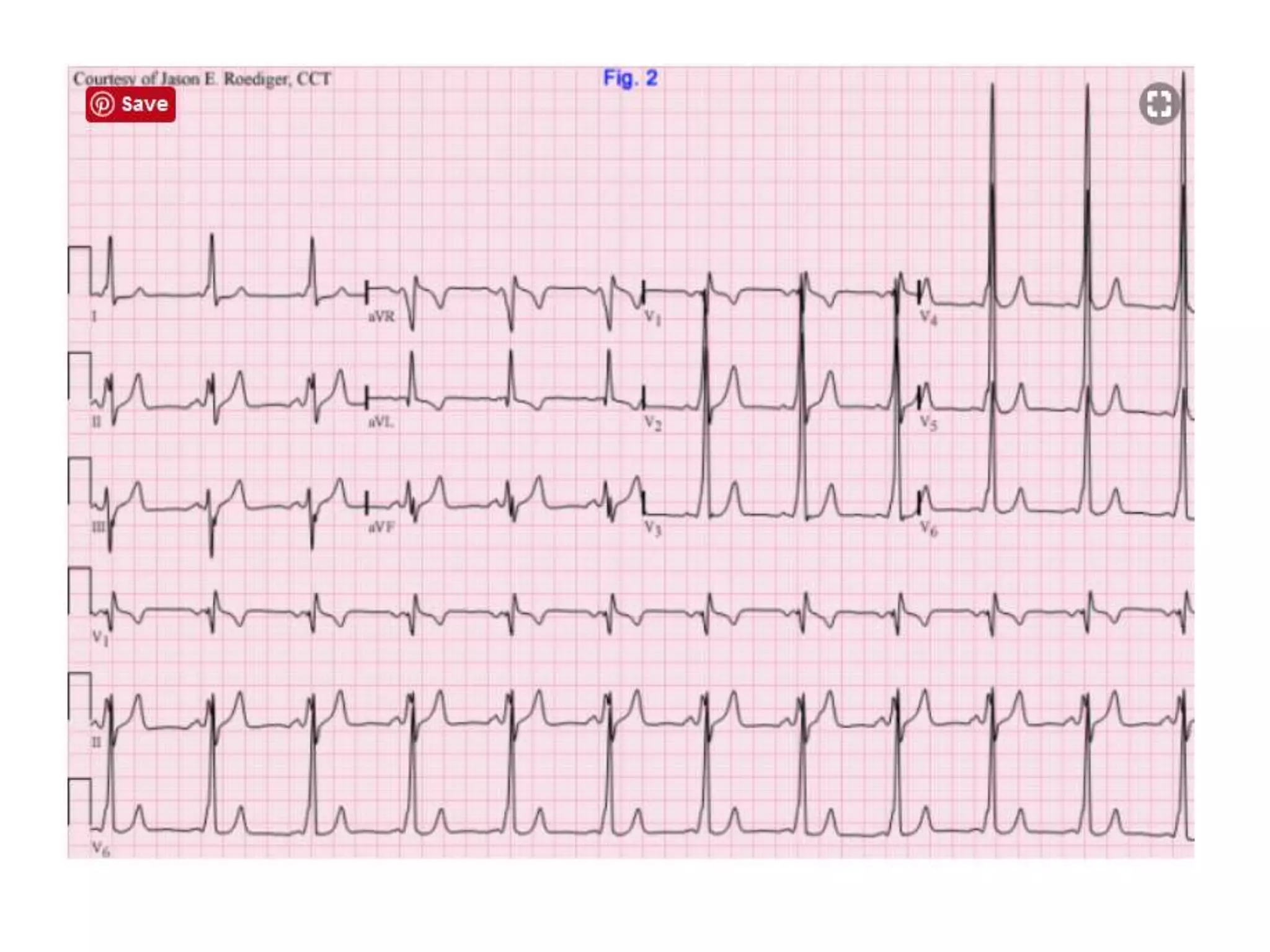
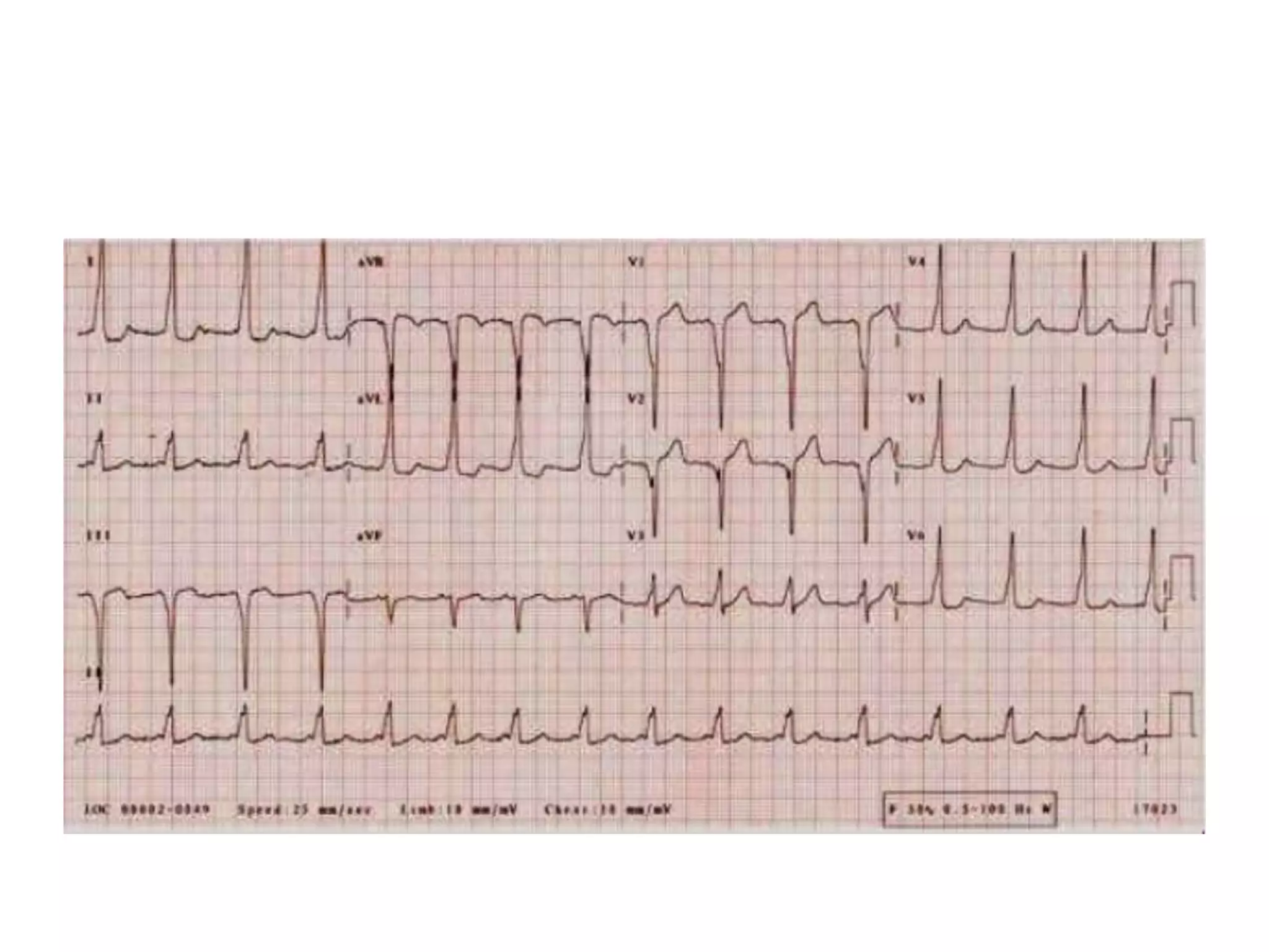
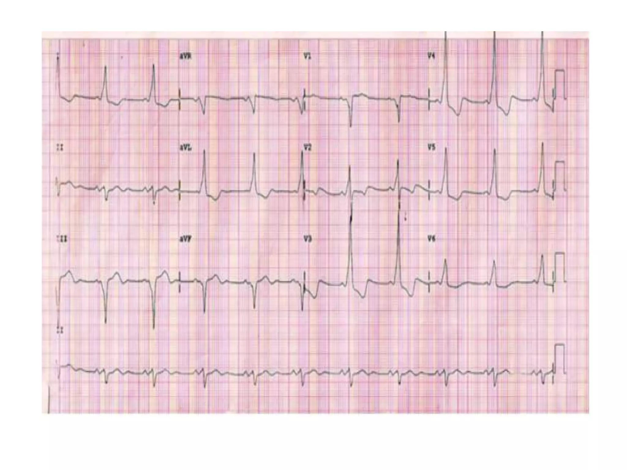
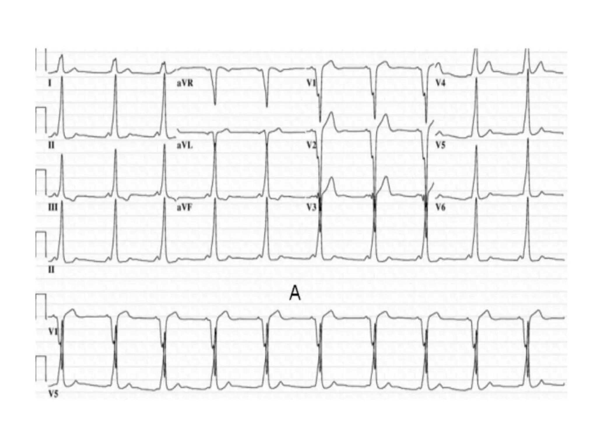
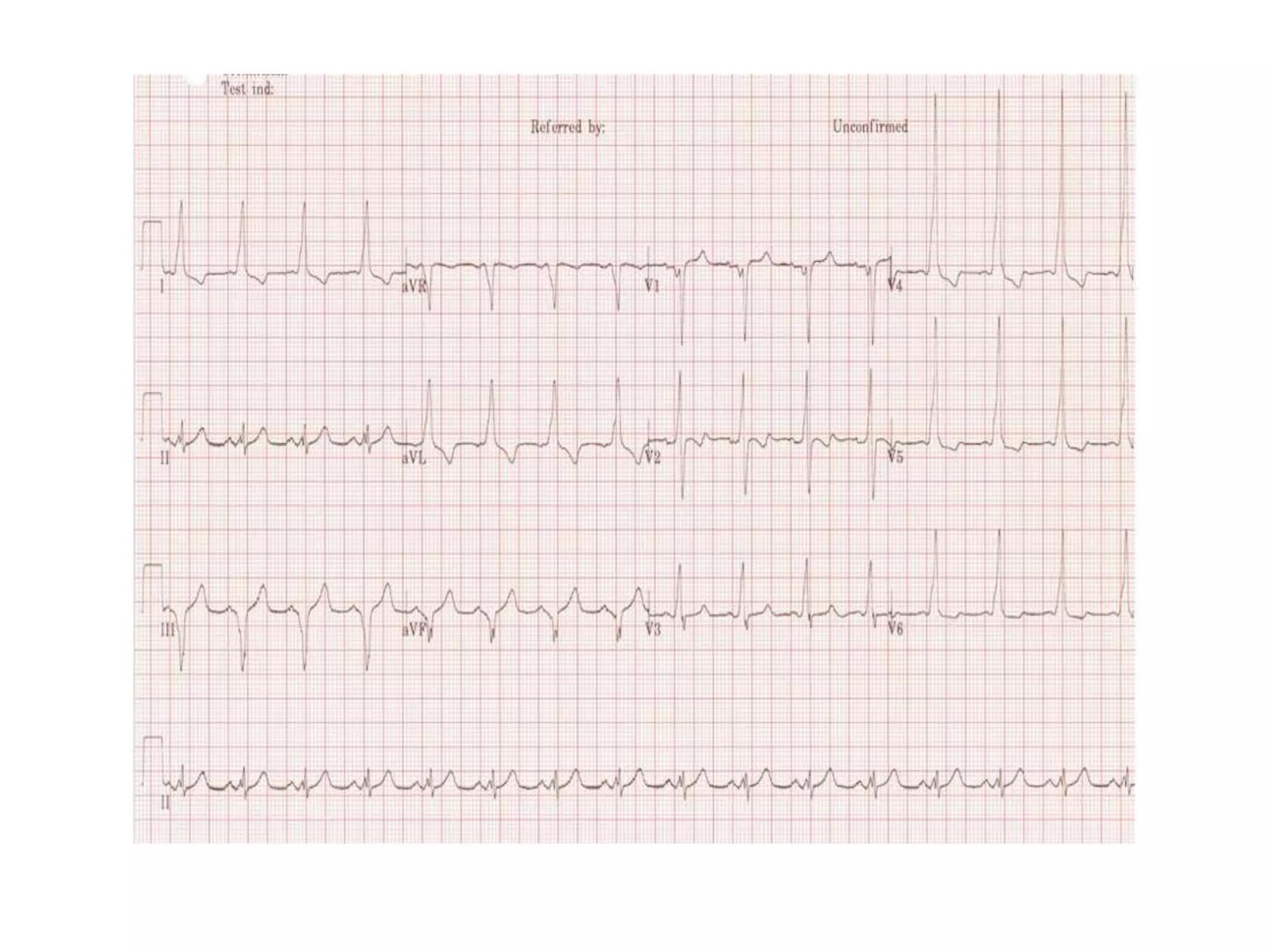
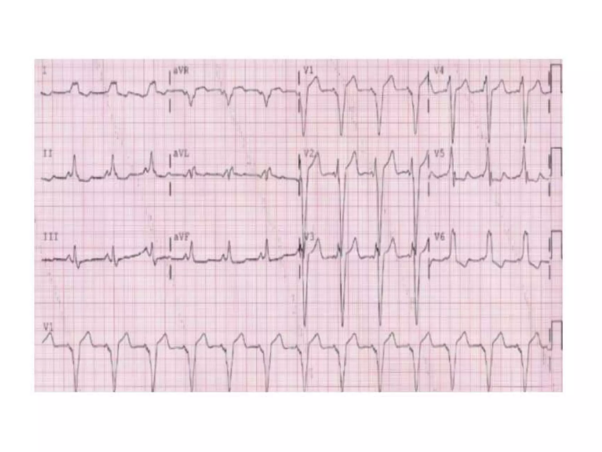


This document discusses localization of accessory pathways using electrocardiography. It describes that accessory pathways can be located in eight anatomical positions along the tricuspid and mitral valve annuli. Several algorithms are proposed to determine the location based on delta wave polarity and amplitude in various leads. The most accurate is the Arruda approach, which uses step-wise analysis of delta wave characteristics in leads I, II, aVL, aVF and V1 to identify the specific accessory pathway location with 90% sensitivity and 99% specificity. Characteristic ECG patterns are presented that help localize right anteroseptal, right posteroseptal, left lateral and right free wall accessory pathways.


