SlideShare a Scribd company logo
THE EISENMENGER SYNDROME
OR
PULMONARY HYPERTENSION WITH
REVERSED CENTRAL SHUNT
-PAUL WOOD
Paper presented by:
DR. MURTAZA KAMAL
MBBS, MD, DNB
DNB-SS FELLOW PEDIATRIC CARDIOLOGY
DOP: 08/02/2018
murtaza.vmmc@gmail.com
1
2
PAUL WOOD3
EISENMENGER’S
SYNDROME
Pulmonary HT due to high PVR with
reversed/ bidirectional shunt at aorto-
pulmonary, ventricular or atrial level
4
CAUSES
 Patent ductus arteriosus
 Aorto-pulmonary septal defect
 Persistent truncus
 Transposition with ventricular septal defect
 Corrected transposition with ventricular septal defect
 Ventricular septal defect
 Single ventricle
 Common atrioventricular canal
 Persistent ostium primum atrial septal defect
 Single atrium and
 Hemianomalous or total anomalous pulmonary venous drainage into the right
side of heart
5
DATA
 127 cases of Eisenmenger syndrome
 Clinical and physiological features analyses
 Special emphasis on distinguishing
characteristics of each type
 53 necropsis from well investigated during life
studied
6
Necropsy Findings
 127 ES
 15 died
 38 additional necropsies
 Total 127
7
MODE OF DEATH
 Cause of death known : 42
 Haemoptysis: MCC, 29%
 Profuse, Death in minutes
 Cause:
Pulmonary infarction from arterial thrombosis
Rupture of a thin walled dilated arteriole immediately
distal to end of a thick muscular artery
Rupture of aneurysm of PA (Most unusual)
8
MODE OF DEATH
 Surgical repair of defect: 26%
 CHF: 17%
 VF: 14%
 IE/ Cerebral abscess/ Cerebral thrombosis/ Pregnancy
induced: 5%
9
Accuracy of Diagnosis
 15 necropsied case: 9 correct, 2 wrong, 4 (Correct in respect
to site of shunt, wrong in respect to nature of defect)
 40% error: Embarrassing
 Same in 38 borrowed cases+ published literatures
 Eg:
Simple OS ASD TAPVD into SVC
Single ventricle with intact IAS Common AV canal with
functionally single atrium + virtually single ventricle
10
SIZE OF THE DEFECT
 No case of PDA with Eisenmenger reaction has ever
been reported with a lumen < 5 mm
 No case of Eisenmenger's complex has ever been
reported with a VSD < 1cm
 Size of an ASD has relatively little bearing on presence or
absence of a high PVR, and varies freely between 3- 8
cm in both uncomplicated and pulmonary hypertensive
groups
11
SIZE OF COMMUNICATION
Normal resistance
usual range
Eisenmnger Range
DUCT .1-1cm .7-1.5 cm
VSD .1-2cm 1.5-3cm
ASD 3-8 cm 3-8 cm
12
SIZES
 Have in common a large communication b/w 2 circulations
 If PVR were normal These would produce Qp/Qs at least
3.5 (4-6)
13
Aorto- pulmonary 0.7 cm
Inter ventricular 1.5 cm
Inter arterial 3 cm
ELASTIC ARTERIES
 In fetus/ neonates: Elastic fibers in PA; Densely packed +long
as in aorta
 As PAP falls Fibers break up, shorten, become loosely
arranged
 Found that sections of PA in EC and PDA with reversed shunt
 Always resembled foetal (aortic ) pattern
 Whereas in ASD with PH and reversed shunt+ all forms of
acquired PH Appearance like a normal PA

14
CONCLUDED
 PH in ES must be present from birth with VSD and PDA,
but must be acquired in ASD
 Function of elastic fibers: To prevent distention from high
pressures
15
MUSCULAR ARTERIES
 In EC, PAs with diameter< 1 mm have a thick muscular
media very similar to their structure in foetus
 It was persistence of foetal type of muscular artery that was
responsible for high resistance, and that this saved infants
with large VSDs from dying as a result of an inadequate
blood flow into aorta (Civin, Edwards)
 In their own words, " the pulmonary vascular bed of patients
with EC who survive infancy maintains its foetal function."
16
MUSCULAR ARTERIES
 They assumed: Thick muscular arteries of foetus were
responsible for high PVR necessary to deflect blood from
RV away from lungs into descending aorta via PDA
 During first 2 years of life intima usually remains normal
 Thereafter with varying degree and tempo from case to
case, progressive intimal fibrosis develops (Edwards, 1957)
17
ARTERIOLES
 Arterioles of lung: Vessels < 100u
 Consist of endothelium, a single elastic layer, and
adventitia; they are not supplied with muscle
 Usually thin-walled and dilated in ES, and that thick-walled
vessels within usually accepted compass of an arteriole
are really muscular arteries in a state of contraction
18
THE OVERRIDING AORTA
 1. VSD is but one of a total of some 12 kinds of
communication between two circulations, all of which may
be complicated by a high PVR and reversed shunt of similar
magnitude
 An overriding aorta or its physiological equivalent is absent
in 7 of these conditions
 2. When PVR is high, SPO2 averages close to 80% in
transposition with VSD, persistent truncus, single ventricle,
Taussig-Bing syndrome and EC with or without apparent
override
19
 3. EC with all classical features may be associated with a
defect in muscular part of IVS as in cases described by
Weiss (1927) and Heath et al. (1956)
 There can be no overriding of the aorta in such instances
20
EXPERIMENTAL PHYSIO-PATHOLOGY
 Reaction of pulmonary circulation in Eisenmenger's
syndrome to pulmonary vasoconstrictor and vasodilator
drugs
 O2:
Adults/ majority children>5: 100% O2 fails to lower
PAP or influence shunt magnitude
Other hand: 10%O2 raises PVR ad increases
reversed shunt (at least in PDA)
Infants< 2 and occasionally small children: When
intimal fibrosis in pulmonary circulation negligible:
100% O2 halves resistance and increases PBF
21
Failure of 100% oxygen to lower PV
pressure in case of EC
22
ACETYLCHOLINE
 When injected direct into PA in a dose of 0.5-1.5 mg.: An ideal
selective pulmonary vasodilator, for this quantity is usually
inactivated by cholinesterase by the time it reaches systemic
circulation (Wood et al., 1957)
 When severe PH was primary, or due to MS or cor pulmonale,
acetylcholine lowered PAP and resistance, and raised CO in
85% of cases (Wood, 1958)
 In 13 cases of Eisenmenger's syndrome, however, no such
effect could be demonstrated in a single instance
23
Failure of acetylcholine to lower PAP in
case of EC
24
 The youngest patient in series, however, was 5 years old, and
it is probable that infants under 2, without intimal fibrosis,
would react differently
 This belief is founded on the different response of these infants
to 100% oxygen, and on the fact that acetylcholine lowers PVR
sharply in foetal lung (Dawes, 1958)
 Negative reactions to tolazoline hydrochloride (" priscol ") and
aminophylline have also been without exception in cases of
Eisenmenger's syndrome in patients over 5
25
PROTECTIVE EFFECT OF
STENOSIS
 Vessels which are protected from extreme pulmonary
hypertension, or which have not had time to develop serious
intimal fibrosis secondary to hypertension, respond briskly to
acetylcholine
 This disposes of the idea that there is anything fundamentally
wrong with pulmonary vessels at birth inES
 Eg: Follows
26
27
Pressure pulses in a case of ES
associated with an AP septal defect and
stenosis of RPA near its origin
Pressure pulse from RPA beyond the
stricture, showing a marked reduction of
pressure following the
injection of acetylcholine. The pulmonary
vessels have been protected by the
stenosis and are functioning normally
 This functional evidence stands solidly side by side with
anatomical observation that when origin of LPA is atresic
in ES, its distal branches and muscular arteries, which are
not subjected to high pressure, are perfectly normal
 Whereas vessels on other side have all features
characteristic of Eisenmenger's complex (McKim and
Wiglesworth, 1954)
28
COURSE & PROGNOSIS
 ES commonly established at birth or during the first two years
of life when shunt is aorto-pulmonary or interventricular
 But is typically acquired when shunt is interatrial
 In present series, cyanosis dated from infancy in 80% of VSD,
but did not start until adult life in 92% ofASD
 Onset of reversed shunt in PDA: More difficult to establish,
because differential cyanosis usually overlooked
 Aorto-pulmonary septal defects, however, behave likeVSDs
29
 General rule: Neither cyanosis nor breathlessness changes
appreciably during childhood or adolescence, but
deterioration sets in sooner or later in adult life
 Onset of recurrent haemoptysis may signal downward trend
which is due in many cases to the development of
thromboobstructive lesions
 Average age of natural death :
33: Aortopulmonary defects
33: VSDs
36: ASDs
30
 Downhill course usually punctuated with haemoptyses,
which when severe sometimes caused sudden collapse and
death within a few minutes
 Bacterial endocarditis: 5% for death
 Cerebral abscess from paradoxical embolism occurred once
and proved fatal
 Cerebral thrombosis in one case, but not fatal
 14% died abruptly, presumably from VF
 26% died from a surgical attempt to repair defect, mostly
from ligation of a patent ductus
 Patients who survive these risks develop CHF sooner or
later, death in 17% of the fatal cases
31
TREATMENT
 Medical treatment: Aims at preventing secondary
thromboobstructive lesions by means of permanent
anticoagulants, such as phenindione (" dindevan ")
 Pulmonary vasodilators (tolazoline, aminophylline,and
reserpine::Fail to lowerPVR, and do not influence course of
disease
 Small venesections, repeated from time to time: May be
helpful in cases with gross polycythaemia
 Heart failure management
32
 Surgical repair of defect: Responsible for more deaths in ES
than any other agent except haemoptysis
 Ellis et al. (1956): Surgical mortality in cases with reversed
shunt through a patent ductus was 56
33
 Direct repair of a defect is justified only when it can be
predicted with confidence that its closure must result in a fall of
pulmonary blood pressure because a hyperkinetic factor of
sufficient magnitude has been eliminated
 In practice this prediction can be made when PBF is at least
twice the systemic flow at rest, and PVR does not exceed 10
units (800 dynes sec./cm.')
 Borderline cases have Qp/Qs 1.75- 2, and resistances of 10 to
12 units
 No case with PBF <1.75 times the systemic flow or with a
resistance over 12 units should have defect repaired in the
ordinary way, whether the shunt is reversed, direct, or
bidirectional
34
SYNTHESIS
 CO of newborn babe is about 0.5 1./min., and is presumably
much same in foetus at term
 0.1 1./min. passes through pulmonary circulation
 Since mean PAP is same as aortic, about 50 mm. Hg at birth,
foetal pulmonary vascular resistance must be about 500 units
(40,000 dynes sec./cm.5)
 During 1st 3 hours after birth a reversed shunt through PDA is
invariable;
 From 3-72 hours after birth shunt may be direct, bidirectional
or reversed and by end of 3rd day duct is usually closed
 It follows that in newborn infants PVR is more or less same
as the systemic-that is, about 100 units
35
 Within a remarkably short time after birth, then, probably
within a few minutes of independent life, a PVR around 500
units falls to about 100
 4 questions clamour for an answer:
 (1) What is the cause of the fantastic PVR in the foetus?
 (2) Why does it drop so precipitously at birth ?
 (3) Why is the rapid fall halted temporarily more or less at
systemic level ?
 (4) What is responsible for the subsequent slower decline to
normal ?
 In the correct answer to these four questions lies the secret
of the Eisenmenger syndrome
36
What is the cause of the fantastic PVR in the
foetus?
 Resistance to flow in a tube is greatly increased by kinking
the vessel
 In the fully developed foetus the pulmonary vessels are as
long as they are a few minutes after birth, but since the lung
is completely collapsed they must be coiled up, or kinked in
innumerable places
 Burton has demonstrated their "gnarled" appearance
 Vessels in such a state may be highly resistant to flow
37
 Answer to the second question:
 As the lungs expand the vessels uncoil: as the kinks
straighten out the resistance falls; as the blood begins to flow
through the lungs the vessels must dilate, and this also lowers
the resistance
 Blood that fills up pulmonary vascular bed acts as a natural
scaffolding and encourages further expansion (Jaykkai, 1957)
 Stepwise, and in a matter of a minute or so, and aided by
lusty cries, the lungs are fully expanded (Lind and Wegelius,
1956)
 Vessels are wholly unfurled, and PVR is at systemic level, at
least five times lower than in utero
38
Why is the rapid fall in resistance at birth
temporarily halted at systemic level ?
 This may well be a function of the thick muscular arteries
just mentioned
 In the foetus these have developed in relation to the
systemic pressure to which they have been subjected, and,
as shown by Dawes (1958), they are also in a state of active
vasoconstriction, which can be released by acetylcholine
 After birth a powerful RV, as big and strong as the left,
pumps its contents into a system of vessels which differs
little from the systemic circulation;it is only natural, therefore,
that pulmonary and systemic resistances should be similar
39
 During 1st of life, then, physiological and anatomical findings
are identical with ES associated with PDA
 2events of great importance
 1st: Relaxing effect of an increased alveolar oxygen tension
on the tone of muscular arteries
 2nd: Closure of PDA
 Before the duct closes, release of vasoconstrictor tone in the
muscular arteries is met by an increased flow from aorta to
PA, and this hyperkinetic factor tends to maintain PA pressure
at systemic level
40
 When duct closes, and this hyperkinetic factor is eliminated,
release of vasoconstrictor tone lowers the pulmonary artery
pressure, and the approach to normal adult physiology
begins
 Just how quickly this goal is reached depends on the speed
of involution of the muscular arteries
 Complete involution takes about three months
41
What is responsible for the subsequent slower
decline to normal ?
 (1) As the pressure falls in the pulmonary circulation
vasoconstrictor tone is diminished, for muscular arteries
react to increased pressure by contracting and to decreased
pressure by relaxing (Bayliss, 1902)
 This local response of muscular arteries to any change in
pressure must increase that change in either direction
 (2) As the resistance declines PBF increases
 This must dilate vessels and so lower the resistance still
further (Williams, 1954)
 (3) As the pressure falls the muscular arteries atrophy and
so diminish their resistance to flow
42
THE EISENMENGER REACTION
 All that is necessary to prevent these natural forces from
lowering PVR to normal is a sufficient increase of flow to
keep the pressure at systemic level
 Any large AP or IV communication will do just this, and the
higher the resistance the less increase of flow necessary to
maintain the pressure
 If the pressure is maintained pulmonary vasoconstrictor tone
must remain high, and the muscular arteries will not atrophy
 This is the situation inES, and it will be understood that it
must be established at birth if the defect is larg enough
43
 If the defect is not quite critical, however, the shunt flow at rest
may not be sufficient to maintain pressure at systemic level,
and the forces acting in the opposite direction may win or
partially win the competition
 Once resistance falls appreciably much larger flows are
necessary to maintain high pressure
44
THANKS45

More Related Content

What's hot

Cath hemodynamics vir
Cath hemodynamics virCath hemodynamics vir
Cath hemodynamics vir
Dr Virbhan Balai
 
Asd echo assessment
Asd echo assessmentAsd echo assessment
Asd echo assessment
Mashiul Alam
 
SEGMENTAL ANALYSIS OF CONGENITAL HEART DISEASE
SEGMENTAL ANALYSIS OF CONGENITAL HEART DISEASE SEGMENTAL ANALYSIS OF CONGENITAL HEART DISEASE
SEGMENTAL ANALYSIS OF CONGENITAL HEART DISEASE
Dr. Murtaza Kamal MD,DNB,DrNB Ped Cardiology
 
Assessment of shunt by cardiac catheterization
Assessment of shunt by cardiac catheterizationAssessment of shunt by cardiac catheterization
Assessment of shunt by cardiac catheterization
Ramachandra Barik
 
Echo assessment of RV function
Echo assessment of RV functionEcho assessment of RV function
Echo assessment of RV function
Dr. Md. Ahasanul Kabir Shahin
 
Echo assessment of cardiomyopathy pdf
Echo assessment of cardiomyopathy pdfEcho assessment of cardiomyopathy pdf
Echo assessment of cardiomyopathy pdf
Nizam Uddin
 
CONTRAST ECHOCARDIOGRAPHY
CONTRAST ECHOCARDIOGRAPHYCONTRAST ECHOCARDIOGRAPHY
CONTRAST ECHOCARDIOGRAPHY
soumenprasad
 
Echocardiographic Evaluation of LV Diastolic Function
Echocardiographic Evaluation of LV Diastolic FunctionEchocardiographic Evaluation of LV Diastolic Function
Echocardiographic Evaluation of LV Diastolic Function
Junhao Koh
 
Echocardiographic evaluation of of coronary arteries
Echocardiographic evaluation of  of coronary arteriesEchocardiographic evaluation of  of coronary arteries
Echocardiographic evaluation of of coronary arteries
Raghu Kishore Galla
 
Echocardiography in mitral stenosis
Echocardiography in mitral stenosisEchocardiography in mitral stenosis
Echocardiography in mitral stenosis
Malleswara rao Dangeti
 
Diastolic Dysfunction 2016
Diastolic Dysfunction 2016Diastolic Dysfunction 2016
Diastolic Dysfunction 2016
drabhishekbabbu
 
Echocardiographic Evaluation of Hypertrophic Cardiomyopathy
Echocardiographic Evaluation of Hypertrophic CardiomyopathyEchocardiographic Evaluation of Hypertrophic Cardiomyopathy
Echocardiographic Evaluation of Hypertrophic Cardiomyopathy
Hatem Soliman Aboumarie
 
Double outlet right ventricle
Double outlet right ventricleDouble outlet right ventricle
Double outlet right ventricle
Himanshu Rana
 
SINUS OF VALSALVA ANEURYSM
SINUS OF VALSALVA ANEURYSMSINUS OF VALSALVA ANEURYSM
SINUS OF VALSALVA ANEURYSM
Jyotindra Singh
 
EBSTEIN ANOMALY
EBSTEIN ANOMALYEBSTEIN ANOMALY
Strain and strain rate
Strain  and strain rateStrain  and strain rate
Strain and strain rate
Malleswara rao Dangeti
 
Echocardiographic assesment of systolic and diastolic dysfunction
Echocardiographic assesment of systolic and diastolic dysfunctionEchocardiographic assesment of systolic and diastolic dysfunction
Echocardiographic assesment of systolic and diastolic dysfunction
Malleswara rao Dangeti
 
Shunt quantification
Shunt quantificationShunt quantification
Shunt quantification
Ankur Gupta
 
Shunt quantification
Shunt quantificationShunt quantification
Shunt quantification
Girish Mishra
 
Asd device closure
Asd device closureAsd device closure
Asd device closure
sruthiMeenaxshiSR
 

What's hot (20)

Cath hemodynamics vir
Cath hemodynamics virCath hemodynamics vir
Cath hemodynamics vir
 
Asd echo assessment
Asd echo assessmentAsd echo assessment
Asd echo assessment
 
SEGMENTAL ANALYSIS OF CONGENITAL HEART DISEASE
SEGMENTAL ANALYSIS OF CONGENITAL HEART DISEASE SEGMENTAL ANALYSIS OF CONGENITAL HEART DISEASE
SEGMENTAL ANALYSIS OF CONGENITAL HEART DISEASE
 
Assessment of shunt by cardiac catheterization
Assessment of shunt by cardiac catheterizationAssessment of shunt by cardiac catheterization
Assessment of shunt by cardiac catheterization
 
Echo assessment of RV function
Echo assessment of RV functionEcho assessment of RV function
Echo assessment of RV function
 
Echo assessment of cardiomyopathy pdf
Echo assessment of cardiomyopathy pdfEcho assessment of cardiomyopathy pdf
Echo assessment of cardiomyopathy pdf
 
CONTRAST ECHOCARDIOGRAPHY
CONTRAST ECHOCARDIOGRAPHYCONTRAST ECHOCARDIOGRAPHY
CONTRAST ECHOCARDIOGRAPHY
 
Echocardiographic Evaluation of LV Diastolic Function
Echocardiographic Evaluation of LV Diastolic FunctionEchocardiographic Evaluation of LV Diastolic Function
Echocardiographic Evaluation of LV Diastolic Function
 
Echocardiographic evaluation of of coronary arteries
Echocardiographic evaluation of  of coronary arteriesEchocardiographic evaluation of  of coronary arteries
Echocardiographic evaluation of of coronary arteries
 
Echocardiography in mitral stenosis
Echocardiography in mitral stenosisEchocardiography in mitral stenosis
Echocardiography in mitral stenosis
 
Diastolic Dysfunction 2016
Diastolic Dysfunction 2016Diastolic Dysfunction 2016
Diastolic Dysfunction 2016
 
Echocardiographic Evaluation of Hypertrophic Cardiomyopathy
Echocardiographic Evaluation of Hypertrophic CardiomyopathyEchocardiographic Evaluation of Hypertrophic Cardiomyopathy
Echocardiographic Evaluation of Hypertrophic Cardiomyopathy
 
Double outlet right ventricle
Double outlet right ventricleDouble outlet right ventricle
Double outlet right ventricle
 
SINUS OF VALSALVA ANEURYSM
SINUS OF VALSALVA ANEURYSMSINUS OF VALSALVA ANEURYSM
SINUS OF VALSALVA ANEURYSM
 
EBSTEIN ANOMALY
EBSTEIN ANOMALYEBSTEIN ANOMALY
EBSTEIN ANOMALY
 
Strain and strain rate
Strain  and strain rateStrain  and strain rate
Strain and strain rate
 
Echocardiographic assesment of systolic and diastolic dysfunction
Echocardiographic assesment of systolic and diastolic dysfunctionEchocardiographic assesment of systolic and diastolic dysfunction
Echocardiographic assesment of systolic and diastolic dysfunction
 
Shunt quantification
Shunt quantificationShunt quantification
Shunt quantification
 
Shunt quantification
Shunt quantificationShunt quantification
Shunt quantification
 
Asd device closure
Asd device closureAsd device closure
Asd device closure
 

Similar to EISENMENGER SYNDROME- PAUL WOOD

7.congenital heart dss
7.congenital heart dss7.congenital heart dss
7.congenital heart dss
Whiteraven68
 
Congenital heart diseases
Congenital heart diseasesCongenital heart diseases
Congenital heart diseases
MWIZERWA JEAN-LUC
 
Congenital heart diseases
Congenital heart diseasesCongenital heart diseases
Congenital heart diseases
Arifa T N
 
Endocardial Cushion Defect / AVSD
Endocardial Cushion Defect / AVSDEndocardial Cushion Defect / AVSD
Endocardial Cushion Defect / AVSD
Harshitha
 
Absent pulmonary valve
Absent pulmonary valveAbsent pulmonary valve
Absent pulmonary valve
Sherif Thabet
 
PDA,AP WINDOW, TA.pptx
PDA,AP WINDOW, TA.pptxPDA,AP WINDOW, TA.pptx
PDA,AP WINDOW, TA.pptx
EDWINjose43
 
Atrial septal defect
Atrial septal defectAtrial septal defect
Atrial septal defect
Dhanesh Bhardwaj
 
Ventricular septal defect, congenital heart disease
Ventricular septal defect,  congenital heart diseaseVentricular septal defect,  congenital heart disease
Ventricular septal defect, congenital heart disease
Shabnam Mohammadzadeh
 
TOTAL ANOMALOUS PULMONARY VENOUS CONNECTION (TAPVC)
TOTAL ANOMALOUS PULMONARY VENOUS CONNECTION (TAPVC)TOTAL ANOMALOUS PULMONARY VENOUS CONNECTION (TAPVC)
TOTAL ANOMALOUS PULMONARY VENOUS CONNECTION (TAPVC)
Vishwanath Hesarur
 
ANESTHESIA FOR CONGENITAL HEART DISEASES
ANESTHESIA FOR CONGENITAL HEART DISEASES ANESTHESIA FOR CONGENITAL HEART DISEASES
ANESTHESIA FOR CONGENITAL HEART DISEASES
Ali Mahareak
 
Transposition of great arteries
Transposition of great arteriesTransposition of great arteries
Transposition of great arteriesPriya Dharshini
 
Tetralogy of Fallot (TOF)
 Tetralogy of Fallot (TOF) Tetralogy of Fallot (TOF)
Tetralogy of Fallot (TOF)
Syed Kamrul Hasan
 
Acyanotic chd
Acyanotic chdAcyanotic chd
Acyanotic chd
Hristo Rahman
 
Total anomalous pulmonary venous connections seminar ppt.
Total anomalous pulmonary venous connections seminar ppt.Total anomalous pulmonary venous connections seminar ppt.
Total anomalous pulmonary venous connections seminar ppt.
Pawan Ola
 
Sami asd work
Sami asd workSami asd work
Sami asd work
sami islam
 
CONGENITAL HEART DISEASES.pptx
CONGENITAL HEART DISEASES.pptxCONGENITAL HEART DISEASES.pptx
CONGENITAL HEART DISEASES.pptx
Manikandan T
 
congenitalheartdiseases-221105151001-9038e702 (1).pdf
congenitalheartdiseases-221105151001-9038e702 (1).pdfcongenitalheartdiseases-221105151001-9038e702 (1).pdf
congenitalheartdiseases-221105151001-9038e702 (1).pdf
jiregnaetichadako
 
Eisenmenger syndrome - physiology and evaulation of eisenmenger’s syndrome
Eisenmenger syndrome - physiology and evaulation of eisenmenger’s syndromeEisenmenger syndrome - physiology and evaulation of eisenmenger’s syndrome
Eisenmenger syndrome - physiology and evaulation of eisenmenger’s syndrome
gshah205
 

Similar to EISENMENGER SYNDROME- PAUL WOOD (20)

7.congenital heart dss
7.congenital heart dss7.congenital heart dss
7.congenital heart dss
 
Congenital heart diseases
Congenital heart diseasesCongenital heart diseases
Congenital heart diseases
 
Congenital heart diseases
Congenital heart diseasesCongenital heart diseases
Congenital heart diseases
 
Endocardial Cushion Defect / AVSD
Endocardial Cushion Defect / AVSDEndocardial Cushion Defect / AVSD
Endocardial Cushion Defect / AVSD
 
Absent pulmonary valve
Absent pulmonary valveAbsent pulmonary valve
Absent pulmonary valve
 
Eao lancet
Eao lancetEao lancet
Eao lancet
 
PDA,AP WINDOW, TA.pptx
PDA,AP WINDOW, TA.pptxPDA,AP WINDOW, TA.pptx
PDA,AP WINDOW, TA.pptx
 
Atrial septal defect
Atrial septal defectAtrial septal defect
Atrial septal defect
 
Ventricular septal defect, congenital heart disease
Ventricular septal defect,  congenital heart diseaseVentricular septal defect,  congenital heart disease
Ventricular septal defect, congenital heart disease
 
TOTAL ANOMALOUS PULMONARY VENOUS CONNECTION (TAPVC)
TOTAL ANOMALOUS PULMONARY VENOUS CONNECTION (TAPVC)TOTAL ANOMALOUS PULMONARY VENOUS CONNECTION (TAPVC)
TOTAL ANOMALOUS PULMONARY VENOUS CONNECTION (TAPVC)
 
ANESTHESIA FOR CONGENITAL HEART DISEASES
ANESTHESIA FOR CONGENITAL HEART DISEASES ANESTHESIA FOR CONGENITAL HEART DISEASES
ANESTHESIA FOR CONGENITAL HEART DISEASES
 
Transposition of great arteries
Transposition of great arteriesTransposition of great arteries
Transposition of great arteries
 
Tetralogy of Fallot (TOF)
 Tetralogy of Fallot (TOF) Tetralogy of Fallot (TOF)
Tetralogy of Fallot (TOF)
 
Acyanotic chd
Acyanotic chdAcyanotic chd
Acyanotic chd
 
Total anomalous pulmonary venous connections seminar ppt.
Total anomalous pulmonary venous connections seminar ppt.Total anomalous pulmonary venous connections seminar ppt.
Total anomalous pulmonary venous connections seminar ppt.
 
Congenital heart disease,anesthetic management
Congenital heart disease,anesthetic managementCongenital heart disease,anesthetic management
Congenital heart disease,anesthetic management
 
Sami asd work
Sami asd workSami asd work
Sami asd work
 
CONGENITAL HEART DISEASES.pptx
CONGENITAL HEART DISEASES.pptxCONGENITAL HEART DISEASES.pptx
CONGENITAL HEART DISEASES.pptx
 
congenitalheartdiseases-221105151001-9038e702 (1).pdf
congenitalheartdiseases-221105151001-9038e702 (1).pdfcongenitalheartdiseases-221105151001-9038e702 (1).pdf
congenitalheartdiseases-221105151001-9038e702 (1).pdf
 
Eisenmenger syndrome - physiology and evaulation of eisenmenger’s syndrome
Eisenmenger syndrome - physiology and evaulation of eisenmenger’s syndromeEisenmenger syndrome - physiology and evaulation of eisenmenger’s syndrome
Eisenmenger syndrome - physiology and evaulation of eisenmenger’s syndrome
 

More from Dr. Murtaza Kamal MD,DNB,DrNB Ped Cardiology

PEDIATRIC SUDDEN CARDIAC DEATH, SYNCOPE, INHERITABLE ARRHYTHMIAS
PEDIATRIC SUDDEN CARDIAC DEATH, SYNCOPE, INHERITABLE ARRHYTHMIASPEDIATRIC SUDDEN CARDIAC DEATH, SYNCOPE, INHERITABLE ARRHYTHMIAS
PEDIATRIC SUDDEN CARDIAC DEATH, SYNCOPE, INHERITABLE ARRHYTHMIAS
Dr. Murtaza Kamal MD,DNB,DrNB Ped Cardiology
 
FETAL CARDIAC SCREENING
FETAL CARDIAC SCREENINGFETAL CARDIAC SCREENING
SYNCOPE, SUDDEN CARDIAC DEATH AND INHERITED ARRHYTHMIAS
SYNCOPE, SUDDEN CARDIAC DEATH AND INHERITED ARRHYTHMIASSYNCOPE, SUDDEN CARDIAC DEATH AND INHERITED ARRHYTHMIAS
SYNCOPE, SUDDEN CARDIAC DEATH AND INHERITED ARRHYTHMIAS
Dr. Murtaza Kamal MD,DNB,DrNB Ped Cardiology
 
PEDIATRIC CARDIOLOGY CASE SCENARIOS
PEDIATRIC CARDIOLOGY CASE SCENARIOSPEDIATRIC CARDIOLOGY CASE SCENARIOS
PEDIATRIC CARDIOLOGY CASE SCENARIOS
Dr. Murtaza Kamal MD,DNB,DrNB Ped Cardiology
 
PERCUTANEOUS DEVICE CLOSURE OF AORTO- PULMONARY WINDOW (RESIDUAL)
PERCUTANEOUS DEVICE CLOSURE OF AORTO- PULMONARY WINDOW (RESIDUAL) PERCUTANEOUS DEVICE CLOSURE OF AORTO- PULMONARY WINDOW (RESIDUAL)
PERCUTANEOUS DEVICE CLOSURE OF AORTO- PULMONARY WINDOW (RESIDUAL)
Dr. Murtaza Kamal MD,DNB,DrNB Ped Cardiology
 
LONG TERM OUTCOMES OF POST OPERATIVE CHILD WITH CONGENITAL HEART DISEASES
LONG TERM OUTCOMES OF POST OPERATIVE CHILD WITH CONGENITAL HEART DISEASESLONG TERM OUTCOMES OF POST OPERATIVE CHILD WITH CONGENITAL HEART DISEASES
LONG TERM OUTCOMES OF POST OPERATIVE CHILD WITH CONGENITAL HEART DISEASES
Dr. Murtaza Kamal MD,DNB,DrNB Ped Cardiology
 
WHEN TO REFER TO A PEDIATRIC CARDIOLOGIST
WHEN TO REFER TO A PEDIATRIC CARDIOLOGISTWHEN TO REFER TO A PEDIATRIC CARDIOLOGIST
WHEN TO REFER TO A PEDIATRIC CARDIOLOGIST
Dr. Murtaza Kamal MD,DNB,DrNB Ped Cardiology
 
PEDIATRIC ECHOCARDIOGRAPHY: APICAL AND PARASTERNAL VIEWS
PEDIATRIC ECHOCARDIOGRAPHY: APICAL AND PARASTERNAL VIEWSPEDIATRIC ECHOCARDIOGRAPHY: APICAL AND PARASTERNAL VIEWS
PEDIATRIC ECHOCARDIOGRAPHY: APICAL AND PARASTERNAL VIEWS
Dr. Murtaza Kamal MD,DNB,DrNB Ped Cardiology
 
WHEN TO REFER A CHILD TO A PEDIATRIC CARDIOLOGIST FOR INTERVENTION
WHEN TO REFER A CHILD TO A PEDIATRIC CARDIOLOGIST FOR INTERVENTIONWHEN TO REFER A CHILD TO A PEDIATRIC CARDIOLOGIST FOR INTERVENTION
WHEN TO REFER A CHILD TO A PEDIATRIC CARDIOLOGIST FOR INTERVENTION
Dr. Murtaza Kamal MD,DNB,DrNB Ped Cardiology
 
PEDAITRIC OBESITY AND HYPERLIPEDEMIA
PEDAITRIC OBESITY AND HYPERLIPEDEMIAPEDAITRIC OBESITY AND HYPERLIPEDEMIA
PEDAITRIC OBESITY AND HYPERLIPEDEMIA
Dr. Murtaza Kamal MD,DNB,DrNB Ped Cardiology
 
PEDIATRIC CARDIAC SERVICES IN INDIA: WHERE DO WE ACTUALLY STAND?
PEDIATRIC CARDIAC SERVICES IN INDIA: WHERE DO WE ACTUALLY STAND?PEDIATRIC CARDIAC SERVICES IN INDIA: WHERE DO WE ACTUALLY STAND?
PEDIATRIC CARDIAC SERVICES IN INDIA: WHERE DO WE ACTUALLY STAND?
Dr. Murtaza Kamal MD,DNB,DrNB Ped Cardiology
 
Examination of Cardio Vascular System (CVS): Pediatrics+ APPROACH TO A CHILD ...
Examination of Cardio Vascular System (CVS): Pediatrics+ APPROACH TO A CHILD ...Examination of Cardio Vascular System (CVS): Pediatrics+ APPROACH TO A CHILD ...
Examination of Cardio Vascular System (CVS): Pediatrics+ APPROACH TO A CHILD ...
Dr. Murtaza Kamal MD,DNB,DrNB Ped Cardiology
 
TACHYPNIC NEOANTE: IS IS A CHD: APPROACH TO A CHILD WITH CONGENITAL HEART DIS...
TACHYPNIC NEOANTE: IS IS A CHD: APPROACH TO A CHILD WITH CONGENITAL HEART DIS...TACHYPNIC NEOANTE: IS IS A CHD: APPROACH TO A CHILD WITH CONGENITAL HEART DIS...
TACHYPNIC NEOANTE: IS IS A CHD: APPROACH TO A CHILD WITH CONGENITAL HEART DIS...
Dr. Murtaza Kamal MD,DNB,DrNB Ped Cardiology
 

More from Dr. Murtaza Kamal MD,DNB,DrNB Ped Cardiology (20)

PEDIATRIC SUDDEN CARDIAC DEATH, SYNCOPE, INHERITABLE ARRHYTHMIAS
PEDIATRIC SUDDEN CARDIAC DEATH, SYNCOPE, INHERITABLE ARRHYTHMIASPEDIATRIC SUDDEN CARDIAC DEATH, SYNCOPE, INHERITABLE ARRHYTHMIAS
PEDIATRIC SUDDEN CARDIAC DEATH, SYNCOPE, INHERITABLE ARRHYTHMIAS
 
FETAL CARDIAC SCREENING
FETAL CARDIAC SCREENINGFETAL CARDIAC SCREENING
FETAL CARDIAC SCREENING
 
SYNCOPE, SUDDEN CARDIAC DEATH AND INHERITED ARRHYTHMIAS
SYNCOPE, SUDDEN CARDIAC DEATH AND INHERITED ARRHYTHMIASSYNCOPE, SUDDEN CARDIAC DEATH AND INHERITED ARRHYTHMIAS
SYNCOPE, SUDDEN CARDIAC DEATH AND INHERITED ARRHYTHMIAS
 
PEDIATRIC CARDIOLOGY CASE SCENARIOS
PEDIATRIC CARDIOLOGY CASE SCENARIOSPEDIATRIC CARDIOLOGY CASE SCENARIOS
PEDIATRIC CARDIOLOGY CASE SCENARIOS
 
PERCUTANEOUS DEVICE CLOSURE OF AORTO- PULMONARY WINDOW (RESIDUAL)
PERCUTANEOUS DEVICE CLOSURE OF AORTO- PULMONARY WINDOW (RESIDUAL) PERCUTANEOUS DEVICE CLOSURE OF AORTO- PULMONARY WINDOW (RESIDUAL)
PERCUTANEOUS DEVICE CLOSURE OF AORTO- PULMONARY WINDOW (RESIDUAL)
 
LONG TERM OUTCOMES OF POST OPERATIVE CHILD WITH CONGENITAL HEART DISEASES
LONG TERM OUTCOMES OF POST OPERATIVE CHILD WITH CONGENITAL HEART DISEASESLONG TERM OUTCOMES OF POST OPERATIVE CHILD WITH CONGENITAL HEART DISEASES
LONG TERM OUTCOMES OF POST OPERATIVE CHILD WITH CONGENITAL HEART DISEASES
 
WHEN TO REFER TO A PEDIATRIC CARDIOLOGIST
WHEN TO REFER TO A PEDIATRIC CARDIOLOGISTWHEN TO REFER TO A PEDIATRIC CARDIOLOGIST
WHEN TO REFER TO A PEDIATRIC CARDIOLOGIST
 
PEDIATRIC ECHOCARDIOGRAPHY: APICAL AND PARASTERNAL VIEWS
PEDIATRIC ECHOCARDIOGRAPHY: APICAL AND PARASTERNAL VIEWSPEDIATRIC ECHOCARDIOGRAPHY: APICAL AND PARASTERNAL VIEWS
PEDIATRIC ECHOCARDIOGRAPHY: APICAL AND PARASTERNAL VIEWS
 
WHEN TO REFER A CHILD TO A PEDIATRIC CARDIOLOGIST FOR INTERVENTION
WHEN TO REFER A CHILD TO A PEDIATRIC CARDIOLOGIST FOR INTERVENTIONWHEN TO REFER A CHILD TO A PEDIATRIC CARDIOLOGIST FOR INTERVENTION
WHEN TO REFER A CHILD TO A PEDIATRIC CARDIOLOGIST FOR INTERVENTION
 
PEDAITRIC OBESITY AND HYPERLIPEDEMIA
PEDAITRIC OBESITY AND HYPERLIPEDEMIAPEDAITRIC OBESITY AND HYPERLIPEDEMIA
PEDAITRIC OBESITY AND HYPERLIPEDEMIA
 
Micronutrient deficiency In Children
Micronutrient deficiency In ChildrenMicronutrient deficiency In Children
Micronutrient deficiency In Children
 
DYSBIOSIS IN CHILDREN BORN BY CAESAREAN SECTION
DYSBIOSIS IN CHILDREN BORN BY CAESAREAN SECTIONDYSBIOSIS IN CHILDREN BORN BY CAESAREAN SECTION
DYSBIOSIS IN CHILDREN BORN BY CAESAREAN SECTION
 
PEDIATRIC CARDIAC SERVICES IN INDIA: WHERE DO WE ACTUALLY STAND?
PEDIATRIC CARDIAC SERVICES IN INDIA: WHERE DO WE ACTUALLY STAND?PEDIATRIC CARDIAC SERVICES IN INDIA: WHERE DO WE ACTUALLY STAND?
PEDIATRIC CARDIAC SERVICES IN INDIA: WHERE DO WE ACTUALLY STAND?
 
Heart diseases in children
Heart diseases in childrenHeart diseases in children
Heart diseases in children
 
ICCU ECGs
ICCU ECGsICCU ECGs
ICCU ECGs
 
CONGENITAL HEART DISEASES: A SIMPLIFIED APPROACH
CONGENITAL HEART DISEASES: A SIMPLIFIED APPROACHCONGENITAL HEART DISEASES: A SIMPLIFIED APPROACH
CONGENITAL HEART DISEASES: A SIMPLIFIED APPROACH
 
Examination of Cardio Vascular System (CVS): Pediatrics+ APPROACH TO A CHILD ...
Examination of Cardio Vascular System (CVS): Pediatrics+ APPROACH TO A CHILD ...Examination of Cardio Vascular System (CVS): Pediatrics+ APPROACH TO A CHILD ...
Examination of Cardio Vascular System (CVS): Pediatrics+ APPROACH TO A CHILD ...
 
TACHYPNIC NEOANTE: IS IS A CHD: APPROACH TO A CHILD WITH CONGENITAL HEART DIS...
TACHYPNIC NEOANTE: IS IS A CHD: APPROACH TO A CHILD WITH CONGENITAL HEART DIS...TACHYPNIC NEOANTE: IS IS A CHD: APPROACH TO A CHILD WITH CONGENITAL HEART DIS...
TACHYPNIC NEOANTE: IS IS A CHD: APPROACH TO A CHILD WITH CONGENITAL HEART DIS...
 
Cath meet 25020202 (TGA, VSD, PS FOR PA PRESSURES)
Cath meet   25020202 (TGA, VSD, PS FOR PA PRESSURES)Cath meet   25020202 (TGA, VSD, PS FOR PA PRESSURES)
Cath meet 25020202 (TGA, VSD, PS FOR PA PRESSURES)
 
Cath meet 03022020 (VSD PAH FOR REVERSIBILITY, PVR)
Cath meet 03022020 (VSD PAH FOR REVERSIBILITY, PVR)Cath meet 03022020 (VSD PAH FOR REVERSIBILITY, PVR)
Cath meet 03022020 (VSD PAH FOR REVERSIBILITY, PVR)
 

Recently uploaded

Couples presenting to the infertility clinic- Do they really have infertility...
Couples presenting to the infertility clinic- Do they really have infertility...Couples presenting to the infertility clinic- Do they really have infertility...
Couples presenting to the infertility clinic- Do they really have infertility...
Sujoy Dasgupta
 
Surat @ℂall @Girls ꧁❤8527049040❤꧂@ℂall @Girls Service Vip Top Model Safe
Surat @ℂall @Girls ꧁❤8527049040❤꧂@ℂall @Girls Service Vip Top Model SafeSurat @ℂall @Girls ꧁❤8527049040❤꧂@ℂall @Girls Service Vip Top Model Safe
Surat @ℂall @Girls ꧁❤8527049040❤꧂@ℂall @Girls Service Vip Top Model Safe
Savita Shen $i11
 
Novas diretrizes da OMS para os cuidados perinatais de mais qualidade
Novas diretrizes da OMS para os cuidados perinatais de mais qualidadeNovas diretrizes da OMS para os cuidados perinatais de mais qualidade
Novas diretrizes da OMS para os cuidados perinatais de mais qualidade
Prof. Marcus Renato de Carvalho
 
Lung Cancer: Artificial Intelligence, Synergetics, Complex System Analysis, S...
Lung Cancer: Artificial Intelligence, Synergetics, Complex System Analysis, S...Lung Cancer: Artificial Intelligence, Synergetics, Complex System Analysis, S...
Lung Cancer: Artificial Intelligence, Synergetics, Complex System Analysis, S...
Oleg Kshivets
 
Superficial & Deep Fascia of the NECK.pptx
Superficial & Deep Fascia of the NECK.pptxSuperficial & Deep Fascia of the NECK.pptx
Superficial & Deep Fascia of the NECK.pptx
Dr. Rabia Inam Gandapore
 
How STIs Influence the Development of Pelvic Inflammatory Disease.pptx
How STIs Influence the Development of Pelvic Inflammatory Disease.pptxHow STIs Influence the Development of Pelvic Inflammatory Disease.pptx
How STIs Influence the Development of Pelvic Inflammatory Disease.pptx
FFragrant
 
ARTHROLOGY PPT NCISM SYLLABUS AYURVEDA STUDENTS
ARTHROLOGY PPT NCISM SYLLABUS AYURVEDA STUDENTSARTHROLOGY PPT NCISM SYLLABUS AYURVEDA STUDENTS
ARTHROLOGY PPT NCISM SYLLABUS AYURVEDA STUDENTS
Dr. Vinay Pareek
 
Non-respiratory Functions of the Lungs.pdf
Non-respiratory Functions of the Lungs.pdfNon-respiratory Functions of the Lungs.pdf
Non-respiratory Functions of the Lungs.pdf
MedicoseAcademics
 
How to Give Better Lectures: Some Tips for Doctors
How to Give Better Lectures: Some Tips for DoctorsHow to Give Better Lectures: Some Tips for Doctors
How to Give Better Lectures: Some Tips for Doctors
LanceCatedral
 
The Normal Electrocardiogram - Part I of II
The Normal Electrocardiogram - Part I of IIThe Normal Electrocardiogram - Part I of II
The Normal Electrocardiogram - Part I of II
MedicoseAcademics
 
Physiology of Special Chemical Sensation of Taste
Physiology of Special Chemical Sensation of TastePhysiology of Special Chemical Sensation of Taste
Physiology of Special Chemical Sensation of Taste
MedicoseAcademics
 
ARTIFICIAL INTELLIGENCE IN HEALTHCARE.pdf
ARTIFICIAL INTELLIGENCE IN  HEALTHCARE.pdfARTIFICIAL INTELLIGENCE IN  HEALTHCARE.pdf
ARTIFICIAL INTELLIGENCE IN HEALTHCARE.pdf
Anujkumaranit
 
Ophthalmology Clinical Tests for OSCE exam
Ophthalmology Clinical Tests for OSCE examOphthalmology Clinical Tests for OSCE exam
Ophthalmology Clinical Tests for OSCE exam
KafrELShiekh University
 
Report Back from SGO 2024: What’s the Latest in Cervical Cancer?
Report Back from SGO 2024: What’s the Latest in Cervical Cancer?Report Back from SGO 2024: What’s the Latest in Cervical Cancer?
Report Back from SGO 2024: What’s the Latest in Cervical Cancer?
bkling
 
Flu Vaccine Alert in Bangalore Karnataka
Flu Vaccine Alert in Bangalore KarnatakaFlu Vaccine Alert in Bangalore Karnataka
Flu Vaccine Alert in Bangalore Karnataka
addon Scans
 
Pharynx and Clinical Correlations BY Dr.Rabia Inam Gandapore.pptx
Pharynx and Clinical Correlations BY Dr.Rabia Inam Gandapore.pptxPharynx and Clinical Correlations BY Dr.Rabia Inam Gandapore.pptx
Pharynx and Clinical Correlations BY Dr.Rabia Inam Gandapore.pptx
Dr. Rabia Inam Gandapore
 
Knee anatomy and clinical tests 2024.pdf
Knee anatomy and clinical tests 2024.pdfKnee anatomy and clinical tests 2024.pdf
Knee anatomy and clinical tests 2024.pdf
vimalpl1234
 
Triangles of Neck and Clinical Correlation by Dr. RIG.pptx
Triangles of Neck and Clinical Correlation by Dr. RIG.pptxTriangles of Neck and Clinical Correlation by Dr. RIG.pptx
Triangles of Neck and Clinical Correlation by Dr. RIG.pptx
Dr. Rabia Inam Gandapore
 
Alcohol_Dr. Jeenal Mistry MD Pharmacology.pdf
Alcohol_Dr. Jeenal Mistry MD Pharmacology.pdfAlcohol_Dr. Jeenal Mistry MD Pharmacology.pdf
Alcohol_Dr. Jeenal Mistry MD Pharmacology.pdf
Dr Jeenal Mistry
 
New Directions in Targeted Therapeutic Approaches for Older Adults With Mantl...
New Directions in Targeted Therapeutic Approaches for Older Adults With Mantl...New Directions in Targeted Therapeutic Approaches for Older Adults With Mantl...
New Directions in Targeted Therapeutic Approaches for Older Adults With Mantl...
i3 Health
 

Recently uploaded (20)

Couples presenting to the infertility clinic- Do they really have infertility...
Couples presenting to the infertility clinic- Do they really have infertility...Couples presenting to the infertility clinic- Do they really have infertility...
Couples presenting to the infertility clinic- Do they really have infertility...
 
Surat @ℂall @Girls ꧁❤8527049040❤꧂@ℂall @Girls Service Vip Top Model Safe
Surat @ℂall @Girls ꧁❤8527049040❤꧂@ℂall @Girls Service Vip Top Model SafeSurat @ℂall @Girls ꧁❤8527049040❤꧂@ℂall @Girls Service Vip Top Model Safe
Surat @ℂall @Girls ꧁❤8527049040❤꧂@ℂall @Girls Service Vip Top Model Safe
 
Novas diretrizes da OMS para os cuidados perinatais de mais qualidade
Novas diretrizes da OMS para os cuidados perinatais de mais qualidadeNovas diretrizes da OMS para os cuidados perinatais de mais qualidade
Novas diretrizes da OMS para os cuidados perinatais de mais qualidade
 
Lung Cancer: Artificial Intelligence, Synergetics, Complex System Analysis, S...
Lung Cancer: Artificial Intelligence, Synergetics, Complex System Analysis, S...Lung Cancer: Artificial Intelligence, Synergetics, Complex System Analysis, S...
Lung Cancer: Artificial Intelligence, Synergetics, Complex System Analysis, S...
 
Superficial & Deep Fascia of the NECK.pptx
Superficial & Deep Fascia of the NECK.pptxSuperficial & Deep Fascia of the NECK.pptx
Superficial & Deep Fascia of the NECK.pptx
 
How STIs Influence the Development of Pelvic Inflammatory Disease.pptx
How STIs Influence the Development of Pelvic Inflammatory Disease.pptxHow STIs Influence the Development of Pelvic Inflammatory Disease.pptx
How STIs Influence the Development of Pelvic Inflammatory Disease.pptx
 
ARTHROLOGY PPT NCISM SYLLABUS AYURVEDA STUDENTS
ARTHROLOGY PPT NCISM SYLLABUS AYURVEDA STUDENTSARTHROLOGY PPT NCISM SYLLABUS AYURVEDA STUDENTS
ARTHROLOGY PPT NCISM SYLLABUS AYURVEDA STUDENTS
 
Non-respiratory Functions of the Lungs.pdf
Non-respiratory Functions of the Lungs.pdfNon-respiratory Functions of the Lungs.pdf
Non-respiratory Functions of the Lungs.pdf
 
How to Give Better Lectures: Some Tips for Doctors
How to Give Better Lectures: Some Tips for DoctorsHow to Give Better Lectures: Some Tips for Doctors
How to Give Better Lectures: Some Tips for Doctors
 
The Normal Electrocardiogram - Part I of II
The Normal Electrocardiogram - Part I of IIThe Normal Electrocardiogram - Part I of II
The Normal Electrocardiogram - Part I of II
 
Physiology of Special Chemical Sensation of Taste
Physiology of Special Chemical Sensation of TastePhysiology of Special Chemical Sensation of Taste
Physiology of Special Chemical Sensation of Taste
 
ARTIFICIAL INTELLIGENCE IN HEALTHCARE.pdf
ARTIFICIAL INTELLIGENCE IN  HEALTHCARE.pdfARTIFICIAL INTELLIGENCE IN  HEALTHCARE.pdf
ARTIFICIAL INTELLIGENCE IN HEALTHCARE.pdf
 
Ophthalmology Clinical Tests for OSCE exam
Ophthalmology Clinical Tests for OSCE examOphthalmology Clinical Tests for OSCE exam
Ophthalmology Clinical Tests for OSCE exam
 
Report Back from SGO 2024: What’s the Latest in Cervical Cancer?
Report Back from SGO 2024: What’s the Latest in Cervical Cancer?Report Back from SGO 2024: What’s the Latest in Cervical Cancer?
Report Back from SGO 2024: What’s the Latest in Cervical Cancer?
 
Flu Vaccine Alert in Bangalore Karnataka
Flu Vaccine Alert in Bangalore KarnatakaFlu Vaccine Alert in Bangalore Karnataka
Flu Vaccine Alert in Bangalore Karnataka
 
Pharynx and Clinical Correlations BY Dr.Rabia Inam Gandapore.pptx
Pharynx and Clinical Correlations BY Dr.Rabia Inam Gandapore.pptxPharynx and Clinical Correlations BY Dr.Rabia Inam Gandapore.pptx
Pharynx and Clinical Correlations BY Dr.Rabia Inam Gandapore.pptx
 
Knee anatomy and clinical tests 2024.pdf
Knee anatomy and clinical tests 2024.pdfKnee anatomy and clinical tests 2024.pdf
Knee anatomy and clinical tests 2024.pdf
 
Triangles of Neck and Clinical Correlation by Dr. RIG.pptx
Triangles of Neck and Clinical Correlation by Dr. RIG.pptxTriangles of Neck and Clinical Correlation by Dr. RIG.pptx
Triangles of Neck and Clinical Correlation by Dr. RIG.pptx
 
Alcohol_Dr. Jeenal Mistry MD Pharmacology.pdf
Alcohol_Dr. Jeenal Mistry MD Pharmacology.pdfAlcohol_Dr. Jeenal Mistry MD Pharmacology.pdf
Alcohol_Dr. Jeenal Mistry MD Pharmacology.pdf
 
New Directions in Targeted Therapeutic Approaches for Older Adults With Mantl...
New Directions in Targeted Therapeutic Approaches for Older Adults With Mantl...New Directions in Targeted Therapeutic Approaches for Older Adults With Mantl...
New Directions in Targeted Therapeutic Approaches for Older Adults With Mantl...
 

EISENMENGER SYNDROME- PAUL WOOD

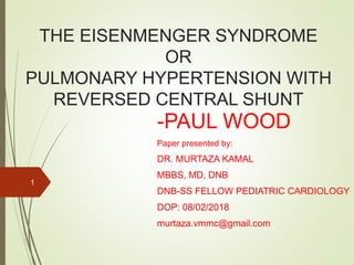
  • 1. THE EISENMENGER SYNDROME OR PULMONARY HYPERTENSION WITH REVERSED CENTRAL SHUNT -PAUL WOOD Paper presented by: DR. MURTAZA KAMAL MBBS, MD, DNB DNB-SS FELLOW PEDIATRIC CARDIOLOGY DOP: 08/02/2018 murtaza.vmmc@gmail.com 1
  • 2. 2
  • 4. EISENMENGER’S SYNDROME Pulmonary HT due to high PVR with reversed/ bidirectional shunt at aorto- pulmonary, ventricular or atrial level 4
  • 5. CAUSES  Patent ductus arteriosus  Aorto-pulmonary septal defect  Persistent truncus  Transposition with ventricular septal defect  Corrected transposition with ventricular septal defect  Ventricular septal defect  Single ventricle  Common atrioventricular canal  Persistent ostium primum atrial septal defect  Single atrium and  Hemianomalous or total anomalous pulmonary venous drainage into the right side of heart 5
  • 6. DATA  127 cases of Eisenmenger syndrome  Clinical and physiological features analyses  Special emphasis on distinguishing characteristics of each type  53 necropsis from well investigated during life studied 6
  • 7. Necropsy Findings  127 ES  15 died  38 additional necropsies  Total 127 7
  • 8. MODE OF DEATH  Cause of death known : 42  Haemoptysis: MCC, 29%  Profuse, Death in minutes  Cause: Pulmonary infarction from arterial thrombosis Rupture of a thin walled dilated arteriole immediately distal to end of a thick muscular artery Rupture of aneurysm of PA (Most unusual) 8
  • 9. MODE OF DEATH  Surgical repair of defect: 26%  CHF: 17%  VF: 14%  IE/ Cerebral abscess/ Cerebral thrombosis/ Pregnancy induced: 5% 9
  • 10. Accuracy of Diagnosis  15 necropsied case: 9 correct, 2 wrong, 4 (Correct in respect to site of shunt, wrong in respect to nature of defect)  40% error: Embarrassing  Same in 38 borrowed cases+ published literatures  Eg: Simple OS ASD TAPVD into SVC Single ventricle with intact IAS Common AV canal with functionally single atrium + virtually single ventricle 10
  • 11. SIZE OF THE DEFECT  No case of PDA with Eisenmenger reaction has ever been reported with a lumen < 5 mm  No case of Eisenmenger's complex has ever been reported with a VSD < 1cm  Size of an ASD has relatively little bearing on presence or absence of a high PVR, and varies freely between 3- 8 cm in both uncomplicated and pulmonary hypertensive groups 11
  • 12. SIZE OF COMMUNICATION Normal resistance usual range Eisenmnger Range DUCT .1-1cm .7-1.5 cm VSD .1-2cm 1.5-3cm ASD 3-8 cm 3-8 cm 12
  • 13. SIZES  Have in common a large communication b/w 2 circulations  If PVR were normal These would produce Qp/Qs at least 3.5 (4-6) 13 Aorto- pulmonary 0.7 cm Inter ventricular 1.5 cm Inter arterial 3 cm
  • 14. ELASTIC ARTERIES  In fetus/ neonates: Elastic fibers in PA; Densely packed +long as in aorta  As PAP falls Fibers break up, shorten, become loosely arranged  Found that sections of PA in EC and PDA with reversed shunt  Always resembled foetal (aortic ) pattern  Whereas in ASD with PH and reversed shunt+ all forms of acquired PH Appearance like a normal PA  14
  • 15. CONCLUDED  PH in ES must be present from birth with VSD and PDA, but must be acquired in ASD  Function of elastic fibers: To prevent distention from high pressures 15
  • 16. MUSCULAR ARTERIES  In EC, PAs with diameter< 1 mm have a thick muscular media very similar to their structure in foetus  It was persistence of foetal type of muscular artery that was responsible for high resistance, and that this saved infants with large VSDs from dying as a result of an inadequate blood flow into aorta (Civin, Edwards)  In their own words, " the pulmonary vascular bed of patients with EC who survive infancy maintains its foetal function." 16
  • 17. MUSCULAR ARTERIES  They assumed: Thick muscular arteries of foetus were responsible for high PVR necessary to deflect blood from RV away from lungs into descending aorta via PDA  During first 2 years of life intima usually remains normal  Thereafter with varying degree and tempo from case to case, progressive intimal fibrosis develops (Edwards, 1957) 17
  • 18. ARTERIOLES  Arterioles of lung: Vessels < 100u  Consist of endothelium, a single elastic layer, and adventitia; they are not supplied with muscle  Usually thin-walled and dilated in ES, and that thick-walled vessels within usually accepted compass of an arteriole are really muscular arteries in a state of contraction 18
  • 19. THE OVERRIDING AORTA  1. VSD is but one of a total of some 12 kinds of communication between two circulations, all of which may be complicated by a high PVR and reversed shunt of similar magnitude  An overriding aorta or its physiological equivalent is absent in 7 of these conditions  2. When PVR is high, SPO2 averages close to 80% in transposition with VSD, persistent truncus, single ventricle, Taussig-Bing syndrome and EC with or without apparent override 19
  • 20.  3. EC with all classical features may be associated with a defect in muscular part of IVS as in cases described by Weiss (1927) and Heath et al. (1956)  There can be no overriding of the aorta in such instances 20
  • 21. EXPERIMENTAL PHYSIO-PATHOLOGY  Reaction of pulmonary circulation in Eisenmenger's syndrome to pulmonary vasoconstrictor and vasodilator drugs  O2: Adults/ majority children>5: 100% O2 fails to lower PAP or influence shunt magnitude Other hand: 10%O2 raises PVR ad increases reversed shunt (at least in PDA) Infants< 2 and occasionally small children: When intimal fibrosis in pulmonary circulation negligible: 100% O2 halves resistance and increases PBF 21
  • 22. Failure of 100% oxygen to lower PV pressure in case of EC 22
  • 23. ACETYLCHOLINE  When injected direct into PA in a dose of 0.5-1.5 mg.: An ideal selective pulmonary vasodilator, for this quantity is usually inactivated by cholinesterase by the time it reaches systemic circulation (Wood et al., 1957)  When severe PH was primary, or due to MS or cor pulmonale, acetylcholine lowered PAP and resistance, and raised CO in 85% of cases (Wood, 1958)  In 13 cases of Eisenmenger's syndrome, however, no such effect could be demonstrated in a single instance 23
  • 24. Failure of acetylcholine to lower PAP in case of EC 24
  • 25.  The youngest patient in series, however, was 5 years old, and it is probable that infants under 2, without intimal fibrosis, would react differently  This belief is founded on the different response of these infants to 100% oxygen, and on the fact that acetylcholine lowers PVR sharply in foetal lung (Dawes, 1958)  Negative reactions to tolazoline hydrochloride (" priscol ") and aminophylline have also been without exception in cases of Eisenmenger's syndrome in patients over 5 25
  • 26. PROTECTIVE EFFECT OF STENOSIS  Vessels which are protected from extreme pulmonary hypertension, or which have not had time to develop serious intimal fibrosis secondary to hypertension, respond briskly to acetylcholine  This disposes of the idea that there is anything fundamentally wrong with pulmonary vessels at birth inES  Eg: Follows 26
  • 27. 27 Pressure pulses in a case of ES associated with an AP septal defect and stenosis of RPA near its origin Pressure pulse from RPA beyond the stricture, showing a marked reduction of pressure following the injection of acetylcholine. The pulmonary vessels have been protected by the stenosis and are functioning normally
  • 28.  This functional evidence stands solidly side by side with anatomical observation that when origin of LPA is atresic in ES, its distal branches and muscular arteries, which are not subjected to high pressure, are perfectly normal  Whereas vessels on other side have all features characteristic of Eisenmenger's complex (McKim and Wiglesworth, 1954) 28
  • 29. COURSE & PROGNOSIS  ES commonly established at birth or during the first two years of life when shunt is aorto-pulmonary or interventricular  But is typically acquired when shunt is interatrial  In present series, cyanosis dated from infancy in 80% of VSD, but did not start until adult life in 92% ofASD  Onset of reversed shunt in PDA: More difficult to establish, because differential cyanosis usually overlooked  Aorto-pulmonary septal defects, however, behave likeVSDs 29
  • 30.  General rule: Neither cyanosis nor breathlessness changes appreciably during childhood or adolescence, but deterioration sets in sooner or later in adult life  Onset of recurrent haemoptysis may signal downward trend which is due in many cases to the development of thromboobstructive lesions  Average age of natural death : 33: Aortopulmonary defects 33: VSDs 36: ASDs 30
  • 31.  Downhill course usually punctuated with haemoptyses, which when severe sometimes caused sudden collapse and death within a few minutes  Bacterial endocarditis: 5% for death  Cerebral abscess from paradoxical embolism occurred once and proved fatal  Cerebral thrombosis in one case, but not fatal  14% died abruptly, presumably from VF  26% died from a surgical attempt to repair defect, mostly from ligation of a patent ductus  Patients who survive these risks develop CHF sooner or later, death in 17% of the fatal cases 31
  • 32. TREATMENT  Medical treatment: Aims at preventing secondary thromboobstructive lesions by means of permanent anticoagulants, such as phenindione (" dindevan ")  Pulmonary vasodilators (tolazoline, aminophylline,and reserpine::Fail to lowerPVR, and do not influence course of disease  Small venesections, repeated from time to time: May be helpful in cases with gross polycythaemia  Heart failure management 32
  • 33.  Surgical repair of defect: Responsible for more deaths in ES than any other agent except haemoptysis  Ellis et al. (1956): Surgical mortality in cases with reversed shunt through a patent ductus was 56 33
  • 34.  Direct repair of a defect is justified only when it can be predicted with confidence that its closure must result in a fall of pulmonary blood pressure because a hyperkinetic factor of sufficient magnitude has been eliminated  In practice this prediction can be made when PBF is at least twice the systemic flow at rest, and PVR does not exceed 10 units (800 dynes sec./cm.')  Borderline cases have Qp/Qs 1.75- 2, and resistances of 10 to 12 units  No case with PBF <1.75 times the systemic flow or with a resistance over 12 units should have defect repaired in the ordinary way, whether the shunt is reversed, direct, or bidirectional 34
  • 35. SYNTHESIS  CO of newborn babe is about 0.5 1./min., and is presumably much same in foetus at term  0.1 1./min. passes through pulmonary circulation  Since mean PAP is same as aortic, about 50 mm. Hg at birth, foetal pulmonary vascular resistance must be about 500 units (40,000 dynes sec./cm.5)  During 1st 3 hours after birth a reversed shunt through PDA is invariable;  From 3-72 hours after birth shunt may be direct, bidirectional or reversed and by end of 3rd day duct is usually closed  It follows that in newborn infants PVR is more or less same as the systemic-that is, about 100 units 35
  • 36.  Within a remarkably short time after birth, then, probably within a few minutes of independent life, a PVR around 500 units falls to about 100  4 questions clamour for an answer:  (1) What is the cause of the fantastic PVR in the foetus?  (2) Why does it drop so precipitously at birth ?  (3) Why is the rapid fall halted temporarily more or less at systemic level ?  (4) What is responsible for the subsequent slower decline to normal ?  In the correct answer to these four questions lies the secret of the Eisenmenger syndrome 36
  • 37. What is the cause of the fantastic PVR in the foetus?  Resistance to flow in a tube is greatly increased by kinking the vessel  In the fully developed foetus the pulmonary vessels are as long as they are a few minutes after birth, but since the lung is completely collapsed they must be coiled up, or kinked in innumerable places  Burton has demonstrated their "gnarled" appearance  Vessels in such a state may be highly resistant to flow 37
  • 38.  Answer to the second question:  As the lungs expand the vessels uncoil: as the kinks straighten out the resistance falls; as the blood begins to flow through the lungs the vessels must dilate, and this also lowers the resistance  Blood that fills up pulmonary vascular bed acts as a natural scaffolding and encourages further expansion (Jaykkai, 1957)  Stepwise, and in a matter of a minute or so, and aided by lusty cries, the lungs are fully expanded (Lind and Wegelius, 1956)  Vessels are wholly unfurled, and PVR is at systemic level, at least five times lower than in utero 38
  • 39. Why is the rapid fall in resistance at birth temporarily halted at systemic level ?  This may well be a function of the thick muscular arteries just mentioned  In the foetus these have developed in relation to the systemic pressure to which they have been subjected, and, as shown by Dawes (1958), they are also in a state of active vasoconstriction, which can be released by acetylcholine  After birth a powerful RV, as big and strong as the left, pumps its contents into a system of vessels which differs little from the systemic circulation;it is only natural, therefore, that pulmonary and systemic resistances should be similar 39
  • 40.  During 1st of life, then, physiological and anatomical findings are identical with ES associated with PDA  2events of great importance  1st: Relaxing effect of an increased alveolar oxygen tension on the tone of muscular arteries  2nd: Closure of PDA  Before the duct closes, release of vasoconstrictor tone in the muscular arteries is met by an increased flow from aorta to PA, and this hyperkinetic factor tends to maintain PA pressure at systemic level 40
  • 41.  When duct closes, and this hyperkinetic factor is eliminated, release of vasoconstrictor tone lowers the pulmonary artery pressure, and the approach to normal adult physiology begins  Just how quickly this goal is reached depends on the speed of involution of the muscular arteries  Complete involution takes about three months 41
  • 42. What is responsible for the subsequent slower decline to normal ?  (1) As the pressure falls in the pulmonary circulation vasoconstrictor tone is diminished, for muscular arteries react to increased pressure by contracting and to decreased pressure by relaxing (Bayliss, 1902)  This local response of muscular arteries to any change in pressure must increase that change in either direction  (2) As the resistance declines PBF increases  This must dilate vessels and so lower the resistance still further (Williams, 1954)  (3) As the pressure falls the muscular arteries atrophy and so diminish their resistance to flow 42
  • 43. THE EISENMENGER REACTION  All that is necessary to prevent these natural forces from lowering PVR to normal is a sufficient increase of flow to keep the pressure at systemic level  Any large AP or IV communication will do just this, and the higher the resistance the less increase of flow necessary to maintain the pressure  If the pressure is maintained pulmonary vasoconstrictor tone must remain high, and the muscular arteries will not atrophy  This is the situation inES, and it will be understood that it must be established at birth if the defect is larg enough 43
  • 44.  If the defect is not quite critical, however, the shunt flow at rest may not be sufficient to maintain pressure at systemic level, and the forces acting in the opposite direction may win or partially win the competition  Once resistance falls appreciably much larger flows are necessary to maintain high pressure 44