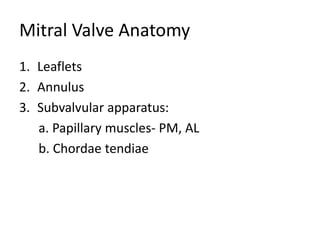
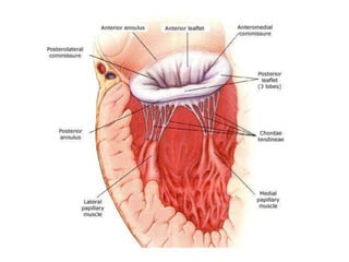
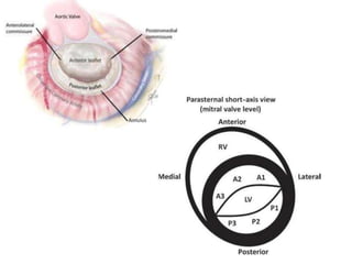
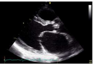
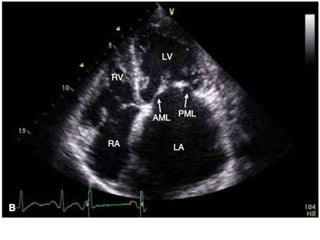
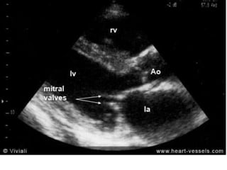
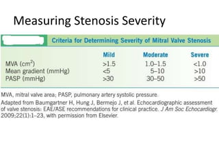
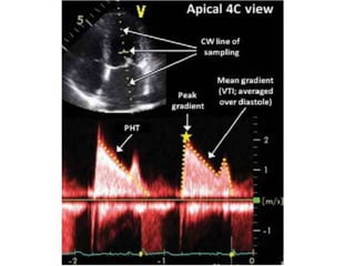
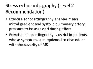
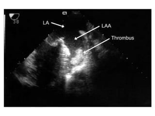
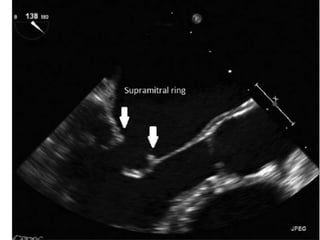
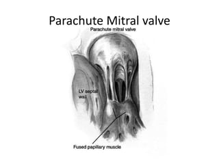
Echocardiographic assessment of
MS
Dr. Md. Mashiul Alam
Resident
University Cardiac Center, BSMMU
Mitral Valve Anatomy
1. Leaflets
2. Annulus
3. Subvalvular apparatus:
a. Papillary muscles- PM, AL
b. Chordae tendiae
Mitral Stenosis etiology
1. Rheumatic: most common cause
2. Severe mitral annular calcification (MAC)
3. Congenital
Parachute mitral valve: single papillary muscle to which chordae to
both leaflets attach;results in mitral stenosis or mitral regurgitation
Supravalvular mitral ring
4. Systemic diseases: can cause valvular fibrosis
Carcinoid
SLE
RA
Healed endocarditis
Rheumatic MS
The main mechanism of rheumatic MS is
commissural fusion.
Other anatomic lesions are chordal shortening
and fusion, and leaflet thickening, and later in
the disease course, superimposed
calcification, which may contribute to the
restriction of leaflet motion
Degenerative MS
The main lesion is annular calcification. It is
frequently observed in the elderly and
associated with HTN, atherosclerotic disease,
and sometimes AS.
This is required to cause restriction of leaflet
motion since there is no commissural fusion.
Valve thickening or calcification predominates at
the base of the leaflets whereas it affects
predominantly the tips in rheumatic MS
Systemic MS
Mainly the consequence of abnormalities of the
subvalvular apparatus.
Leaflet thickening and restriction are common
here, while commissures are rarely fused.
Congenital MS
Methods of echo assessment of MS
1. 2D, M and doppler mode echo
2. 3 D echo
3. Transesophageal echo
4. Stress echo
M mode, 2D and Doppler study
1. Initial diagnosis
2. Determination of severity
3. Evaluation of suitability for PTMC
4. Identification of concomitant Valve lesions
Views
1. PLAX
2. PSAX at MV level
3. A4CV
M mode
1. Dense echo on MV
2. Decreased D-E excursion
3. Poor leaflet seperation
4. Anterior motion of posterior leaflet
5. Decreased E-F slope
2 Dimensional (Initial Assessment)
• Restricted motion and diastolic motion of
leaflets ( hockey stick sign)
• Thickening and calcification of leaflets and
chordae
Measuring Stenosis Severity
Pressure gradient (Level 1
Recommendation).
CW doppler signal is obtained in A4CV and
traced
Caution:
1. Gradient is influenced by HR, cardiac output,
MR, VSD
2. In patients with atrial fibrillation, mean
gradient should be calculated as the average
of five cycles with the least variation of R–R
intervals and as close as possible to normal
heart rate
Measuring valve area
1. MVA Planimetry (Level 1 Recommendation).
planimetry is considered as the reference
measurement of MVA
• Careful scanning from the apex to the base of
the LV PSAX view at the level of mitral valveis
required to ensure that the CSA is measured
at the leaflet tips
• Gain setting should be just sufficient to
visualize the whole contour of the mitral
orifice. Excessive gain setting may cause
underestimation of valve area, in particular
when leaflet tips are dense or calcified.
• Image magnification, using the zoom mode, is
useful to better delineate the contour of the
mitral orifice.
• The optimal timing of the cardiac cycle to
measure planimetry is mid-diastole. This is best
performed using the cineloop mode on a frozen
image.
• Recent reports suggested that real-time 3D echo
and 3D-guided biplane imaging is useful in
optimizing the positioning of the measurement
plane and, therefore, improving reproducibility
• In the particular case of degenerative MS,
planimetry is difficult and mostly not reliable
because of the orifice geometry and
calcification present
Measuring valve area
2. Pressure half-time (Level 1 Recommendation)
• MVA 220 ⁄ T½ or 750/DT
T1/2 is obtained by tracing the deceleration slope
of the E-wave on Doppler spectral display of
transmitral flow and valve area is automatically
calculated by the integrated software of currently
used echo machines
PHT = 29% of total deceleration time (DT)
• In patients with atrial fibrillation, tracing
should avoid mitral flow from short diastoles
and average different cardiac cycles.
• the use of T1/2 in degenerative calcific MS
may be unreliable and should be avoided.
• Impaired LV diastolic function is a likely
explanation of the lower reliability of T1/2 to
assess MVA in the elderly.
Measuring valve area
3. Continuity equation (Level 2
Recommendation)
• The continuity equation cannot be used in
cases of atrial fibrillation or associated
significant MR or AR.
4. Proximal isovelocity surface area or PISA
method (Level 2 Recommendation)
Measuring PASP pressure
• Calculation of TR in A4CV
• RA pressure in subcostal view
PASP = 4 (max TR velocity)² + RA pressure
RA pressure:
IVC ≥2.1 cm with resp. variation > 50% = 0-5mmHg
IVC >2.1 cm with resp. variation <50% = 10-20 mmHg
Does not fit in any category = 5-10 mmHg
Evaluation of suitability for PTMC
• Several scoring system. e.g., Wilkins score,
Cormier score
• Wilkins score less than 8 predicts successful
PTMC
• No score is accurate
Identification of concomitant lesions.
• The quantitation of left atrial enlargement
favours 2D echocardiography enabling left atrial
area or volume to be evaluated.
• Left atrial spontaneous contrast as assessed by
TEE is a better predictor of the thromboembolic
risk than left atrial size.
• Associated MR has important implications for the
choice of intervention
• mediate severity since more than mild
regurgitation is a relative contraindication for
balloon mitral commissurotomy
• Other valve diseases are frequently associated
with rheumatic MS. The severity of AS may be
underestimated because decreased SV due to MS
reduces aortic gradient, thereby highlighting the
need for the estimation of AVA. In cases of severe
AR, the T1/2 method for assessment of MS is not
valid. The analysis of the tricuspid valve should
look for signs of involvement of the rheumatic
process. More frequently, associated tricuspid
disease is functional tricuspid regurgitation (TR).
Stress echocardiography (Level 2
Recommendation)
• Exercise echocardiography enables mean
mitral gradient and systolic pulmonary artery
pressure to be assessed during effort.
• Exercise echocardiography is useful in patients
whose symptoms are equivocal or discordant
with the severity of MS
Transoesophageal echocardiography
Recommended only when the transthoracic
approach is of poor quality, or to detect left
atrial thrombosis before balloon mitral
commissurotomy or following a
thromboembolic event.
Supravalvular mitral ring
Parachute Mitral valve
THANK YOU

Echo Mitral Stenosis

  • 1.
    Echocardiographic assessment of MS Dr.Md. Mashiul Alam Resident University Cardiac Center, BSMMU
  • 2.
    Mitral Valve Anatomy 1.Leaflets 2. Annulus 3. Subvalvular apparatus: a. Papillary muscles- PM, AL b. Chordae tendiae
  • 5.
    Mitral Stenosis etiology 1.Rheumatic: most common cause 2. Severe mitral annular calcification (MAC) 3. Congenital Parachute mitral valve: single papillary muscle to which chordae to both leaflets attach;results in mitral stenosis or mitral regurgitation Supravalvular mitral ring 4. Systemic diseases: can cause valvular fibrosis Carcinoid SLE RA Healed endocarditis
  • 6.
    Rheumatic MS The mainmechanism of rheumatic MS is commissural fusion. Other anatomic lesions are chordal shortening and fusion, and leaflet thickening, and later in the disease course, superimposed calcification, which may contribute to the restriction of leaflet motion
  • 7.
    Degenerative MS The mainlesion is annular calcification. It is frequently observed in the elderly and associated with HTN, atherosclerotic disease, and sometimes AS. This is required to cause restriction of leaflet motion since there is no commissural fusion. Valve thickening or calcification predominates at the base of the leaflets whereas it affects predominantly the tips in rheumatic MS
  • 8.
    Systemic MS Mainly theconsequence of abnormalities of the subvalvular apparatus. Leaflet thickening and restriction are common here, while commissures are rarely fused. Congenital MS
  • 9.
    Methods of echoassessment of MS 1. 2D, M and doppler mode echo 2. 3 D echo 3. Transesophageal echo 4. Stress echo
  • 10.
    M mode, 2Dand Doppler study 1. Initial diagnosis 2. Determination of severity 3. Evaluation of suitability for PTMC 4. Identification of concomitant Valve lesions
  • 11.
    Views 1. PLAX 2. PSAXat MV level 3. A4CV
  • 12.
    M mode 1. Denseecho on MV 2. Decreased D-E excursion 3. Poor leaflet seperation 4. Anterior motion of posterior leaflet 5. Decreased E-F slope
  • 14.
    2 Dimensional (InitialAssessment) • Restricted motion and diastolic motion of leaflets ( hockey stick sign) • Thickening and calcification of leaflets and chordae
  • 18.
  • 19.
    Pressure gradient (Level1 Recommendation). CW doppler signal is obtained in A4CV and traced
  • 21.
    Caution: 1. Gradient isinfluenced by HR, cardiac output, MR, VSD 2. In patients with atrial fibrillation, mean gradient should be calculated as the average of five cycles with the least variation of R–R intervals and as close as possible to normal heart rate
  • 22.
    Measuring valve area 1.MVA Planimetry (Level 1 Recommendation). planimetry is considered as the reference measurement of MVA • Careful scanning from the apex to the base of the LV PSAX view at the level of mitral valveis required to ensure that the CSA is measured at the leaflet tips
  • 23.
    • Gain settingshould be just sufficient to visualize the whole contour of the mitral orifice. Excessive gain setting may cause underestimation of valve area, in particular when leaflet tips are dense or calcified. • Image magnification, using the zoom mode, is useful to better delineate the contour of the mitral orifice.
  • 24.
    • The optimaltiming of the cardiac cycle to measure planimetry is mid-diastole. This is best performed using the cineloop mode on a frozen image. • Recent reports suggested that real-time 3D echo and 3D-guided biplane imaging is useful in optimizing the positioning of the measurement plane and, therefore, improving reproducibility
  • 25.
    • In theparticular case of degenerative MS, planimetry is difficult and mostly not reliable because of the orifice geometry and calcification present
  • 30.
    Measuring valve area 2.Pressure half-time (Level 1 Recommendation) • MVA 220 ⁄ T½ or 750/DT T1/2 is obtained by tracing the deceleration slope of the E-wave on Doppler spectral display of transmitral flow and valve area is automatically calculated by the integrated software of currently used echo machines PHT = 29% of total deceleration time (DT)
  • 33.
    • In patientswith atrial fibrillation, tracing should avoid mitral flow from short diastoles and average different cardiac cycles. • the use of T1/2 in degenerative calcific MS may be unreliable and should be avoided. • Impaired LV diastolic function is a likely explanation of the lower reliability of T1/2 to assess MVA in the elderly.
  • 35.
    Measuring valve area 3.Continuity equation (Level 2 Recommendation) • The continuity equation cannot be used in cases of atrial fibrillation or associated significant MR or AR. 4. Proximal isovelocity surface area or PISA method (Level 2 Recommendation)
  • 36.
    Measuring PASP pressure •Calculation of TR in A4CV • RA pressure in subcostal view PASP = 4 (max TR velocity)² + RA pressure RA pressure: IVC ≥2.1 cm with resp. variation > 50% = 0-5mmHg IVC >2.1 cm with resp. variation <50% = 10-20 mmHg Does not fit in any category = 5-10 mmHg
  • 37.
    Evaluation of suitabilityfor PTMC • Several scoring system. e.g., Wilkins score, Cormier score • Wilkins score less than 8 predicts successful PTMC • No score is accurate
  • 40.
    Identification of concomitantlesions. • The quantitation of left atrial enlargement favours 2D echocardiography enabling left atrial area or volume to be evaluated. • Left atrial spontaneous contrast as assessed by TEE is a better predictor of the thromboembolic risk than left atrial size. • Associated MR has important implications for the choice of intervention • mediate severity since more than mild regurgitation is a relative contraindication for balloon mitral commissurotomy
  • 41.
    • Other valvediseases are frequently associated with rheumatic MS. The severity of AS may be underestimated because decreased SV due to MS reduces aortic gradient, thereby highlighting the need for the estimation of AVA. In cases of severe AR, the T1/2 method for assessment of MS is not valid. The analysis of the tricuspid valve should look for signs of involvement of the rheumatic process. More frequently, associated tricuspid disease is functional tricuspid regurgitation (TR).
  • 46.
    Stress echocardiography (Level2 Recommendation) • Exercise echocardiography enables mean mitral gradient and systolic pulmonary artery pressure to be assessed during effort. • Exercise echocardiography is useful in patients whose symptoms are equivocal or discordant with the severity of MS
  • 47.
    Transoesophageal echocardiography Recommended onlywhen the transthoracic approach is of poor quality, or to detect left atrial thrombosis before balloon mitral commissurotomy or following a thromboembolic event.
  • 49.
  • 52.
  • 54.