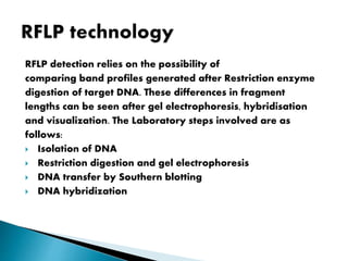
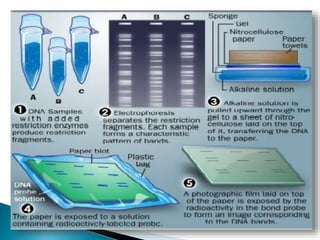
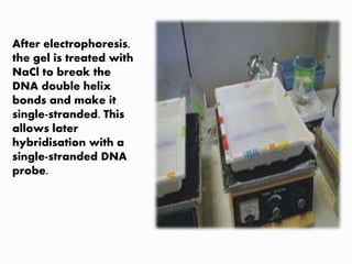
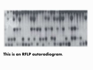
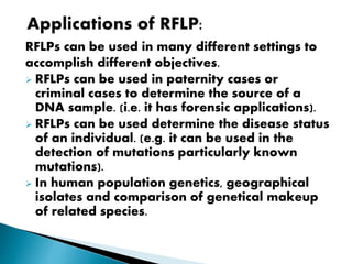
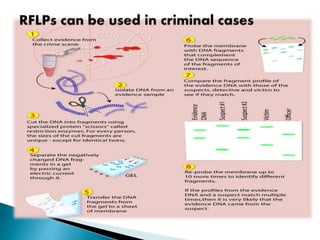
2. Restriction Fragment Length Polymorphism (RFLP) is a
technique in which organisms may be differentiated
by analysis of patterns derived from cleavage of
their DNA.
If two organisms differ in the distance between
sites of cleavage of a particular restriction
endonuclease, the length of the fragments
produced will differ when the DNA is digested with
a restriction enzyme.
The similarity of the patterns generated can be
used to differentiate species (and even strains)
from one another.
A restriction fragment length polymorphism (RFLP) is
a genetic variant that can be examined by cleaving
the DNA into fragments (restriction fragments) with a
restriction enzyme.
3. A restriction enzyme cuts the DNA molecules at every
occurrence of a particular sequence, called
restriction site.
For example, HindII enzyme cuts at GTGCAC or
GTTAAC.
If we apply a restriction enzyme on DNA, it is cut at
every occurrence of the restriction site into a million
restriction fragments each a few thousands
nucleotides long.
4. Any mutation of a single nucleotide may destroy
(CTGCAC or CTTAAC for HindII) and alter the length
of the corresponding fragment.
The term polymorphism refers to the slight
differences between individuals, in base pair
sequences of common genes.
or
A polymorphism is a clinically harmless DNA
variation that does not affect the phenotype.
RFLP analysis is the detection of the change in the
length of the restriction fragments.
5. RFLP detection relies on the possibility of
comparing band profiles generated after Restriction enzyme
digestion of target DNA. These differences in fragment
lengths can be seen after gel electrophoresis, hybridisation
and visualization. The Laboratory steps involved are as
follows:
Isolation of DNA
Restriction digestion and gel electrophoresis
DNA transfer by Southern blotting
DNA hybridization
6.
7. Isolating DNA is the first step for many DNA-based
technologies. DNA is found either in nuclear
chromosomes or in organelles (mitochondria and
chloroplasts).
To extract DNA from its location, several laboratory
procedures are needed to break the cell wall and
nuclear membrane, and so appropriately separate
the DNA from other cell components.
When doing so, care must be taken to ensure the
process does not damage the DNA molecule and that
it is recovered in the form of a long thread.
8. Extracted DNA is digested with specific, carefully
chosen, restriction enzymes.
Each restriction enzyme, under appropriate conditions,
will recognize and cut DNA in a predictable way,
resulting in a reproducible set of DNA fragments
(‘restriction fragments’) of different lengths.
The millions of restriction fragments produced are
commonly separated by electrophoresis on agarose
gels. Because the fragments would be seen as a
continuous ‘smear’ if stained with ethidium bromide,
staining alone cannot detect the polymorphisms.
Hybridisation must therefore be used to detect specific
fragments.
9. DNA transfer is called ‘Southern blotting’, after
E.M. Southern (1975), who invented the technique.
In this method, the gel is first denatured in a basic
solution and placed in a tray. A porous nylon or
nitrocellulose membrane is laid over the gel, and
the whole weighted down.
All the DNA restriction fragments in the gel transfer
as single strands by capillary action to the
membrane. All fragments retain the same pattern
on the membrane as on the gel.
10. The membrane with the target DNA is incubated with the DNA
probe. Incubation conditions are such that if strands on the
membrane are complementary to those of the probe, hybridisation
will occur and labeled duplexes formed.
The DNA probe is a single-stranded molecule, conveniently
labeled, using any standard method (e.g. a radioisotope or
digoxygenin), and hybridized with the target DNA, which is stuck
to the membrane.
Where conditions are highly stringent, hybridisation with distantly
related or non-homologous DNA does not happen.
Thus, the DNA probe picks up sequences that are complementary
and 'ideally‘ homologous to itself among the thousands or millions
of undetected fragments that migrate through the gel.
Desired fragments may be detected after simultaneous exposure
of the hybridized membrane and a photographic film.
11. RFLP technology in pictures
After agarose has been
poured into the gel
mould, combs are
immediately inserted to
form wells and left until
the gel hardens. The
combs are then removed
and the gel placed in an
electrophoresis chamber.
13. After electrophoresis,
the gel is treated with
NaCl to break the
DNA double helix
bonds and make it
single-stranded. This
allows later
hybridisation with a
single-stranded DNA
probe.
14. The blotting tray is
first prepared by
saturating sponges
with NaOH. Safety
glasses and gloves
are required, and a
laboratory coat
recommended.
15. Absorbent paper is placed on top of the
sponges to prevent direct contact with
the gel.
16. Bubbles between the absorbent paper and sponges are removed
by rolling a pipette or a glass rod across the paper. This ensures a
complete transfer of the solution all through
the gel.
18. Membrane is cut
into the appropriate
size.
The membrane is
placed on top of the
gel, then covered
with a piece of
absorbent paper.
19. The entire set-up is
topped with a weight
(here, a bottle of water
standing on a piece of
glass) to promote good
transfer. After some
hours the transfer is
complete, the blotting
paper is taken away, and
the membrane stored
until hybridisation with
the probe.
20. The process of hybridisation begins. A DNA is boiled
to denature it to single strands.
21. The labeled probe is
added to the container
with the hybridisation
solution and membrane,
and incubated overnight
in an oven. The
following day, the
membrane is removed
from the hybridisation
set up, and washed with
the appropriate
stringency solution.
22. The membrane is then
blotted dry and put into
a cassette for holding X-
ray film.
The cassette is wrapped,
or sealed with tape, and
stored in a freezer until
the film is
sufficiently exposed,
usually 1 to 4 days.
24. RFLPs can be used in many different settings to
accomplish different objectives.
RFLPs can be used in paternity cases or
criminal cases to determine the source of a
DNA sample. (i.e. it has forensic applications).
RFLPs can be used determine the disease status
of an individual. (e.g. it can be used in the
detection of mutations particularly known
mutations).
In human population genetics, geographical
isolates and comparison of genetical makeup
of related species.
25.
26. Highly robust methodology with good
transferability between laboratories.
No sequence information required.
highly recommended for phylogenetic analysis
between related species.
Well suited for constructing genetic linkage maps.
Simplicity—given the availability of suitable
probes, the technique can readily be applied to
any plant.
27. Large amounts of DNA required.
Automation not possible.
Low levels of polymorphism in some species.
Time consuming, especially with single-copy
probes
Costly.
Moderately demanding technically.
30. Brian Kennedy, president and CEO of the Buck
Institute for Research on Ageing in the US and
lead author of the study, said by ’removing 238
gene responsible for ageing in yeast cell can
significantly extend the lifespan of yeast cells
by as much as 60% in laboratory testing’.
http://www.sciencealert.com