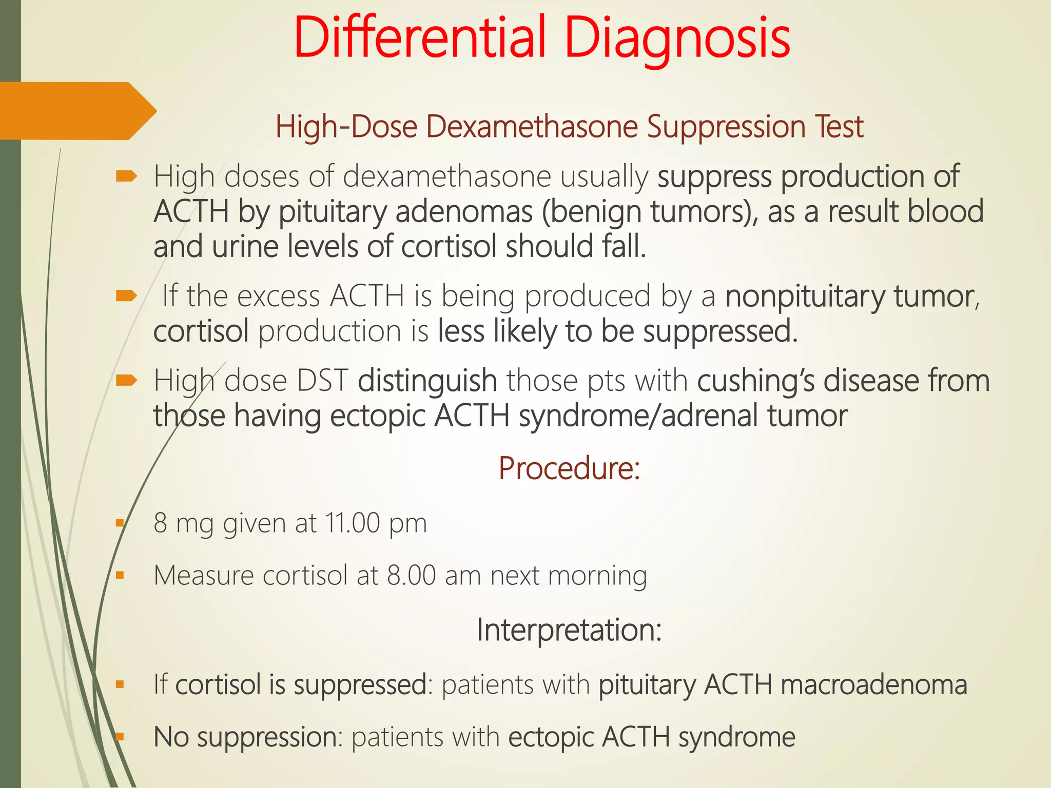
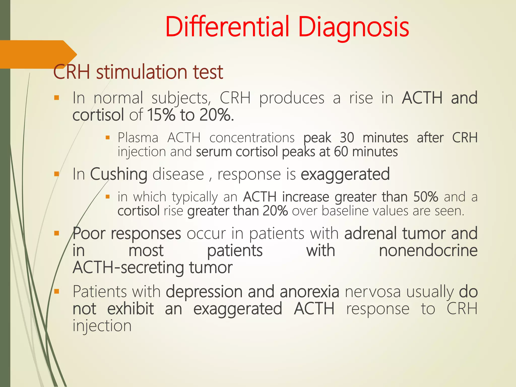
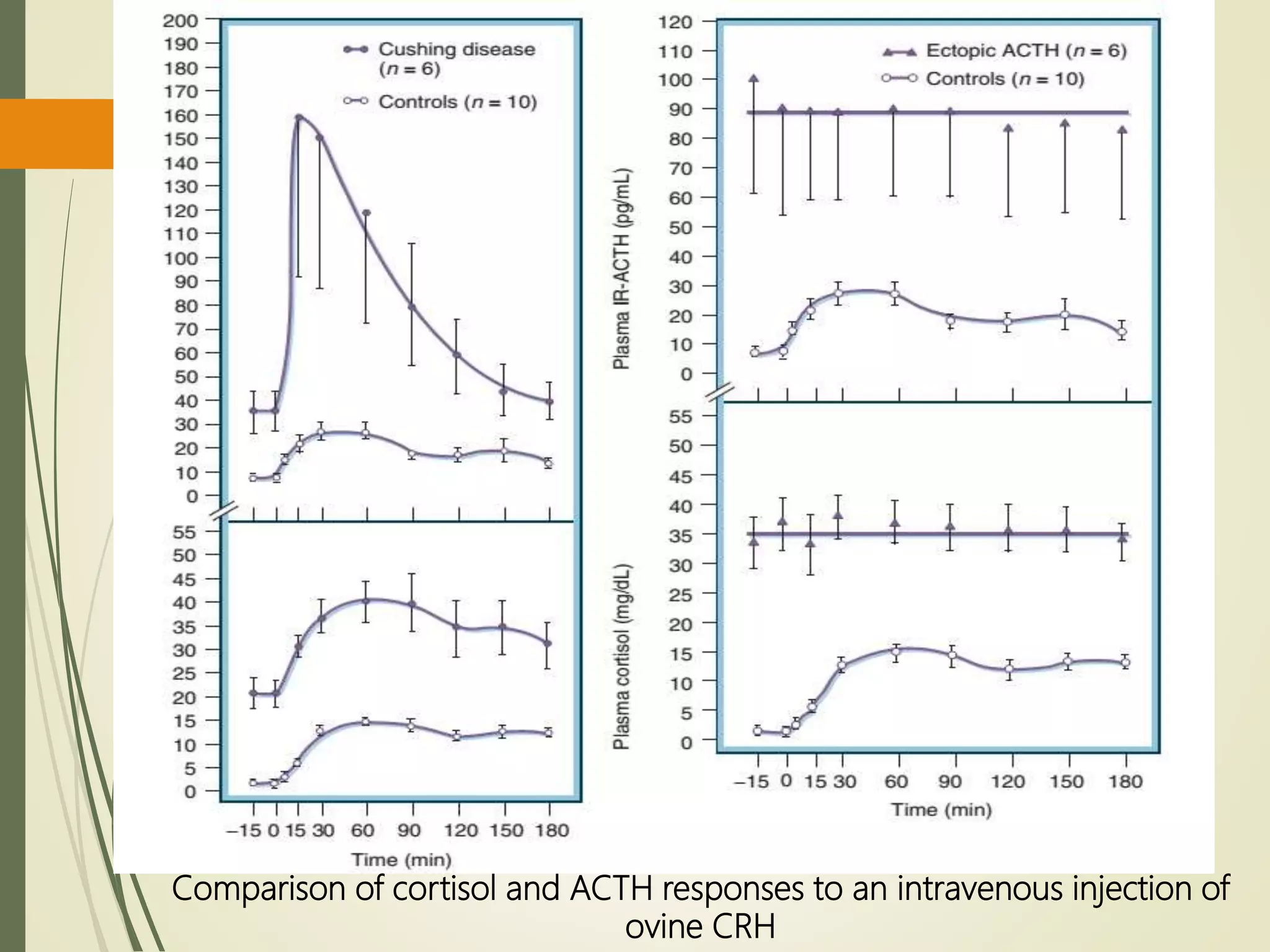
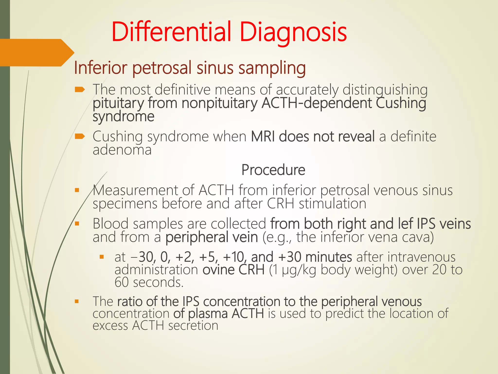
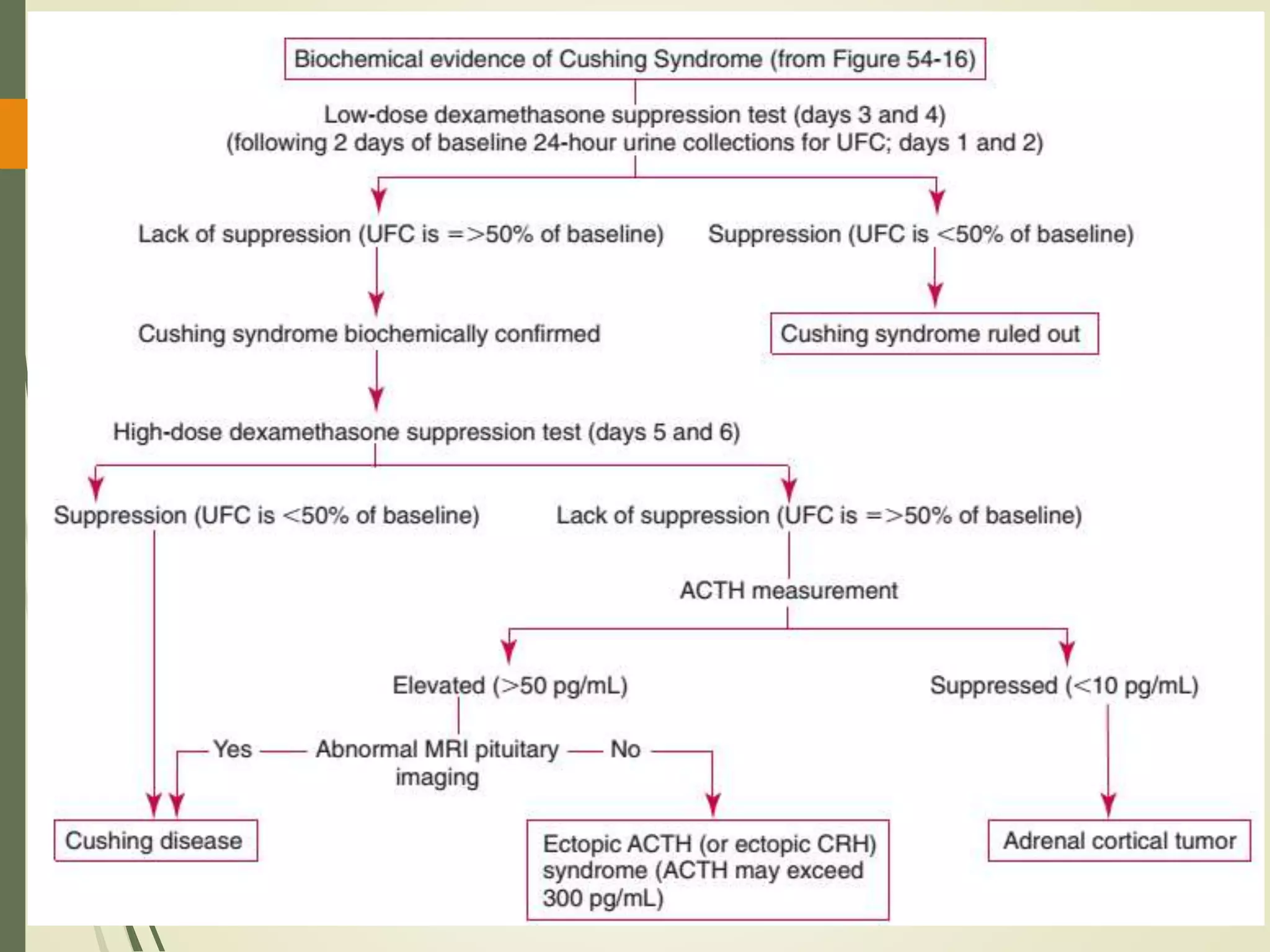
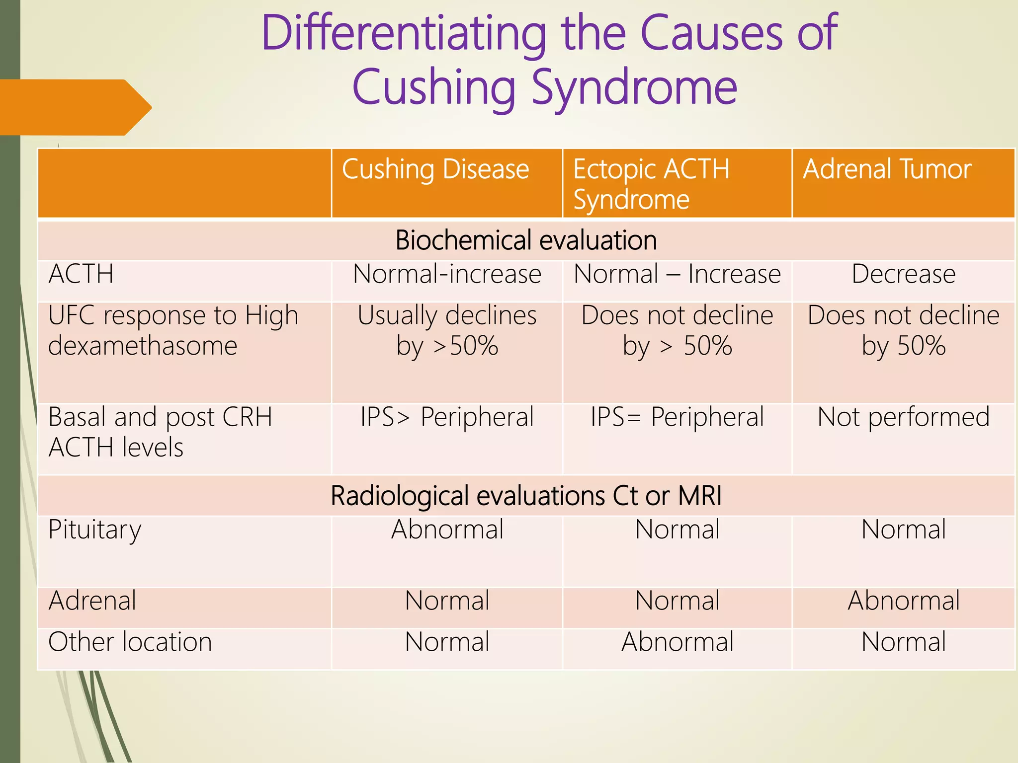
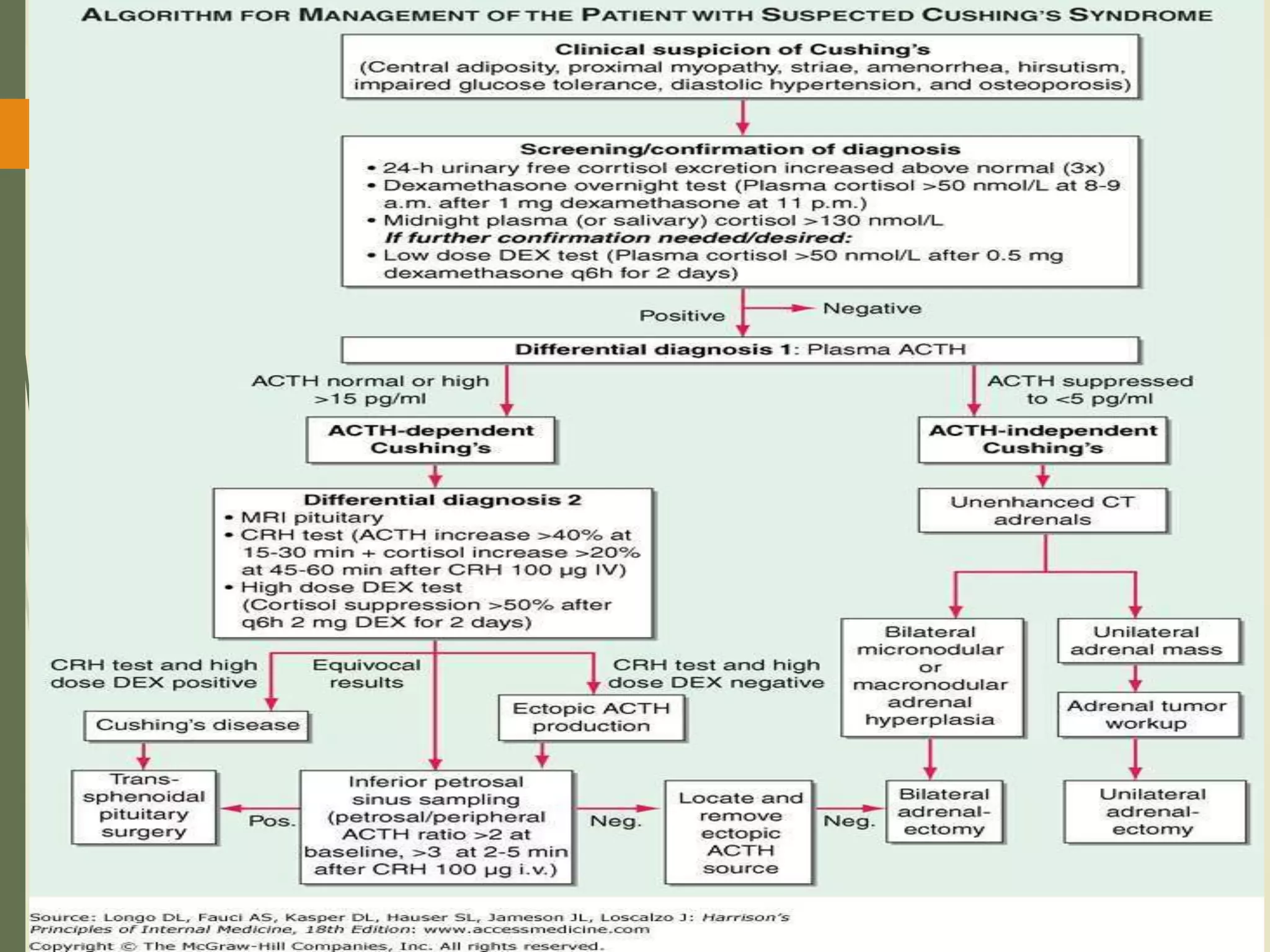
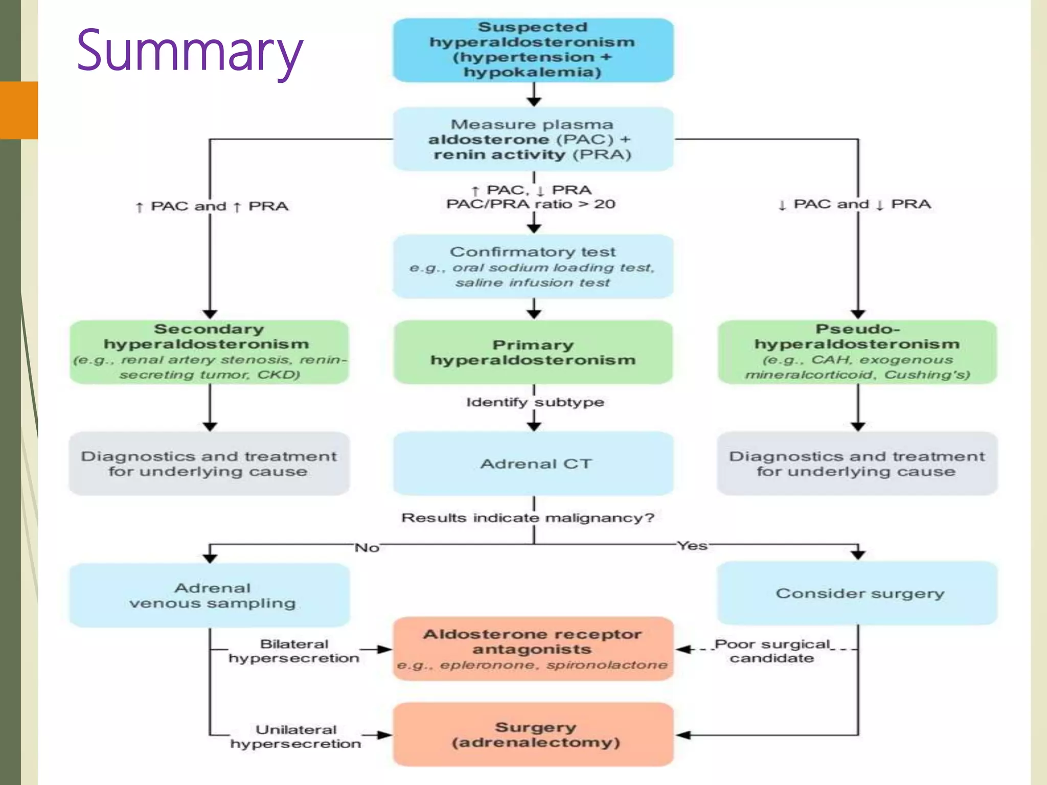
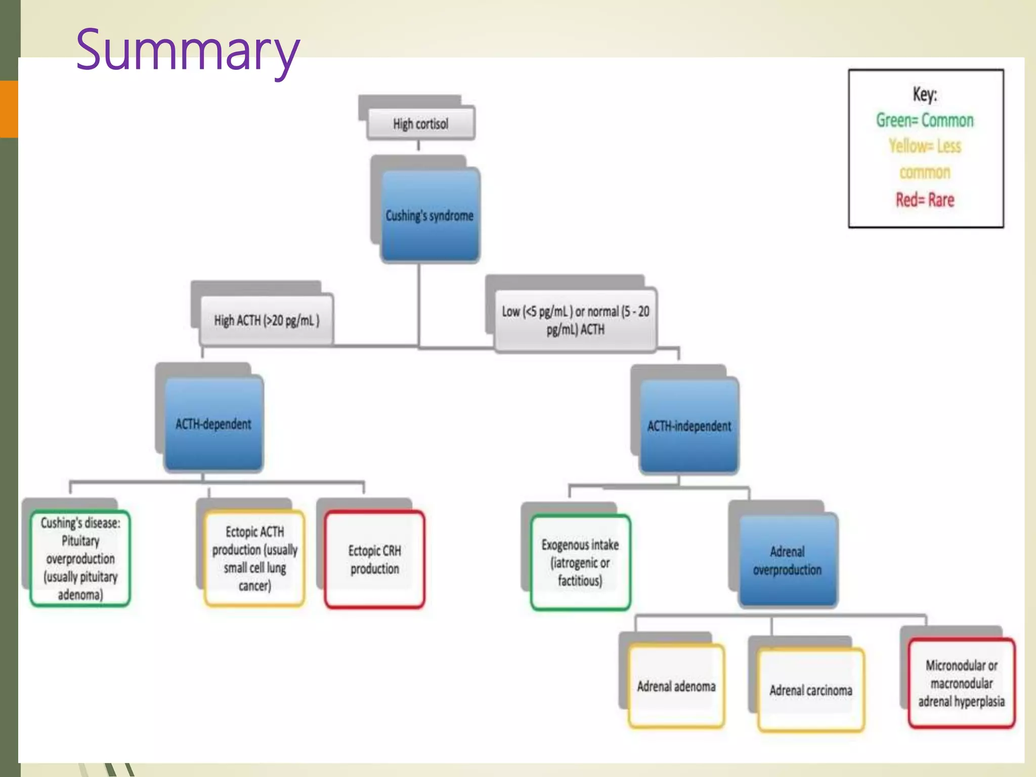
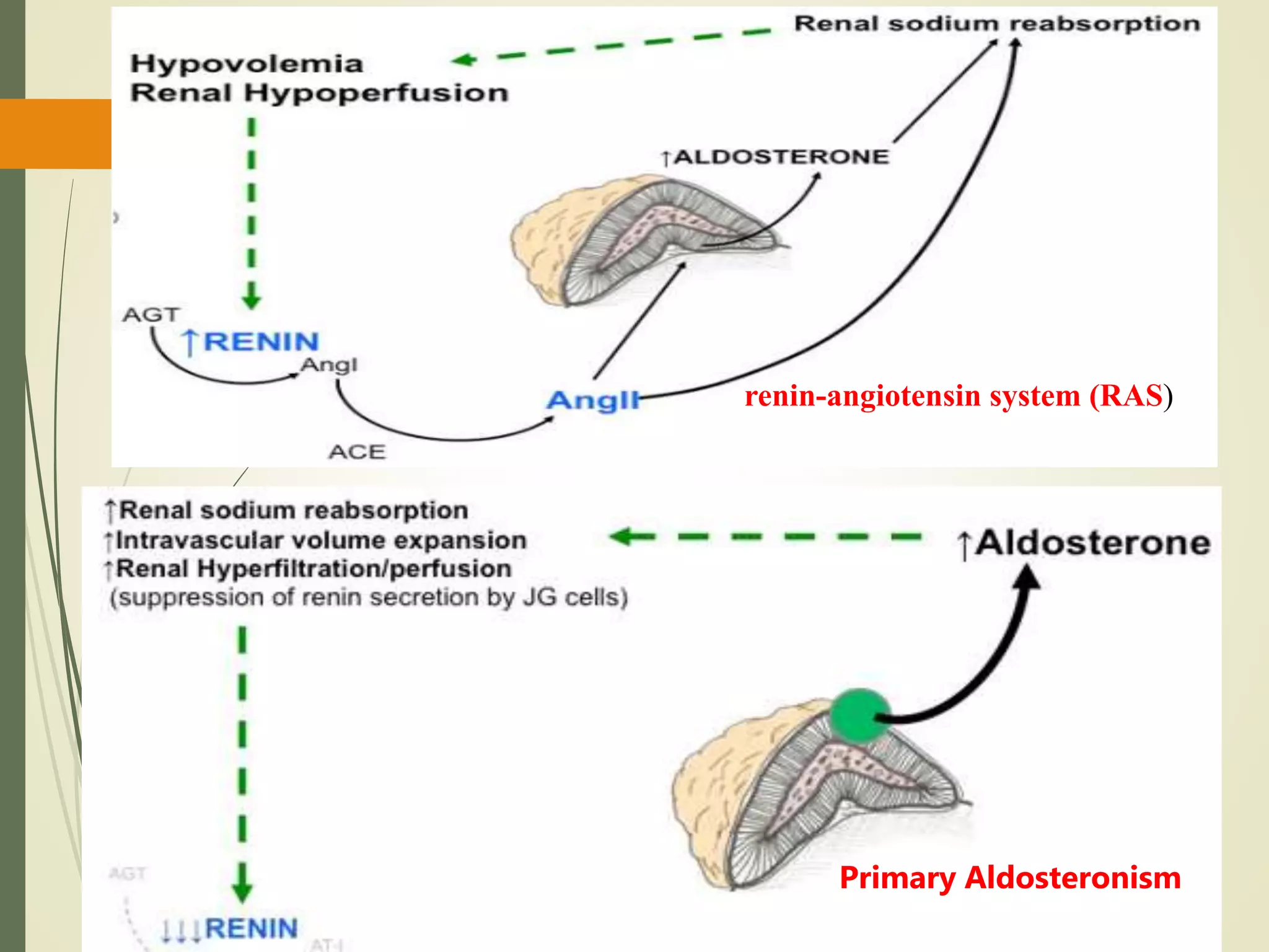
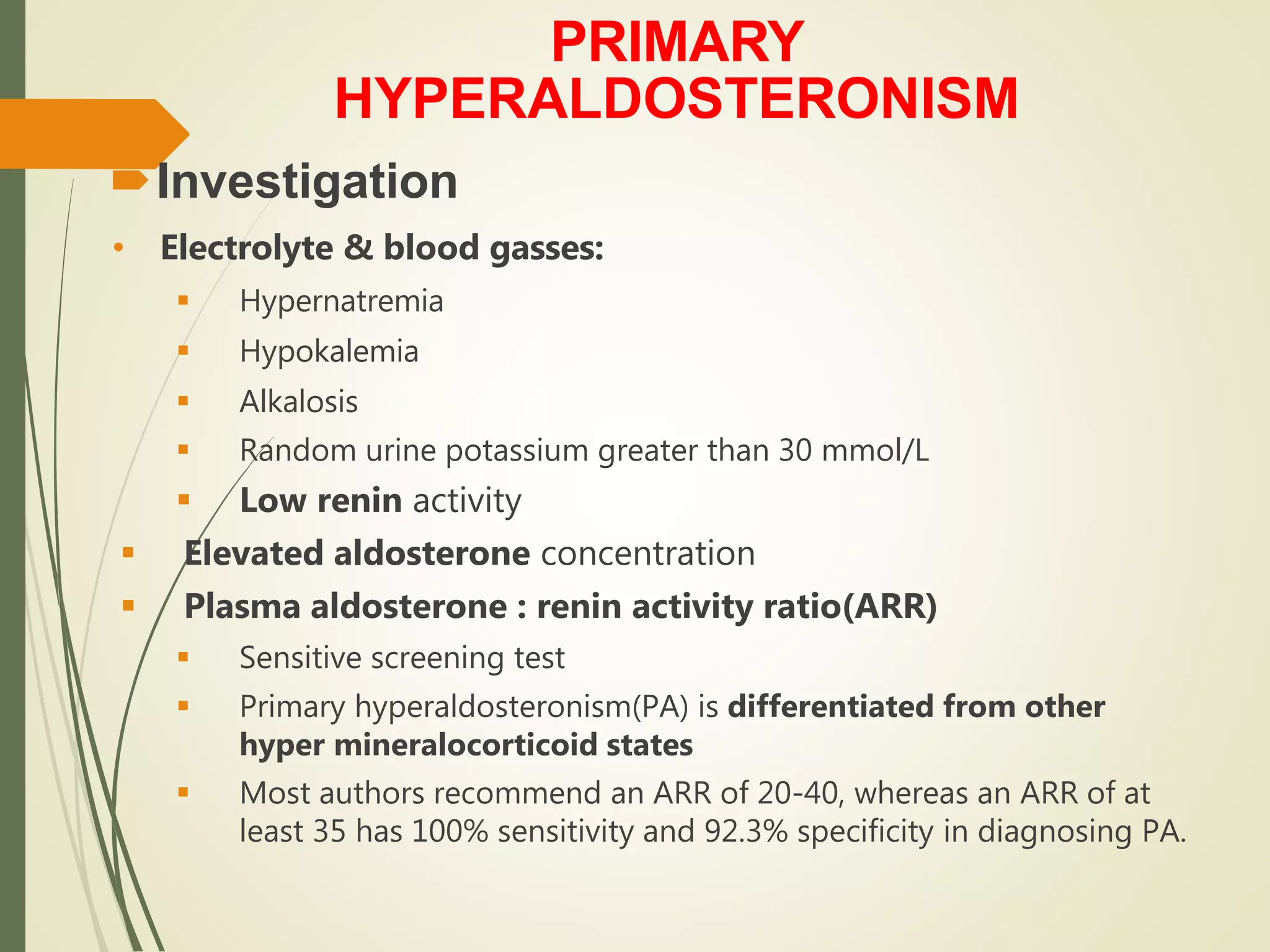
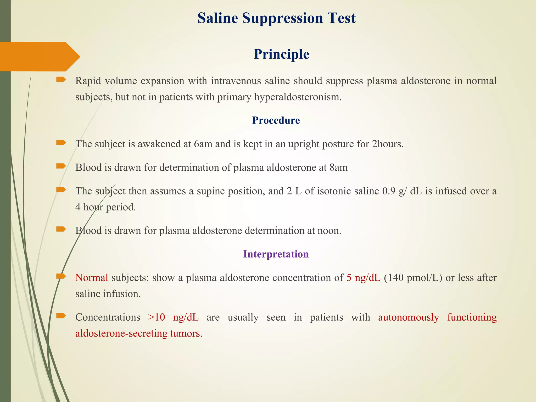
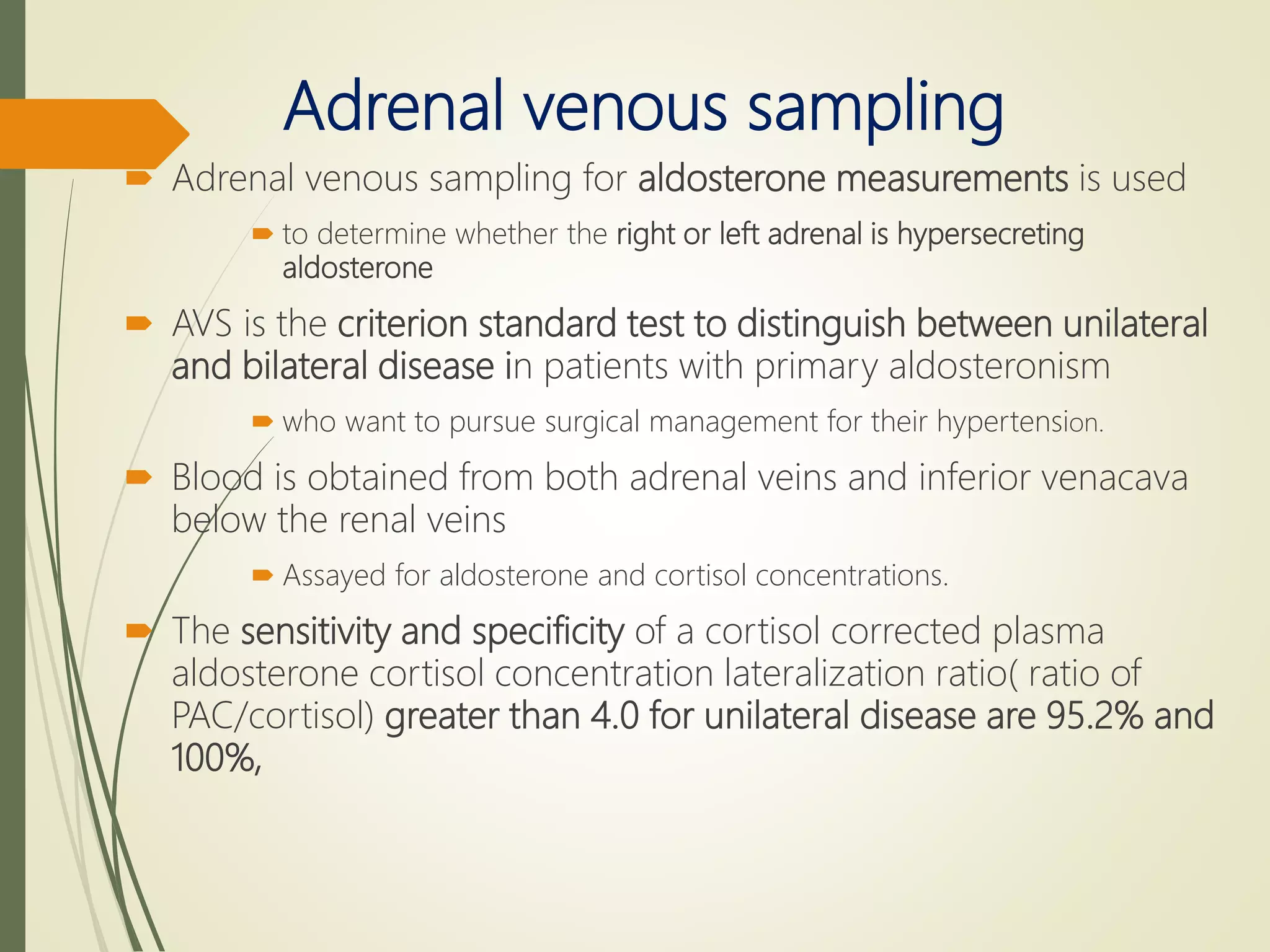
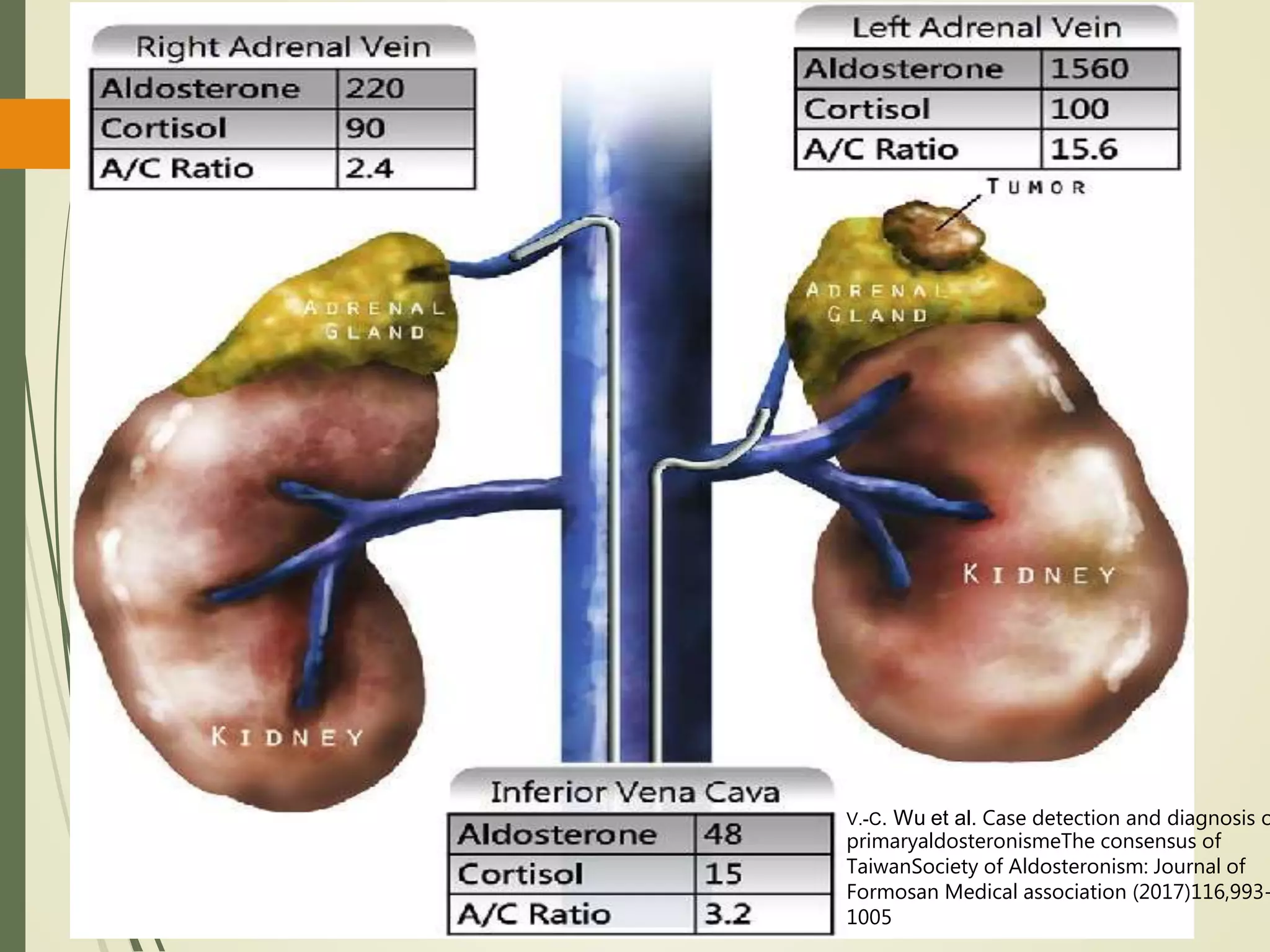
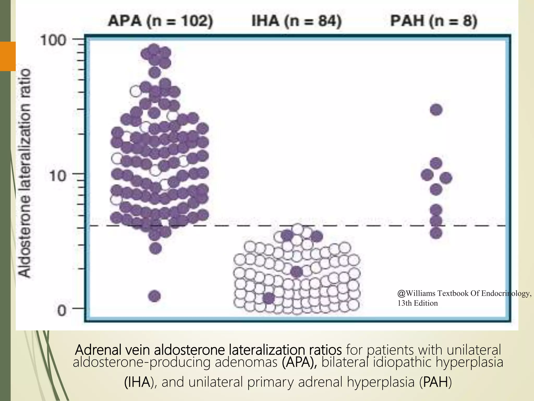
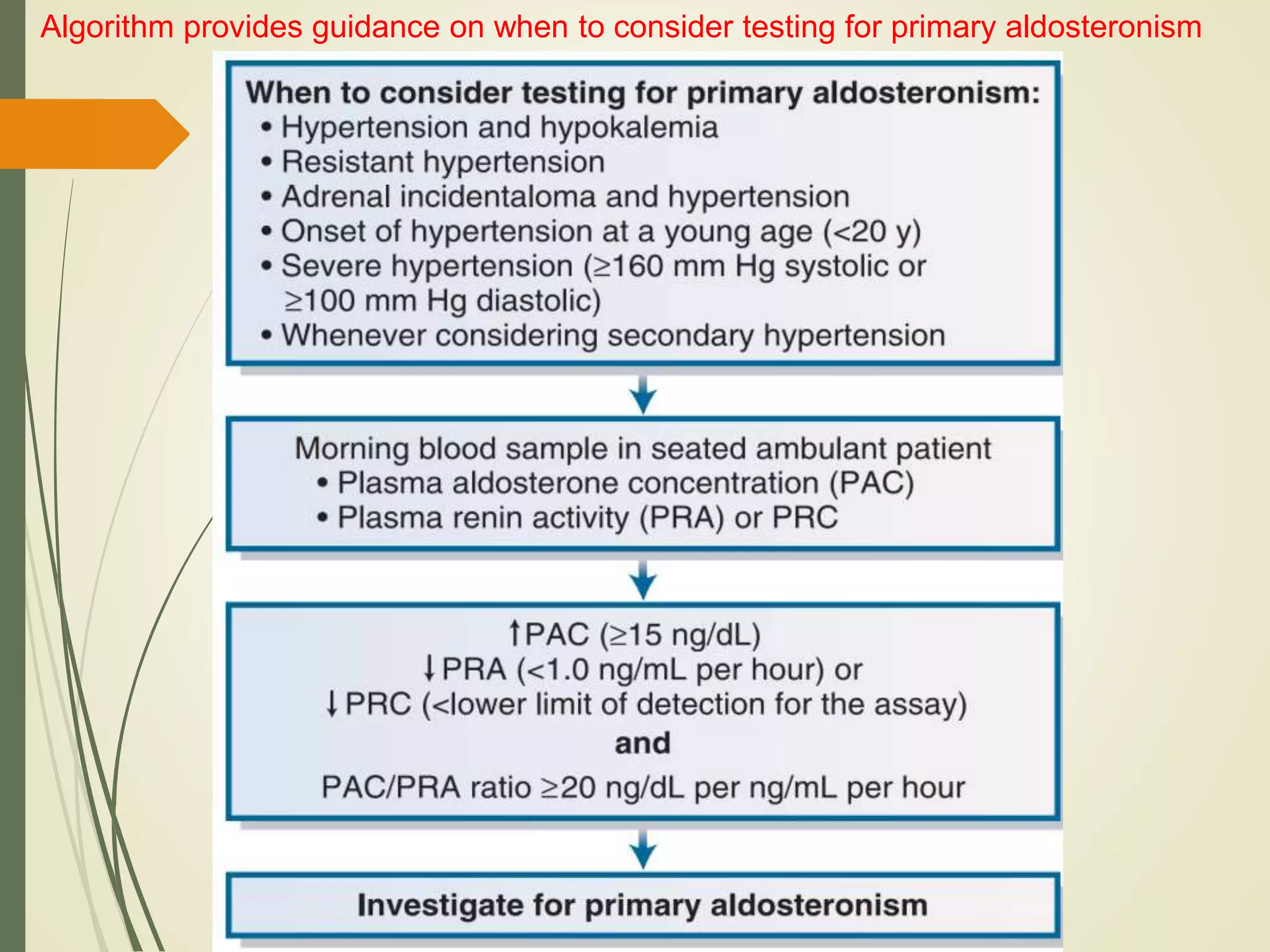
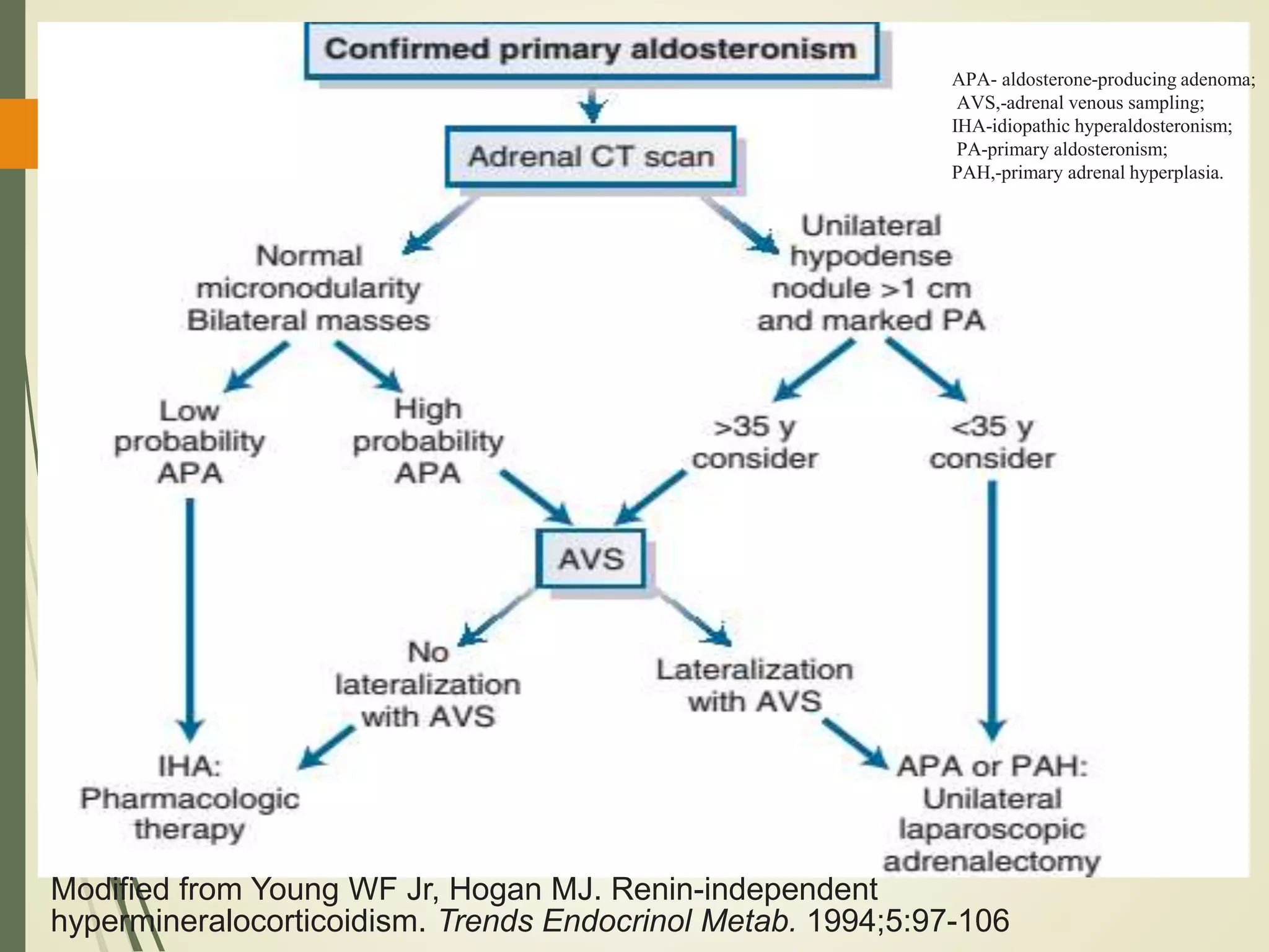
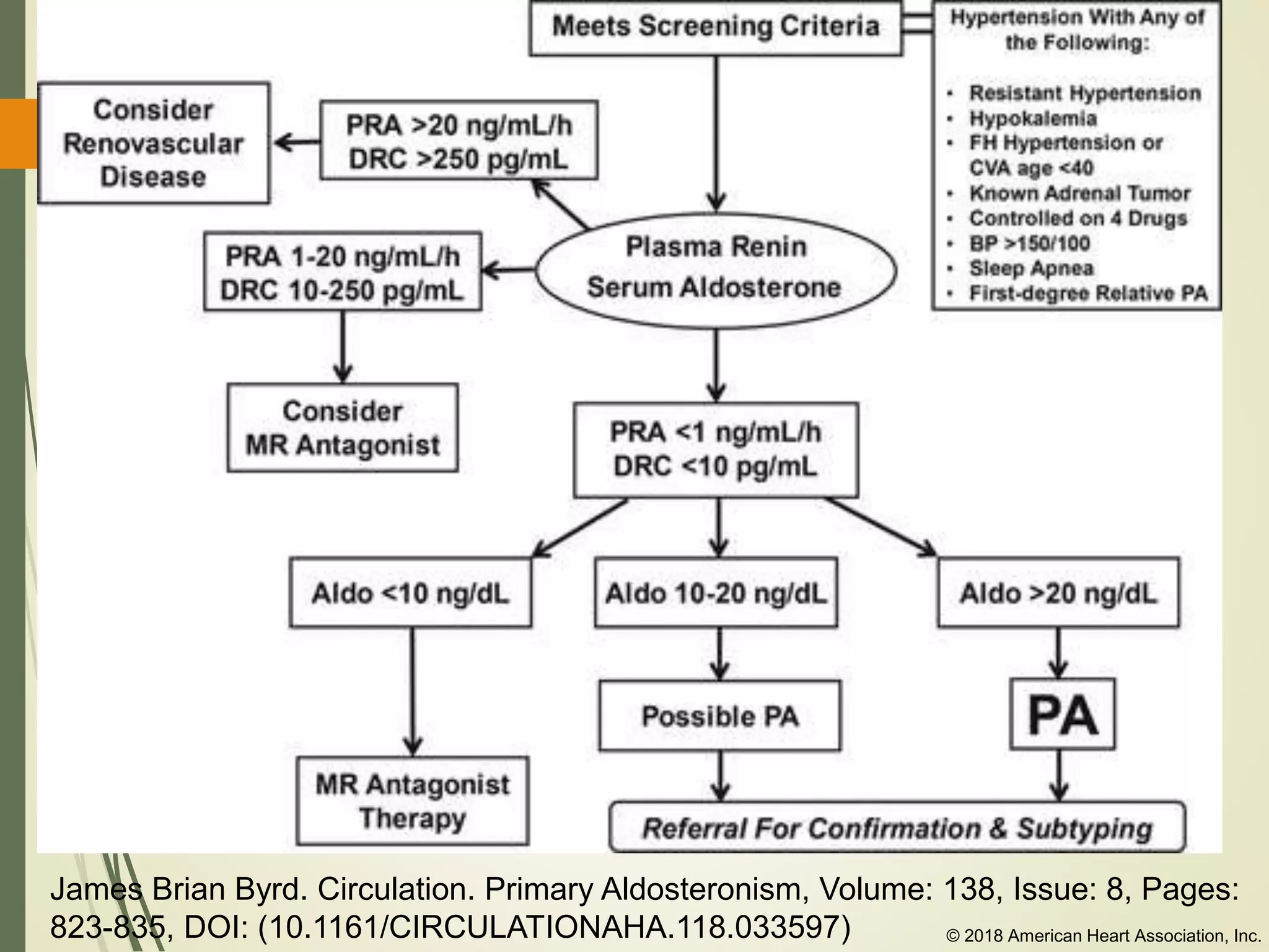
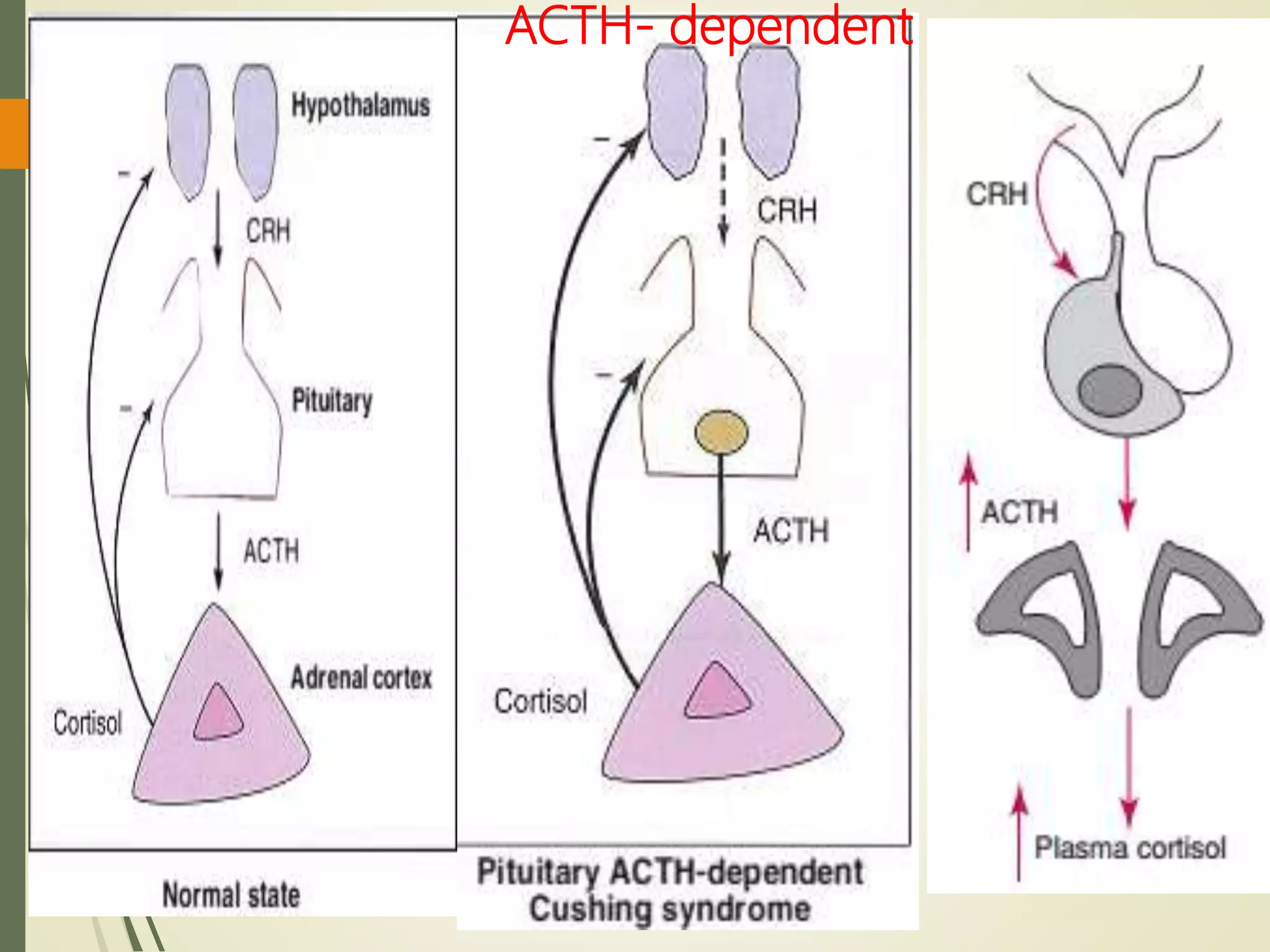
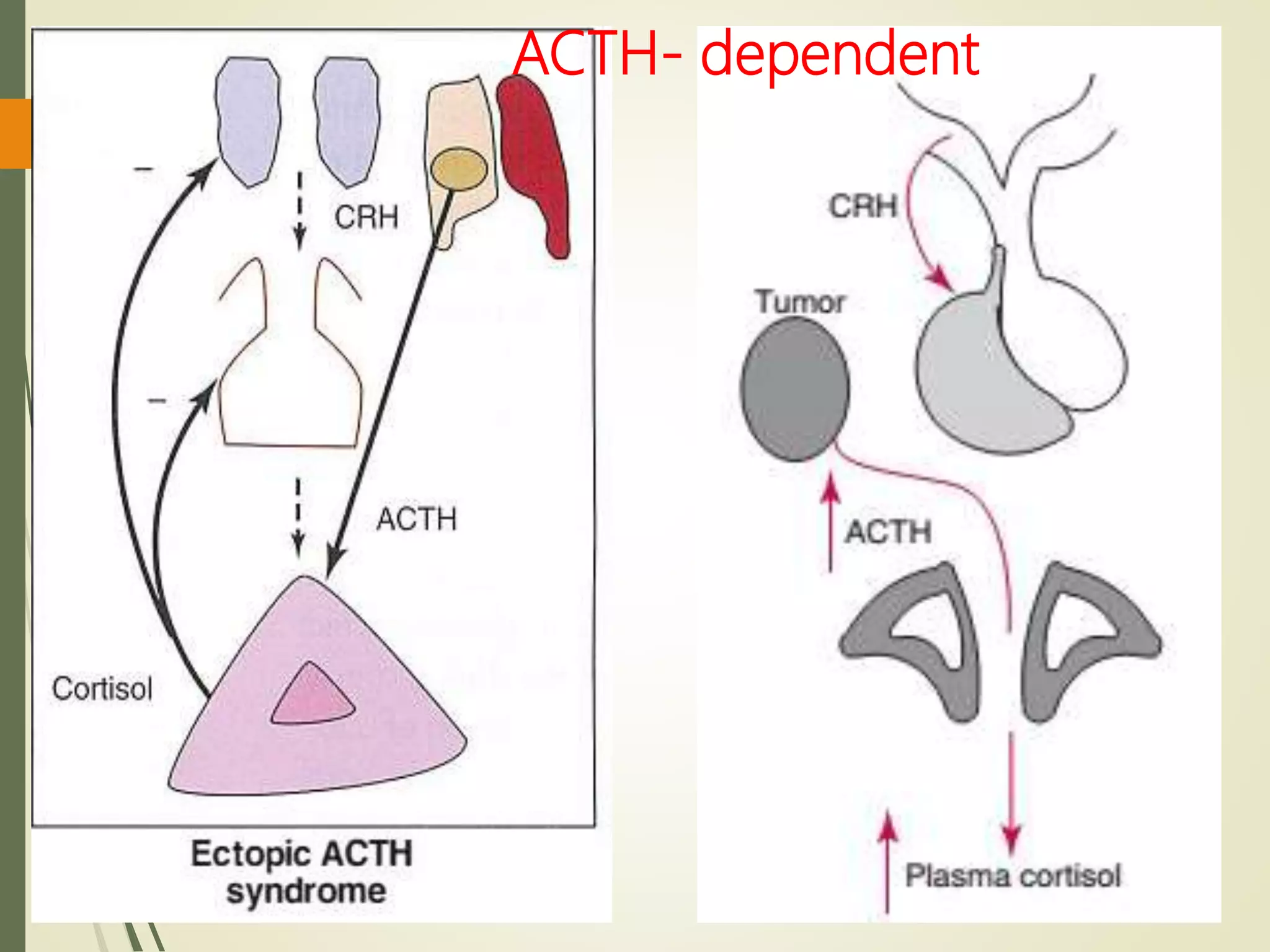
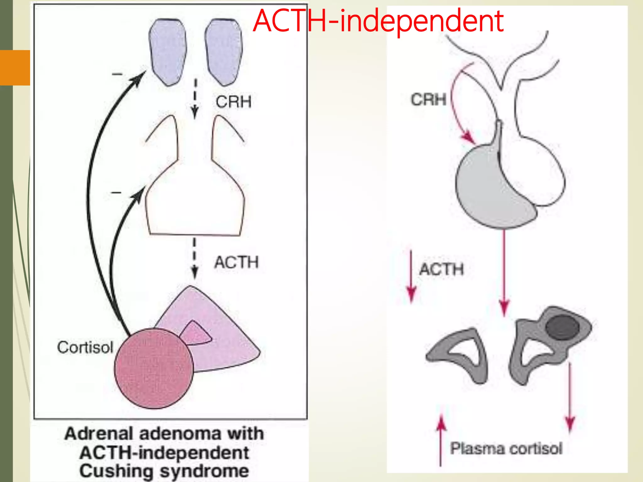
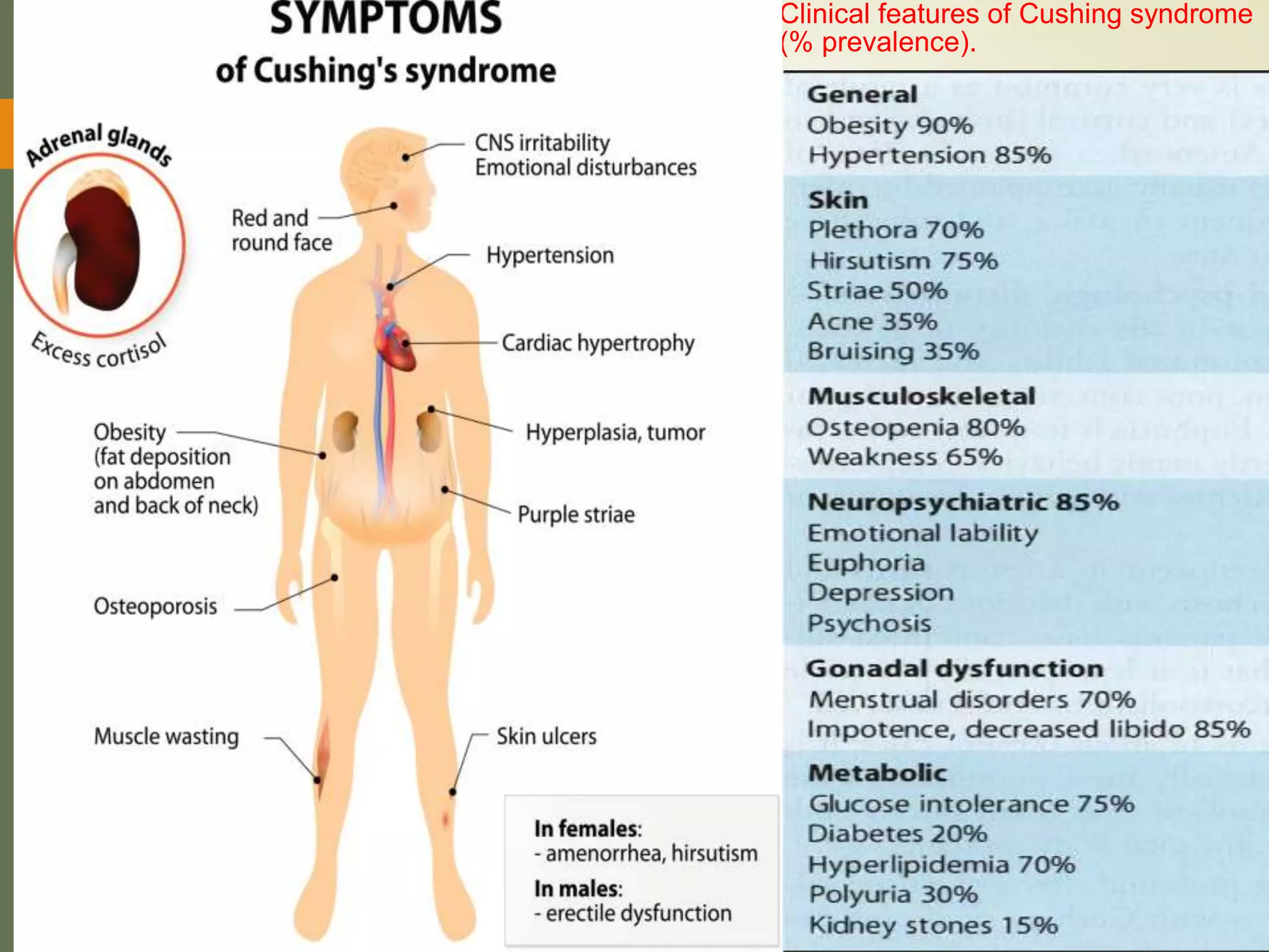
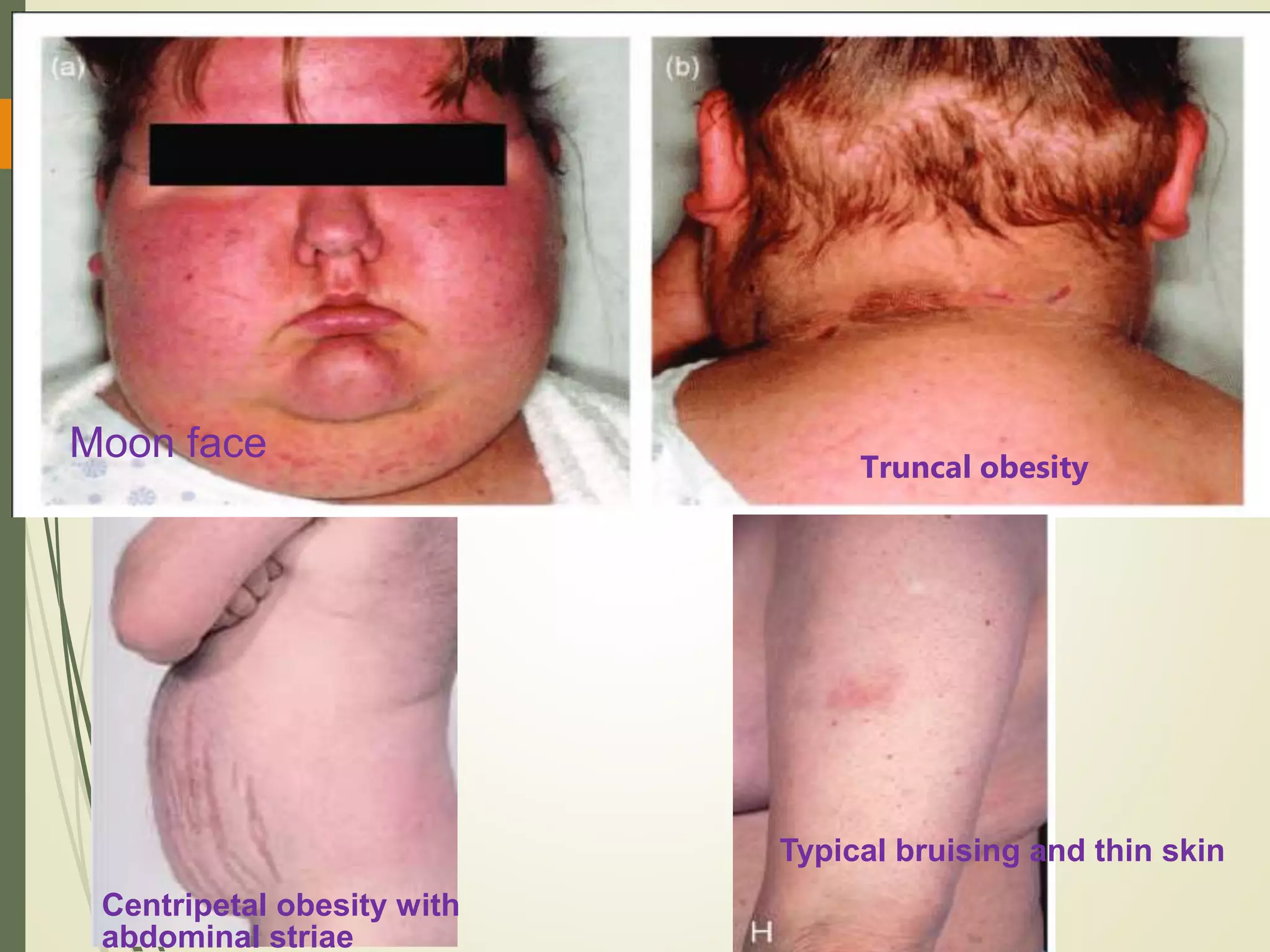
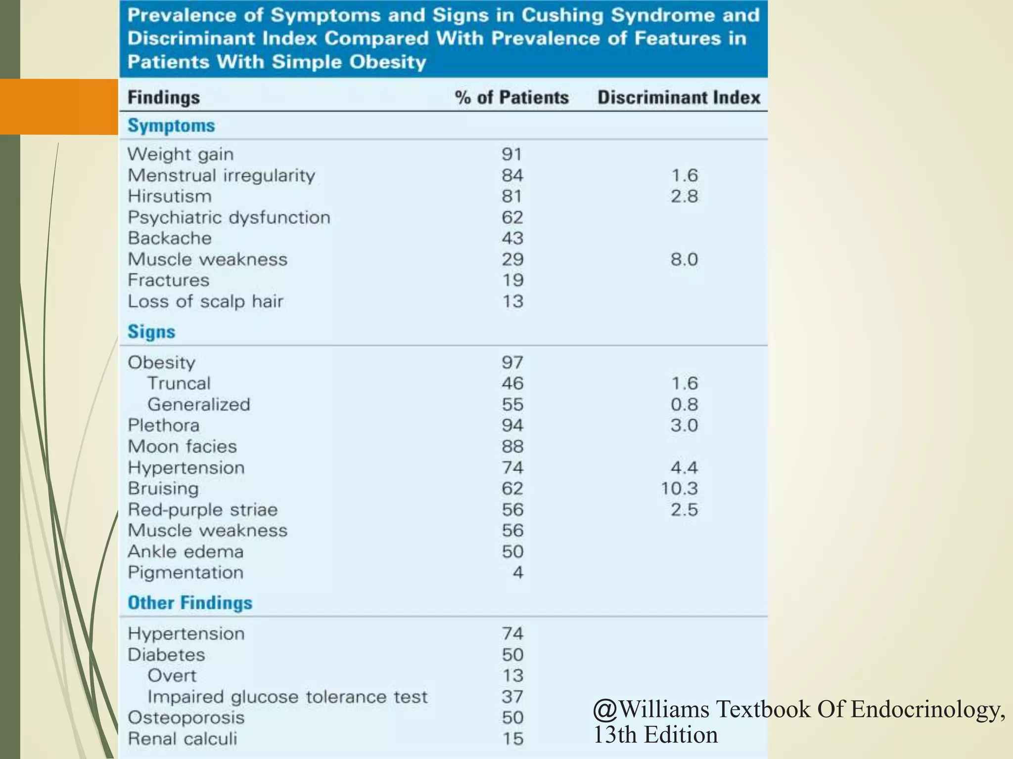
Hyperfunction of the adrenal cortex can cause excess production of mineralocorticoids, glucocorticoids, or androgens. Primary hyperaldosteronism is characterized by autonomous overproduction of aldosterone by the adrenal glands, often leading to hypertension and hypokalemic alkalosis. It can be caused by aldosterone-producing adenomas or bilateral adrenal hyperplasia. Tests like the saline suppression test and adrenal vein sampling are used in the diagnosis. Cushing's syndrome results from prolonged exposure to high levels of cortisol and can be caused by pituitary or adrenal tumors or ectopic ACTH secretion. Signs include central obesity, moon face, and fragile skin
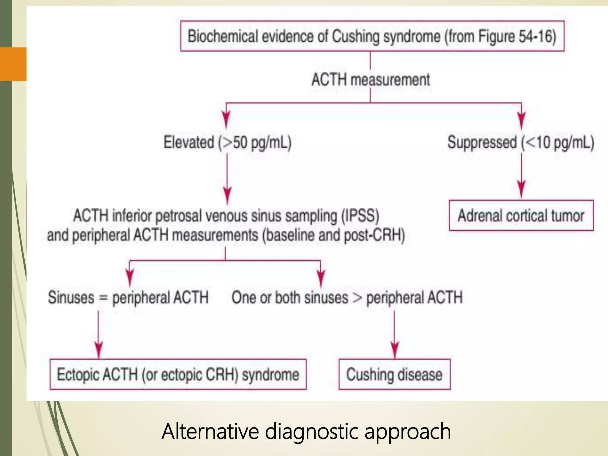
2. Adrenocortical Hyperfunction
Hyper function of the adrenal cortex produces clinical
syndromes of mineralocorticoid excess, glucocorticoid excess,
and androgen excess.
Hyperaldosteronism:
A medical condition where too much aldosterone is produced
by the adrenal glands, which can lead to sodium retention
and potassium loss.
Types:
Primary hyperaldosteronism
Secondary hyperaldosteronism
3. PRIMARY
HYPERALDOSTERONISM
Also known as hyporeninemic hyperaldosteronism / Conn’s
Syndrome
was first described in 1955 by Conn in conjunction with an
aldosterone-producing adrenal adenoma (APA)
Characterized by autonomous excessive production of
aldosterone by adrenal glands
Presents with :
hypertension ,
hypokalemic alkalosis
renal K+ wasting
4. PRIMARY
HYPERALDOSTERONISM
Causes:
Aldosterone-producing adenoma (APA) ≈30% of cases
Bilateral idiopathic hyperplasia (IHA) ≈60% of cases
Unilateral (primary) adrenal hyperplasia ≈2% of cases
Aldosterone-producing adrenocortical carcinoma <1 % of cases
Familial hyperaldosteronism (FH)
Glucocorticoid-remediable aldosteronism (FH type I) <1%of cases
FH type II (APA or IHA) <6% of cases
FH type III(germline KCNJ5 mutations)—<1% of cases
6. PRIMARY
HYPERALDOSTERONISM
• Clinical features:
Hypertension
aldosterone induced Na retention with increase in Extra
cellular fluid volume
Muscle weakness and cramping
Due to decrease K+
Muscle paralysis:
severe hypokalemia
Mild metabolic alkalosis
Because of increased urinary hydrogen excretion mediated both by
hypokalemia and by the direct stimulatory effect of aldosterone on
distal renal tubule acidification.
Polydipsia, polyuria and nocturia:
due to hypokalemia-induced renal concentrating defect
7. PRIMARY
HYPERALDOSTERONISM
Investigation
• Electrolyte & blood gasses:
Hypernatremia
Hypokalemia
Alkalosis
Random urine potassium greater than 30 mmol/L
Low renin activity
Elevated aldosterone concentration
Plasma aldosterone : renin activity ratio(ARR)
Sensitive screening test
Primary hyperaldosteronism(PA) is differentiated from other
hyper mineralocorticoid states
Most authors recommend an ARR of 20-40, whereas an ARR of at
least 35 has 100% sensitivity and 92.3% specificity in diagnosing PA.
8. Saline Suppression Test
Principle
Rapid volume expansion with intravenous saline should suppress plasma aldosterone in normal
subjects, but not in patients with primary hyperaldosteronism.
Procedure
The subject is awakened at 6am and is kept in an upright posture for 2hours.
Blood is drawn for determination of plasma aldosterone at 8am
The subject then assumes a supine position, and 2 L of isotonic saline 0.9 g/ dL is infused over a
4 hour period.
Blood is drawn for plasma aldosterone determination at noon.
Interpretation
Normal subjects: show a plasma aldosterone concentration of 5 ng/dL (140 pmol/L) or less after
saline infusion.
Concentrations >10 ng/dL are usually seen in patients with autonomously functioning
aldosterone-secreting tumors.
9. Adrenal venous sampling
Adrenal venous sampling for aldosterone measurements is used
to determine whether the right or left adrenal is hypersecreting
aldosterone
AVS is the criterion standard test to distinguish between unilateral
and bilateral disease in patients with primary aldosteronism
who want to pursue surgical management for their hypertension.
Blood is obtained from both adrenal veins and inferior venacava
below the renal veins
Assayed for aldosterone and cortisol concentrations.
The sensitivity and specificity of a cortisol corrected plasma
aldosterone cortisol concentration lateralization ratio( ratio of
PAC/cortisol) greater than 4.0 for unilateral disease are 95.2% and
100%,
10. V.-C. Wu et al. Case detection and diagnosis o
primaryaldosteronismeThe consensus of
TaiwanSociety of Aldosteronism: Journal of
Formosan Medical association (2017)116,993-
1005
11. Adrenal vein aldosterone lateralization ratios for patients with unilateral
aldosterone-producing adenomas (APA), bilateral idiopathic hyperplasia
(IHA), and unilateral primary adrenal hyperplasia (PAH)
@Williams Textbook Of Endocrinology,
13th Edition
15. Secondary hyperaldosteronism
Is increased adrenal production of aldosterone in response to
stimulus outside the adrenal gland activates the renin-angiotensin
system.
Increase renin secretion
– hyperreninemic hyperaldosteronism
Common than primary hyperaldosteronism
Secondary hyperaldosteronism is suspected in patients
with volume depletion, edema, and hypokalemic alkalosis.
Hypertension is usually absent in secondary
hyperaldosteronism
unless the patient has a renal artery stenosis.
17. Responses of the renin-aldosterone volume control loop
in primary versus secondary aldosteronism
*Initiating event
18. Other Forms of
Mineralocorticoid Excess
Unusual conditions that suggest aldosterone excess but do not
involve disorders of renin, angiotensin II, or aldosterone include
(1) Apparent Mineralocorticoid Excess,
(2) Type 1 Pseudohyperaldosteronism (Liddle syndrome),
(3) Type 2 Pseudohyperaldosteronism, and
(4) cortisol resistance.
19. Apparent mineralocorticoid
excess (AME)
Results from 11-beta-hydroxysteroid dehydrogenase-2 (HSD11B2)
deficiency or inhibition.
Impaired conversion of cortisol to cortisone allows cortisol
to bind to the MR, producing a mineralocorticoid effect
in the absence of elevations in aldosterone or DOC.
11-beta-hydroxysteroid dehydrogenase-2 (HSD11B2) deficiency is
described as an “apparent” excess of mineralocorticoid
Because patients can manifest hypertension, hypokalemia,
and alkalosis without elevation of aldosterone nor DOC.
Apparent mineralocorticoid excess is diagnosed by an increased
ratio of cortisol to cortisone
20. Type 1 pseudohyperaldosteronism
(Liddle syndrome )
Results from gain-of-function mutations in the beta or gamma
subunit of the amiloride-sensitive Epithelial sodium channel
(ENaC)
Consequences include
excessive sodium reabsorption,
expanded blood volume,
hypertension,
hypokalemia,
alkalosis.
Because the activity of ENaC is autonomous, both renin and
aldosterone are suppressed.
Type 2 pseudohyperaldosteronism
is a gain-of- function mutation in the MR
21. Cortisol Resistance
Loss-of-function mutations in the GR cause cortisol
resistance
In response to decreased cortisol action, CRH and
ACTH concentrations rise,
Increasing the circulating cortisol to a sufficient
concentration that functional cortisol deficiency is not
present.
Protective action of 11-beta-hydroxysteroid dehydrogenase-
2 (HSD11B2) in converting cortisol to cortisone is
overwhelmed,
resulting in increased binding of cortisol to the MR
and excessive mineralocorticoid effects.
24. CUSHING’S SYNDROME
• Adrenal cortex hyperfunction
• Any condition resulting from overproduction of
cortisol
• Cushing syndrome is caused by
• abnormalities of the pituitary or adrenal gland or may
occur as a consequence of ACTH or CRH secretion by
nonpituitary tumors
• Cushing disease is defined as the specific type of
Cushing syndrome due to excessive pituitary ACTH
secretion from a pituitary tumor.
25. Classification Of Causes of Cushing
Syndrome
Cushing syndrome is most readily classified into
ACTH dependent Causes
Cushing disease (pituitary-dependent)≈ 70% of cases
Adenoma (90%)
Hyperplasia (10 %)
Ectopic ACTH syndrome ≈ 15% of cases
Ectopic CRH syndrome
ACTH-independent causes
Iatrogenic (glucocorticoid, Megestrol acetate)
Adrenal neoplasm (adenoma, carcinoma) ≈ 5% of cases
Nodular adrenal hyperplasia
Primary pigmented nodular adrenal disease (PPNAD)
Massive macronodular adrenonodular hyperplasia
29. Other Syndromes of Hypercortisolemia
When there are clinical and biochemical features of Cushing
syndrome, but when hypercortisolemia is secondary to other factors
it is often referred to as pseudo-Cushing syndrome.
Causes
Alcoholism
Abnormalities are all reversible when alcohol abuse by the
patient is stopped.
Severe depression
Abnormalities are reversible on correction of the
psychiatric condition.
Obesity
Patients with obesity have mildly increased cortisol
secretion rates
Urinary free cortisol concentrations are either normal or
only slightly elevated
33. Diagnosis
Routine Laboratory Analysis
Although there are no routine laboratory investigations specific
to CS, each laboratory test may provide some clue with regard
to the diagnosis, including findings such as:
Increased neutrophil
Decreased lymphocyte and eosinophil counts
Hypokalemia
Metabolic alkalosis;
Hyperglycemia; and
Hypercholesterolemia,
Useful for the follow-up of treated patients
34. Diagnosis
Exclude exogenous steroid use.
Screening tests:-
24 hr urinary free cortisol excretion
Dexamethasone overnight test
Midnight plasma / Salivary cortisol
Low dose dexamethasone suppression test
If test are positive then measure plasma ACTH levels
:-
ACTH normal/high ACTH dependent Cushing’s
ACTH low ACTH independent Cushing’s
35. Diagnosis
ACTH dependent Cushing’s:-
High dose dexamethasone suppression test
CRH stimulation test
MRI pituitary
ACTH independent Cushing’s:-
Unenhanced CT adrenals
36. Demonstration of increased cortisol
Assessment of circadian rhythm in cortisol secretion
24-Hour urinary free cortisol excretion
Overnight / low dose dexamethasone suppression test
Assessment of circadian rhythm in cortisol secretion.
Measure 8 am and 11 pm serum cortisol level
Normal : Serum value than value @ 8 am -3 to 20 µg/dL (80-550
nmol/L) average 10 to 12 µg /dl
@ 4 PM are approximately half of morning values.
@ 10 PM to 2 AM, the plasma cortisol concentrations less than 3 µg
/dL
Cushing’s syndrome : rhythm is loss
Pseudo-Cushing : normal circadian
37. Screening tests
24-Hour urinary free cortisol excretion
less than 2% of secreted cortisol appears in urine as free
cortisol.
Diagnostic accuracy is greater than 90%
In general, a 24 hour urinary free cortisol concentration less
than 50 µg/d
excludes the diagnosis of Cushing syndrome
Overnight Low dose Dexamethasone suppression test
Dexamethasone suppresses ACTH and cortisol production in
normal subjects, but not in patients with Cushing syndrome.
Differentiates those who have Cushing syndrome from those
who do not have.
38. Overnight Low dose Dexamethasone
suppression test
Procedure:
1 mg of dexamethasone taken orally between 10 pm and 12 am
Blood drawn at 8am next morning- for determination of serum
cortisol
Interpretation:
In normal subjects cortisol: < 2µg/dl: excludes cushings
syndrome.
Most patients with Cushing syndrome do not show adequate
suppression, and 0800 hours cortisol concentrations are
usually 10 μg/dL or greater.
39. Overnight Low dose
Dexamethasone suppression test
Serum cortisol greater than 2 μg/dL may be seen also in cases of
stress,
obesity,
acute or chronic illness,
alcohol abuse,
severe depression,
pregnancy,
estrogen therapy,
failure to take dexamethasone, or treatment with phenytoin or
phenobarbital (which can enhance dexamethasone
metabolism).
40. Differential Diagnosis
Morning Plasma ACTH
Differentiates ACTH-dependent from
ACTH-independent causes.
In Cushing disease
50% of patients have a 9 AM ACTH
level within the normal reference
range (2 to
11 pmol/L [9 to 52 pg/mL]);
in the remainder- modestly
elevated.
In the ectopic ACTH syndrome
ACTH levels are high (usually >20
pmol/L [>90 pg/mL]);
Overlap values are seen in
Cushing disease in 30% of cases
In patients with adrenal tumors, plasma
ACTH is invariably undetectable (<1
pmol/L).
41. Differential Diagnosis
High-Dose Dexamethasone Suppression Test
High doses of dexamethasone usually suppress production of
ACTH by pituitary adenomas (benign tumors), as a result blood
and urine levels of cortisol should fall.
If the excess ACTH is being produced by a nonpituitary tumor,
cortisol production is less likely to be suppressed.
High dose DST distinguish those pts with cushing’s disease from
those having ectopic ACTH syndrome/adrenal tumor
Procedure:
8 mg given at 11.00 pm
Measure cortisol at 8.00 am next morning
Interpretation:
If cortisol is suppressed: patients with pituitary ACTH macroadenoma
No suppression: patients with ectopic ACTH syndrome
42. Differential Diagnosis
CRH stimulation test
In normal subjects, CRH produces a rise in ACTH and
cortisol of 15% to 20%.
Plasma ACTH concentrations peak 30 minutes after CRH
injection and serum cortisol peaks at 60 minutes
In Cushing disease , response is exaggerated
in which typically an ACTH increase greater than 50% and a
cortisol rise greater than 20% over baseline values are seen.
Poor responses occur in patients with adrenal tumor and
in most patients with nonendocrine
ACTH-secreting tumor
Patients with depression and anorexia nervosa usually do
not exhibit an exaggerated ACTH response to CRH
injection
44. Differential Diagnosis
Inferior petrosal sinus sampling
The most definitive means of accurately distinguishing
pituitary from nonpituitary ACTH-dependent Cushing
syndrome
Cushing syndrome when MRI does not reveal a definite
adenoma
Procedure
Measurement of ACTH from inferior petrosal venous sinus
specimens before and after CRH stimulation
Blood samples are collected from both right and lef IPS veins
and from a peripheral vein (e.g., the inferior vena cava)
at −30, 0, +2, +5, +10, and +30 minutes after intravenous
administration ovine CRH (1 µg/kg body weight) over 20 to
60 seconds.
The ratio of the IPS concentration to the peripheral venous
concentration of plasma ACTH is used to predict the location of
excess ACTH secretion
45. Anatomy of the venous drainage of the pituitary gland through the inferior
@Williams Textbook Of Endocrinolog
13th Edition
46. Inferior petrosal sinus sampling
Interpretation
An inferior petrosal sinus to peripheral (IPS-P) ratio greater
than 2.0 is consistent with a pituitary lesion (Cushing
disease)
as the cause of Cushing syndrome,
Ratio less than 1.4 to 1.7 supports the diagnosis of an ectopic
ACTH syndrome
50. Cushing Disease Ectopic ACTH
Syndrome
Adrenal Tumor
Biochemical evaluation
ACTH Normal-increase Normal – Increase Decrease
UFC response to High
dexamethasome
Usually declines
by >50%
Does not decline
by > 50%
Does not decline
by 50%
Basal and post CRH
ACTH levels
IPS> Peripheral IPS= Peripheral Not performed
Radiological evaluations Ct or MRI
Pituitary Abnormal Normal Normal
Adrenal Normal Normal Abnormal
Other location Normal Abnormal Normal
Differentiating the Causes of
Cushing Syndrome
The picture demonstrates left A/C ratio is 15.6 and right A/C ratio is 2.4. Left side divide to right side is 15.6/2.4 is 6.5. Thus, aldosterone secretion is significantly predominant at left side. Furthermore, the picture demonstrates right A/C ratio is 2.4 and peripheral A/C ratio is 3.2. In other words, right side presents a phenomenon of contralateral suppression. Therefore, the AVS shows left lateralization.
PRA: 1-20ng/ml/per hour PRC: 20-250pg/ml
side-to-side aldosterone-to-cortisol ratios of 4 or greater are consistent with aldosteronoma, whereas side to-side ratios of 3 or less are consistent with IAH.
Whereas hypertension is present in primary hyperaldosteronism Measurements of renin and aldosterone concentrations areseldom needed in cases of secondary hyperaldosteronism,
glucocorticoid-remediable hyperaldosteronismg (GRH) DOComa (DOC-secreting adrenal adenoma) AME, Apparent mineralocorticoid excess, bilateral idiopathic adrenal hyperplasia (IAH), 18e( 18 hydroxycorticosterone )
If the concentration of renin is elevated, diagnostic considerations include a juxtaglomerular renin-secreting tumor (a reninoma) and renovascular hypertension (assuming that intrinsic renal disease has been ruled out, which can cause elevated renin and aldosterone, leading to hypertension). If renin is suppressed and aldosterone is elevated (an aldosterone-torenin ratio >20 to 25), imaging and venography can help separate unilateral disease without a discrete mass (rare unilateral adrenal hyperplasia), unilateral disease with a discrete mass (aldosteronoma), bilateral idiopathic adrenal hyperplasia (IAH), or the rare condition of glucocorticoid-remediable hyperaldosteronism, with no masses or hyperplasia (see text for details). If aldosterone is not elevated, 11-desoxycorticosterone (DOC) and cortisol should be assayed, allowing the discrimination of several entities: Cushing syndrome or cortisol resistance (normal DOC and elevated cortisol), DOComa (DOC-secreting adrenal adenoma) or CYP11B2 (aldosterone synthase) defiiency (increased DOC and normal cortisol), defiiency of CYP17 (17-hydroxylase defiiency) (increased DOC and decreased cortisol), and end-organ disorders (normal DOC and cortisol).* In familial hyperaldosteronism type 2, aldosterone-producing adrenal adenoma (APA), IAH, or combined disease can be observed. AME, Apparent mineralocorticoid excess; decr, decrease; incr, increase; Nl, normal.
Mineralocorticoid and androgen may also be excessive
HIV, anorexia nervosa, and depression are associated with elevated serum cortisol concentration. poorly controlled diabetes mellitus, and pregnancy
salivary cortisol <0.112 µg/dL)
concentrations >9 µg/dL) suggest loss of diurnal variation.
A postdexamethasone 0800 hours cortisol cutoff of less than 5 μg/dL is more sensitive for the detection of Cushing syndrome but is less specific. <2 µg/dL)as the cutoff point, this test is reported to have a 97% to 100% true-positive rate and a false-positive rate of less than 1%
Skin pigmentation is rare in Cushing disease but common in the ectopic ACTH syndrome.
Blood leaves the anterior lobe of the pituitary and drains into the cavernous sinuses, which then empty into the inferior petrosal sinuses and subsequently into the jugular bulb and vein