V.SNEGA
I M.sc BIOTECHNOLOGY
BON SECOURS COLLEGE FOR WOMEN
THANJAVUR
 A probe is normally a short sequence of nucleotide bases
that will bind to specific regions of a target sequence of
nucleotides.
 The degree of homology between target and probe results in
stable hybridization.
 Probes can range in size from as short as 10 nuicleotide
bases (molecular weight of 3,300) to as long as 10,000
bases or more (molecular weight of 3,300,000).
 The most common size range for most probes is
between 14 and 40 bases. For statistical uniqueness, a
minimum of 20 nucleotide bases are usually needed
for a probe.
Which probe is best????
Short or long?????
 Short probes tend to hybridize nucleic acids at very
high rates (in minutes).
 where as longer probes may require reaction times of
hours to achieve a stable hybridization.
 Short probes do have some disadvantages.
 They are subject to more nonspecific hybridizations ,
are limited in specificity , and are more difficult to
label.
 Long probes hybridize more stably than short probes
at high temperatures and low salt concentrations(low
stringency).
- A nucleic acid probe
Is a short , single- stranded molecule of
radioactively labeled or fluorescently labeled DNA or
RNA.
 The base sequence of the probe and the conditions
under which the probe is used determine its specificity.
 What ever label is used, it has to be attached to, or
incorporated into, the nucleic acid probe. Here are 5
methods of labelling probes.
1) Nick translation
2) Primer extension
3) RNA polymerase
4) End labelling
5) Direct labelling
 It involves introducing single- strand breaks (nicks by
endonuclease) in the DNA, leaving exposed 3’ hydroxyl
termini and 5’ phosphate termini.
 Nick serves as a start point for introducing new
nucleotides at the 3’ hydroxyl side using DNA
polymerase.
 As a result, the nick will be moved progressively along
the DNA (translated’) in the 5’ 3’ direction.
 The synthesis reaction allows the incorporation of
labelled nucleotides in place of the previously existing
unlabeled.
 Based on hybridization of a mixture of all possible
hexanucleotides.
 The starting DNA is denatured and then cooled slowly
so that the individual hexanucleotides can bind to
suitably complementary sequences within the DNA
strands.
 DNA synthesis occurs in the presence of the four
dNTPs, at least one of which has a labelled group.
 DNA of interest is denatured to give single-strands.
 Random primer sequences are added (or sequences
unique to a particular sequences of interest).
 DNA polymerase is added together with labelled
nucleotides.
 Complementary DNA strands are synthesized starting
from the primer sequences and incorporating the
labelled nucleotides. There is partial filling in of the
gaps between the primers.
 DNA is then denatured to release the labelled probe
molecules.
 Single-stranded oligonucleotides are usually end-labelled
using polynucleotide kinase ( kinase end- labelling).
 Label is provided in the from of a 32P at the gama-
phosphate position of ATP and the polynucleotide kinase
catalyses an exchange reaction with the 5’-terminal
phosphates.
 The same procedure can also be used for labelling double-
stranded DNA.
 Here fragments carrying label at one end only can then be
generated by cleavage at an internal restriction site,
generating two differently sized fragments which can be
separated by gel electrophoresis and purified.
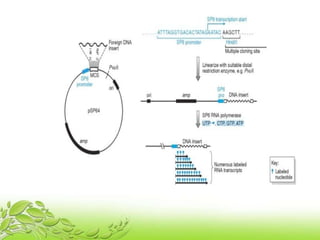
o The preparation of labelled RNA probes (riboprobes) is
most easily achieved by in vitro transcription of insert DNA
cloned in a suitable plasmid vector.
o The vector is designed so that adjacent to the multiple
cloning site is a phage promoter sequence , which can be
recognized by the corresponding phage RNA polymerase.
o By using a mix of NTPs , high specific activity radiolabeled
transcripts can be generated.
o Labelled sense and antisense riboprobes can be generated
from any gene cloned in such vectors and are widely used
in tissue in situ hybridization.
o DNA template + RNA polymerase + labelled
ribonucleotides ======= labelled RNA probe.
 Radiolabels
 Non-radioactive labels
 Chemiluminescence
 Fluorescence
 Antibodies
 Probe nucleic acid can be labelled using radioactive isotopes,
e.g 32p, 35S ,125I ,3 H.
 Detection is by autoradiography or geiger-mullur counters.
 Radiolabelled probes used to be the most commen type but
are less popular today because of safety considerations.
 However,radiolabelled probes are the most sensitive ,e.g .32p
labelled probes can detect single –copy genes in only 0.5 mg
of DNA.
 High sensitivity means that low concentrations of probe –
target hybrid can be detected.
 These are harmless than radiolabels and do not require
dedicated rooms , glassware and equipment or staff
monitoring , etc.but they are not generally as sensitive.
 Some examples:
 Biotin this labels can be detected using avidin or
streptavidin which have high affinities for biotin .
 Enzymes the enzymes is attached to the probe and its
presence usually detected by reaction with a substrate that
changes colour .Used in this way the enzymes is sometimes
referred to as a “reporter group”.
 Examples of enzymes used include alkaline phosphatase
and horseradish peroxidase.
 In this method chemiluminescent chemicals attached
to the probe are detected by their light emission
using a luminometer.
 Fluorescence chemicals attached to probe fluorescence
under UV light. This type of label is especially useful for
the direct examination of microbiological or cytological
specimens under the microscope- a technique known
as fluorescent in situ hybridization (FISH).
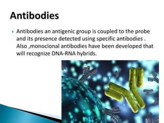
 Antibodies an antigenic group is coupled to the probe
and its presence detected using specific antibodies .
Also ,monoclonal antibodies have been developed that
will recognize DNA-RNA hybrids.
 Probe and target hybridize with each other but how
this is brought about can vary. There are 4 main
formats;
 Target (usually) is bound to a solid support such as a
microtitre tray or filter membrane.
 Probe is added in solution and binds to target (if
present ) on solid support.
 After washing to remove unbound probe, hybridization
is detected on the solid support using whatever
method is appropriate for the probe label.
 Both the probe and the target are in solution. Because
both are free to move , the changes of reaction are
maximized and , therefore , this format is generally
faster than others.
 In this format probe solution is added to fixed tissues,
sections or smears which are then usually examined under
the microscope.
 The probe label, e.g a fluorescent marker, produces a visible
change in the specimen if the target sequence is present and
hybridization has occurred.
 However, the sensitivity may be low if the amount of target
nucleic acid present in the specimen is low.This can be used
for the gene mapping of chromosomes, and for the detection
of microorgnisms in specimens.
 After size fractionation of nucleic acids by electrophoresis,
they are transferred to a filter membrane which is then
probed.
 The presence of target is confirmed by detection of probe on
the filter membrane, e.g radiolabelled probe can be detected
by autoradiography and the bands in the original gel
determined.
1.Southern blots:
Detection of gel –fractionated DNA molecules
transferred to a membrane. This includes restriction
fragment length polymorphism (RFLP) analysis.
2.Northern blots:
As above but used for RNA.
3.Dot blots:
Detection of unfractionated nucleic acid
immobilized on a membrane.
4. Colony and plaque blots:
Detection of immobilized nucleic acid on a
membrane that has been released from lysed bacteria
or phages.
THANK YOU