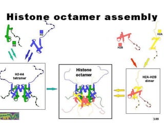
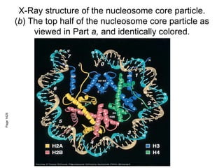
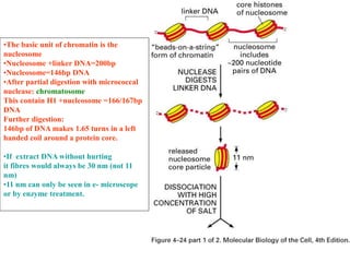
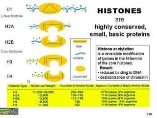
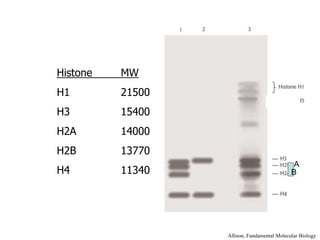
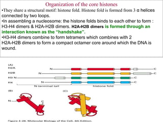
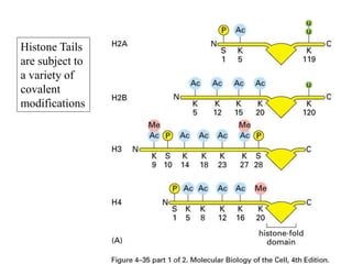
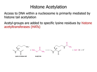
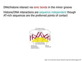
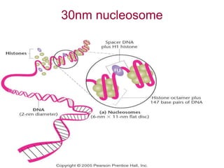
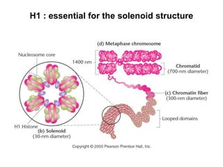
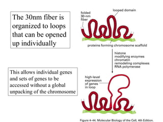
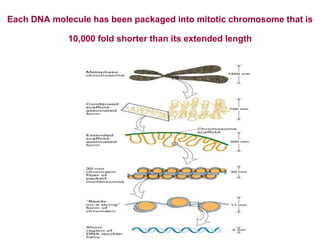
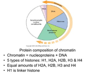
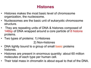
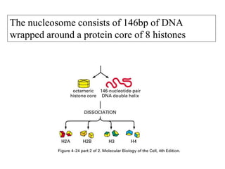
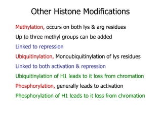
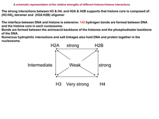
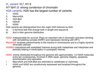
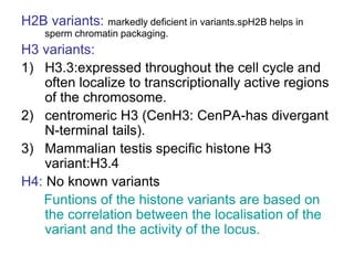
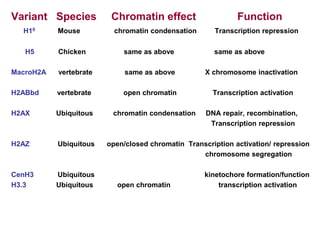
Chromatin Structure
X-Ray structure of the nucleosome core particle.
(b) The top half of the nucleosome core particle as
viewed in Part a, and identically colored.
Page
1426
•The basic unit of chromatin is the
nucleosome
•Nucleosome +linker DNA=200bp
•Nucleosome=146bp DNA
•After partial digestion with micrococcal
nuclease: chromatosome
This contain H1 +nucleosome =166/167bp
DNA
Further digestion:
146bp of DNA makes 1.65 turns in a left
handed coil around a protein core.
•If extract DNA without hurting
it fibres would always be 30 nm (not 11
nm)
•11 nm can only be seen in e- microscope
or by enzyme treatment.
Histone MW
H1 21500
H3 15400
H2A 14000
H2B 13770
H4 11340
Allison, Fundamental Molecular Biology
A
B
Organization of the core histones
•They share a structural motif: histone fold. Histone fold is formed from 3 α helices
connected by two loops.
•In assembling a nucleosome: the histone folds binds to each other to form :
H3-H4 dimers & H2A-H2B dimers. H2A-H2B dimers is formed through an
interaction known as the “handshake”.
•H3-H4 dimers combine to form tetramers which combines with 2
H2A-H2B dimers to form a compact octamer core around which the DNA is
wound.
http://www.ncbi.nlm.nih.gov/books/bv.fcgi?rid=mboc4.figgrp.643
Histones & Their Tails
The Histone tails are a critical determinant of chromatin structure
Each histone has a flexible N-terminus tail that extends out
of the nucleosome interacting with adjacent nucleosomes
Histone Tails
are subject to
a variety of
covalent
modifications
Histone Acetylation
Access to DNA within a nucleosome is primarily mediated by
histone tail acetylation
Acetyl-groups are added to specific lysine residues by histone
acetyltransferases (HATs)
DNA/histone interact via ionic bonds in the minor groove
Histone/DNA interactions are sequence independent though
AT-rich sequences are the preferred points of contact
http://www.ncbi.nlm.nih.gov/books/bv.fcgi?rid=mboc4.figgrp.638
30nm nucleosome
H1 : essential for the solenoid structure
The 30nm fiber is
organized to loops
that can be opened
up individually
This allows individual genes
and sets of genes to be
accessed without a global
unpacking of the chromosome
Each DNA molecule has been packaged into mitotic chromosome that is
10,000 fold shorter than its extended length
Protein composition of chromatin
• Chromatin = nucleoproteins + DNA
• 5 types of histones: H1, H2A, H2B, H3 & H4
• Equal amounts of H2A, H2B, H3 and H4
• H1 is linker histone
Histones
• Histones makes the most basic level of chromosome
organization, the nucleosome.
• Nucleosomes are the basic unit of eukaryotic chromosome
structure.
• They are repeating units of DNA & histones composed of
146bp of DNA wrapped around a core particle of 8 histone
proteins.
• Two types of proteins: 1) Histones
2) Non-histones
• DNA tightly bound to a group of small basic proteins
histones.
• Histones are present in enormous quantity: about 60 million
molecules of each type per human cell.
• Their total mass in chromatin is about equal to that of the DNA.
• Can exist in different forms due to post-translational modifications.
• Important in packaging DNA.
• Found in all eukaryotic nuclei.
• High content of + charged side chains (lysine and arginine).
• H2A, H2B, H3 & H4 highly conserved among species.
• H1 more variable in different species
• Histone play fundamental role in DNA packaging and are highly
conserved eukaryotic proteins.
• Histones regulate transcription through two mechanisms:
1) Modification of histone tails
2) Nucleosome positioning and spacing
The nucleosome consists of 146bp of DNA
wrapped around a protein core of 8 histones
Histone Deacetylation
Removal of acetyl-groups can cause nucleosomes to stabilize
Histone deacetylases (HDACs) are present in complexes
known as corepressors
Corepressors are known to be present at methylated DNA &
leads to transcriptional repression
• Human genome is approximately 3.2x10⁹
nucleotides distributed over 24 different
chromosomes.Approx: 30,000 genes.
• The Nuclear DNA of a cell is organized into
discrete units termed chromosomes.
• Each chromosome is a single DNA molecule.
• The DNA is not bare. It is coated and organized
by a wide variety of proteins.
• Collectively, the DNA and protein assemblage
that constitutes the chromosomes is referred to
as chromatin.
Other Histone Modifications
Methylation, occurs on both lys & arg residues
Up to three methyl groups can be added
Linked to repression
Ubiquitinylation, Monoubiquitinylation of lys residues
Linked to both activation & repression
Ubiquitinylation of H1 leads to it loss from chromation
Phosphorylation, generally leads to activation
Phosphorylation of H1 leads to it loss from chromation
A schematic representation of the relative strengths of different histone-histone interactions
The strong interactions between H3 & H4, and H2A & H2B supports that histone core is composed of:
(H3:H4)2 tetramer and (H2A:H2B) oligomer
The interface between DNA and histone is extensive: 142 hydrogen bonds are formed between DNA
and the histone core in each nucleosome.
Bonds are formed between the aminoacid backbone of the histones and the phosphodiaster backbone
of the DNA.
Numerous hydrophilic interactions and salt linkages also hold DNA and protein together in the
nucleosome.
H2A strong H2B
Intermediate Weak strong
H3 Very strong H4
H1 variant: H1
0, H1.5
H10 &H1.5: strong condensor of chromatin
H2A variants: H2A has the largest number of variants:
1. H2AZ
2. MacroH2A
3. H2A Bbd
4. H2AvD
5. H2AX
H2A variants are distinguished from the major H2A histones by their:
a) C-terminal tails that diverge both in length and sequence
b) And in their genome distribution.
H2AZ: Indespensible for survival. Plays an important role in chromatin activation (binding
with remodelling complex SWR1) and repression (binding with HP1).
MacroH2A localizes predominantly to the inactive X chromosome and is a highly dynamic
chromatin silencer.
H2ABbd colocalized with acetylated histones during both metaphase and interphase and
possibly helping in transcription in synergistic manner.
H2AX: evenly distributed throughout the genome with approximately 1-2 H2AX molecules
every ten nucleosomes. Involved in DNA repair, genome integrity, apoptosis,
recombination and replication.
• MacroH2A and H2A-Bbd are restricted to vertebrates or mammals.
• H2AX and H2AZ are constitutively expressed and localized throughout the
genome.
H2B variants: markedly deficient in variants.spH2B helps in
sperm chromatin packaging.
H3 variants:
1) H3.3:expressed throughout the cell cycle and
often localize to transcriptionally active regions
of the chromosome.
2) centromeric H3 (CenH3: CenPA-has divergant
N-terminal tails).
3) Mammalian testis specific histone H3
variant:H3.4
H4: No known variants
Funtions of the histone variants are based on
the correlation between the localisation of the
variant and the activity of the locus.
Variant Species Chromatin effect Function
H10 Mouse chromatin condensation Transcription repression
H5 Chicken same as above same as above
MacroH2A vertebrate same as above X chromosome inactivation
H2ABbd vertebrate open chromatin Transcription activation
H2AX Ubiquitous chromatin condensation DNA repair, recombination,
Transcription repression
H2AZ Ubiquitous open/closed chromatin Transcription activation/ repression
chromosome segregation
CenH3 Ubiquitous kinetochore formation/function
H3.3 Ubiquitous open chromatin transcription activation

Chromatin Structure2015.ppt

  • 1.
  • 3.
    X-Ray structure ofthe nucleosome core particle. (b) The top half of the nucleosome core particle as viewed in Part a, and identically colored. Page 1426
  • 4.
    •The basic unitof chromatin is the nucleosome •Nucleosome +linker DNA=200bp •Nucleosome=146bp DNA •After partial digestion with micrococcal nuclease: chromatosome This contain H1 +nucleosome =166/167bp DNA Further digestion: 146bp of DNA makes 1.65 turns in a left handed coil around a protein core. •If extract DNA without hurting it fibres would always be 30 nm (not 11 nm) •11 nm can only be seen in e- microscope or by enzyme treatment.
  • 6.
    Histone MW H1 21500 H315400 H2A 14000 H2B 13770 H4 11340 Allison, Fundamental Molecular Biology A B
  • 7.
    Organization of thecore histones •They share a structural motif: histone fold. Histone fold is formed from 3 α helices connected by two loops. •In assembling a nucleosome: the histone folds binds to each other to form : H3-H4 dimers & H2A-H2B dimers. H2A-H2B dimers is formed through an interaction known as the “handshake”. •H3-H4 dimers combine to form tetramers which combines with 2 H2A-H2B dimers to form a compact octamer core around which the DNA is wound.
  • 8.
    http://www.ncbi.nlm.nih.gov/books/bv.fcgi?rid=mboc4.figgrp.643 Histones & TheirTails The Histone tails are a critical determinant of chromatin structure Each histone has a flexible N-terminus tail that extends out of the nucleosome interacting with adjacent nucleosomes
  • 9.
    Histone Tails are subjectto a variety of covalent modifications
  • 10.
    Histone Acetylation Access toDNA within a nucleosome is primarily mediated by histone tail acetylation Acetyl-groups are added to specific lysine residues by histone acetyltransferases (HATs)
  • 11.
    DNA/histone interact viaionic bonds in the minor groove Histone/DNA interactions are sequence independent though AT-rich sequences are the preferred points of contact http://www.ncbi.nlm.nih.gov/books/bv.fcgi?rid=mboc4.figgrp.638
  • 12.
  • 13.
    H1 : essentialfor the solenoid structure
  • 14.
    The 30nm fiberis organized to loops that can be opened up individually This allows individual genes and sets of genes to be accessed without a global unpacking of the chromosome
  • 15.
    Each DNA moleculehas been packaged into mitotic chromosome that is 10,000 fold shorter than its extended length
  • 16.
    Protein composition ofchromatin • Chromatin = nucleoproteins + DNA • 5 types of histones: H1, H2A, H2B, H3 & H4 • Equal amounts of H2A, H2B, H3 and H4 • H1 is linker histone
  • 17.
    Histones • Histones makesthe most basic level of chromosome organization, the nucleosome. • Nucleosomes are the basic unit of eukaryotic chromosome structure. • They are repeating units of DNA & histones composed of 146bp of DNA wrapped around a core particle of 8 histone proteins. • Two types of proteins: 1) Histones 2) Non-histones • DNA tightly bound to a group of small basic proteins histones. • Histones are present in enormous quantity: about 60 million molecules of each type per human cell. • Their total mass in chromatin is about equal to that of the DNA.
  • 18.
    • Can existin different forms due to post-translational modifications. • Important in packaging DNA. • Found in all eukaryotic nuclei. • High content of + charged side chains (lysine and arginine). • H2A, H2B, H3 & H4 highly conserved among species. • H1 more variable in different species • Histone play fundamental role in DNA packaging and are highly conserved eukaryotic proteins. • Histones regulate transcription through two mechanisms: 1) Modification of histone tails 2) Nucleosome positioning and spacing
  • 19.
    The nucleosome consistsof 146bp of DNA wrapped around a protein core of 8 histones
  • 20.
    Histone Deacetylation Removal ofacetyl-groups can cause nucleosomes to stabilize Histone deacetylases (HDACs) are present in complexes known as corepressors Corepressors are known to be present at methylated DNA & leads to transcriptional repression
  • 21.
    • Human genomeis approximately 3.2x10⁹ nucleotides distributed over 24 different chromosomes.Approx: 30,000 genes. • The Nuclear DNA of a cell is organized into discrete units termed chromosomes. • Each chromosome is a single DNA molecule. • The DNA is not bare. It is coated and organized by a wide variety of proteins. • Collectively, the DNA and protein assemblage that constitutes the chromosomes is referred to as chromatin.
  • 22.
    Other Histone Modifications Methylation,occurs on both lys & arg residues Up to three methyl groups can be added Linked to repression Ubiquitinylation, Monoubiquitinylation of lys residues Linked to both activation & repression Ubiquitinylation of H1 leads to it loss from chromation Phosphorylation, generally leads to activation Phosphorylation of H1 leads to it loss from chromation
  • 23.
    A schematic representationof the relative strengths of different histone-histone interactions The strong interactions between H3 & H4, and H2A & H2B supports that histone core is composed of: (H3:H4)2 tetramer and (H2A:H2B) oligomer The interface between DNA and histone is extensive: 142 hydrogen bonds are formed between DNA and the histone core in each nucleosome. Bonds are formed between the aminoacid backbone of the histones and the phosphodiaster backbone of the DNA. Numerous hydrophilic interactions and salt linkages also hold DNA and protein together in the nucleosome. H2A strong H2B Intermediate Weak strong H3 Very strong H4
  • 24.
    H1 variant: H1 0,H1.5 H10 &H1.5: strong condensor of chromatin H2A variants: H2A has the largest number of variants: 1. H2AZ 2. MacroH2A 3. H2A Bbd 4. H2AvD 5. H2AX H2A variants are distinguished from the major H2A histones by their: a) C-terminal tails that diverge both in length and sequence b) And in their genome distribution. H2AZ: Indespensible for survival. Plays an important role in chromatin activation (binding with remodelling complex SWR1) and repression (binding with HP1). MacroH2A localizes predominantly to the inactive X chromosome and is a highly dynamic chromatin silencer. H2ABbd colocalized with acetylated histones during both metaphase and interphase and possibly helping in transcription in synergistic manner. H2AX: evenly distributed throughout the genome with approximately 1-2 H2AX molecules every ten nucleosomes. Involved in DNA repair, genome integrity, apoptosis, recombination and replication. • MacroH2A and H2A-Bbd are restricted to vertebrates or mammals. • H2AX and H2AZ are constitutively expressed and localized throughout the genome.
  • 25.
    H2B variants: markedlydeficient in variants.spH2B helps in sperm chromatin packaging. H3 variants: 1) H3.3:expressed throughout the cell cycle and often localize to transcriptionally active regions of the chromosome. 2) centromeric H3 (CenH3: CenPA-has divergant N-terminal tails). 3) Mammalian testis specific histone H3 variant:H3.4 H4: No known variants Funtions of the histone variants are based on the correlation between the localisation of the variant and the activity of the locus.
  • 26.
    Variant Species Chromatineffect Function H10 Mouse chromatin condensation Transcription repression H5 Chicken same as above same as above MacroH2A vertebrate same as above X chromosome inactivation H2ABbd vertebrate open chromatin Transcription activation H2AX Ubiquitous chromatin condensation DNA repair, recombination, Transcription repression H2AZ Ubiquitous open/closed chromatin Transcription activation/ repression chromosome segregation CenH3 Ubiquitous kinetochore formation/function H3.3 Ubiquitous open chromatin transcription activation