
Recommended
More Related Content
What's hot
What's hot (20)
Similar to DIGESTIVE SYSTEM_RDP_ANATOMY OF ALIMENTARY TRACT.pdf
Similar to DIGESTIVE SYSTEM_RDP_ANATOMY OF ALIMENTARY TRACT.pdf (20)
More from rishi2789
More from rishi2789 (20)
Recently uploaded
Recently uploaded (20)
DIGESTIVE SYSTEM_RDP_ANATOMY OF ALIMENTARY TRACT.pdf
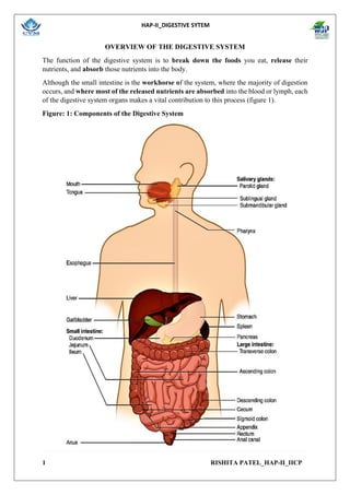
- 1. HAP-II_DIGESTIVE SYTEM 1 RISHITA PATEL_HAP-II_IICP OVERVIEW OF THE DIGESTIVE SYSTEM The function of the digestive system is to break down the foods you eat, release their nutrients, and absorb those nutrients into the body. Although the small intestine is the workhorse of the system, where the majority of digestion occurs, and where most of the released nutrients are absorbed into the blood or lymph, each of the digestive system organs makes a vital contribution to this process (figure 1). Figure: 1: Components of the Digestive System
- 2. HAP-II_DIGESTIVE SYTEM 2 RISHITA PATEL_HAP-II_IICP All digestive organs play integral roles in the life-sustaining process of digestion. DIGESTIVE SYSTEM ORGANS The digestive system is to divide its organs into two main categories. The first group is the organs that make up the alimentary canal. Accessory digestive organs comprise the second group and are critical for orchestrating the breakdown of food and the assimilation of its nutrients into the body. Accessory digestive organs, despite their name, are critical to the function of the digestive system. ALIMENTARY CANAL ORGANS Also called the gastrointestinal (GI) tract or gut, the alimentary canal (aliment- = “to nourish”) is a one-way tube about 7.62 meters (25 feet) in length during life and closer to 10.67 meters (35 feet) in length when measured after death, once smooth muscle tone is lost. The main function of the organs of the alimentary canal is to nourish the body. This tube begins at the mouth and terminates at the anus. Between those two points, the canal is modified as the pharynx, oesophagus, stomach, and small and large intestines to fit the functional needs of the body.
- 3. HAP-II_DIGESTIVE SYTEM 3 RISHITA PATEL_HAP-II_IICP Both the mouth and anus are open to the external environment; thus, food and wastes within the alimentary canal are technically considered to be outside the body. Only through the process of absorption do the nutrients in food enter into and nourish the body’s “inner space.” ACCESSORY STRUCTURES Each accessory digestive organ aids in the breakdown of food. Within the mouth, the teeth and tongue begin mechanical digestion, whereas the salivary glands begin chemical digestion. Once food products enter the small intestine, the gallbladder, liver, and pancreas release secretions—such as bile and enzymes—essential for digestion to continue. Together, these are called accessory organs because they sprout from the lining cells of the developing gut (mucosa) and augment its function; indeed, you could not live without their vital contributions, and many significant diseases result from their malfunction. Even after development is complete, they maintain a connection to the gut by way of ducts. HISTOLOGY OF THE ALIMENTARY CANAL The mucosa is referred to as a mucous membrane, because mucus production is a characteristic feature of gut epithelium. In addition, the mucosa has a thin, smooth muscle layer, called the muscularis mucosae (not to be confused with the muscularis layer, described below). Epithelium—In the mouth, pharynx, oesophagus, and anal canal, the epithelium is primarily a non-keratinized, stratified squamous epithelium.
- 4. HAP-II_DIGESTIVE SYTEM 4 RISHITA PATEL_HAP-II_IICP In the stomach and intestines, it is a simple columnar epithelium. Notice that the epithelium is in direct contact with the lumen, the space inside the alimentary canal. Interspersed among its epithelial cells are goblet cells, which secrete mucus and fluid into the lumen, and enteroendocrine cells, which secrete hormones into the interstitial spaces between cells. 1. Epithelial cells have a very brief lifespan, averaging from only a couple of days (in the mouth) to about a week (in the gut). This process of rapid renewal helps preserve the health of the alimentary canal, despite the wear and tear resulting from continued contact with foodstuffs. 2. Lamina propria—In addition to loose connective tissue, the lamina propria contains numerous blood and lymphatic vessels that transport nutrients absorbed through the alimentary canal to other parts of the body. The lamina propria also serves an immune function by housing clusters of lymphocytes, making up the mucosa-associated lymphoid tissue (MALT). These lymphocyte clusters are particularly substantial in the distal ileum where they are known as Peyer’s patches. When you consider that the alimentary canal is exposed to foodborne bacteria and other foreign matter, it is not hard to appreciate why the immune system has evolved a means of defending against the pathogens encountered within it. 3. Muscularis mucosae—This thin layer of smooth muscle is in a constant state of tension, pulling the mucosa of the stomach and small intestine into undulating folds. These folds dramatically increase the surface area available for digestion and absorption. As its name implies, the submucosa lies immediately beneath the mucosa. A broad layer of dense connective tissue, it connects the overlying mucosa to the underlying muscularis. • It includes blood and lymphatic vessels (which transport absorbed nutrients), and a scattering of submucosal glands that release digestive secretions. ` • Additionally, it serves as a conduit for a dense branching network of nerves, the submucosal plexus, which functions as described below. The third layer of the alimentary canal is the muscularis (also called the muscularis externa). The muscularis in the small intestine is made up of a double layer of smooth muscle: an inner circular layer and an outer longitudinal layer. • The contractions of these layers promote mechanical digestion, expose more of the food to digestive chemicals, and move the food along the canal. • In the most proximal and distal regions of the alimentary canal, including the mouth, pharynx, anterior part of the esophagus, and external anal sphincter, the muscularis is made up of skeletal muscle, which gives you voluntary control over swallowing and defecation.
- 5. HAP-II_DIGESTIVE SYTEM 5 RISHITA PATEL_HAP-II_IICP The serosa is the portion of the alimentary canal superficial to the muscularis. Present only in the region of the alimentary canal within the abdominal cavity, it consists of a layer of visceral peritoneum overlying a layer of loose connective tissue. Instead of serosa, the mouth, pharynx, and esophagus have a dense sheath of collagen fibres called the adventitia. These tissues serve to hold the alimentary canal in place near the ventral surface of the vertebral column. Nerve Supply Intrinsic innervation of much of the alimentary canal is provided by the enteric nervous system, which runs from the oesophagus to the anus, and contains approximately 100 million motors, sensory, and interneurons (unique to this system compared to all other parts of the peripheral nervous system). T these enteric neurons are grouped into two plexuses. The myenteric plexus (plexus of Auerbach) lies in the muscularis layer of the alimentary canal and is responsible for motility, especially the rhythm and force of the contractions of the muscularis. The submucosal plexus (plexus of Meissner) lies in the submucosal layer and is responsible for regulating digestive secretions and reacting to the presence of food. Blood Supply The blood vessels serving the digestive system have two functions. • They transport the protein and carbohydrate nutrients absorbed by mucosal cells after food is digested in the lumen. • Lipids are absorbed via lacteals, tiny structures of the lymphatic system. • The blood vessels’ second function is to supply the organs of the alimentary canal with the nutrients and oxygen needed to drive their cellular processes. DIGESTIVE SYSTEM PROCESSES AND REGULATION Functions of the Digestive Organs Organ Major functions Other functions Mouth • Ingests food • Chews and mixes food • Begins chemical breakdown of carbohydrates • Moves food into the pharynx • Begins breakdown of lipids via lingual lipase • Moistens and dissolves food, allowing you to taste it • Cleans and lubricates the teeth and oral cavity • Has some antimicrobial activity
- 6. HAP-II_DIGESTIVE SYTEM 6 RISHITA PATEL_HAP-II_IICP Pharynx • Propels food from the oral cavity to the oesophagus • Lubricates food and passageways Oesophagus • Propels food to the stomach • Lubricates food and passageways Stomach • Mixes and churns food with gastric juices to form chyme • Begins chemical breakdown of proteins • Releases food into the duodenum as chyme • Absorbs some fat-soluble substances (for example, alcohol, aspirin) • Possesses antimicrobial functions • Stimulates protein-digesting enzymes • Secretes intrinsic factor required for vitamin B12 absorption in small intestine Small intestine • Mixes chyme with digestive juices • Propels food at a rate slow enough for digestion and absorption • Absorbs breakdown products of carbohydrates, proteins, lipids, and nucleic acids, along with vitamins, minerals, and water • Performs physical digestion via segmentation • Provides optimal medium for enzymatic activity Accessory organs • Liver: produces bile salts, which emulsify lipids, aiding their digestion and absorption • Gallbladder: stores, concentrates, and releases bile • Pancreas: produces digestive enzymes and bicarbonate • Bicarbonate-rich pancreatic juices help neutralize acidic chyme and provide optimal environment for enzymatic activity Large intestine • Further breaks down food residues • Absorbs most residual water, electrolytes, and vitamins produced by enteric bacteria • Propels feces toward rectum • Food residue is concentrated and temporarily stored prior to defecation • Mucus eases passage of feces through colon
- 7. HAP-II_DIGESTIVE SYTEM 7 RISHITA PATEL_HAP-II_IICP • Eliminates feces Digestive Processes The processes of digestion include six activities: Ingestion, Propulsion, Mechanical or Physical Digestion, Chemical Digestion, Absorption, And Defecation. The first of these processes, ingestion, refers to the entry of food into the alimentary canal through the mouth. There, the food is chewed and mixed with saliva, which contains enzymes that begin breaking down the carbohydrates in the food plus some lipid digestion via lingual lipase. Chewing increases the surface area of the food and allows an appropriately sized bolus to be produced. Food leaves the mouth when the tongue and pharyngeal muscles propel it into the oesophagus. This act of swallowing, the last voluntary act until defecation, is an example of propulsion, which refers to the movement of food through the digestive tract. It includes both the voluntary process of swallowing and the involuntary process of peristalsis. Peristalsis consists of sequential, alternating waves of contraction and relaxation of alimentary wall smooth muscles, which act to propel food along. These waves also play a role in mixing food with digestive juices. Peristalsis is so powerful that foods and liquids you swallow enter your stomach even if you are standing on your head.
- 8. HAP-II_DIGESTIVE SYTEM 8 RISHITA PATEL_HAP-II_IICP Digestion includes both mechanical and chemical processes. Mechanical digestion is a purely physical process that does not change the chemical nature of the food. Instead, it makes the food smaller to increase both surface area and mobility. It includes mastication, or chewing, as well as tongue movements that help break food into smaller bits and mix food with saliva. Although there may be a tendency to think that mechanical digestion is limited to the first steps of the digestive process, it occurs after the food leaves the mouth, as well. The mechanical churning of food in the stomach serves to further break it apart and expose more of its surface area to digestive juices, creating an acidic “soup” called chyme. Segmentation, which occurs mainly in the small intestine, consists of localized contractions of circular muscle of the muscularis layer of the alimentary canal. These contractions isolate small sections of the intestine, moving their contents back and forth while continuously subdividing, breaking up, and mixing the contents. By moving food back and forth in the intestinal lumen, segmentation mixes food with digestive juices and facilitates absorption. In chemical digestion, starting in the mouth, digestive secretions break down complex food molecules into their chemical building blocks (for example, proteins into separate amino acids). These secretions vary in composition, but typically contain water, various enzymes, acids, and salts. The process is completed in the small intestine. Food that has been broken down is of no value to the body unless it enters the bloodstream and its nutrients are put to work. This occurs through the process of absorption, which takes place primarily within the small intestine. There, most nutrients are absorbed from the lumen of the alimentary canal into the bloodstream through the epithelial cells that make up the mucosa. Lipids are absorbed into lacteals and are transported via the lymphatic vessels to the bloodstream (the subclavian veins near the heart). The details of these processes will be discussed later. In defecation, the final step in digestion, undigested materials are removed from the body as feces.
- 9. HAP-II_DIGESTIVE SYTEM 9 RISHITA PATEL_HAP-II_IICP THE MOUTH, PHARYNX, AND ESOPHAGUS The Mouth The cheeks, tongue, and palate frame the mouth, which is also called the oral cavity (or buccal cavity). At the entrance to the mouth are the lips, or labia (singular = labium). Their outer covering is skin, which transitions to a mucous membrane in the mouth proper. Lips are very vascular with a thin layer of keratin; hence, the reason they are "red."
- 10. HAP-II_DIGESTIVE SYTEM 10 RISHITA PATEL_HAP-II_IICP The labial frenulum is a midline fold of mucous membrane that attaches the inner surface of each lip to the gum. The cheeks make up the oral cavity’s sidewalls. While their outer covering is skin, their inner covering is mucous membrane. This membrane is made up of non-keratinized, stratified squamous epithelium. Between the skin and mucous membranes are connective tissue and buccinator muscles. The pocket-like part of the mouth that is framed on the inside by the gums and teeth, and on the outside by the cheeks and lips is called the oral vestibule. Moving farther into the mouth, the opening between the oral cavity and throat (oropharynx) is called the fauces (like the kitchen "faucet"). The main open area of the mouth, or oral cavity proper, runs from the gums and teeth to the fauces. The palate is a wall or septum that separates the oral cavity from the nasal cavity, forming the roof of the mouth. This important structure makes it possible to chew and breathe at the same time.
- 11. HAP-II_DIGESTIVE SYTEM 11 RISHITA PATEL_HAP-II_IICP The hard palate—the anterior portion of the roof of the mouth—is formed by the maxillae and palatine bones and is covered by a mucous membrane; it forms a bony partition between the oral and nasal cavities. The soft palate, which forms the posterior portion of the roof of the mouth, is an arch-shaped muscular partition between the oropharynx and nasopharynx that is lined with mucous membrane. Hanging from the free border of the soft palate is a conical muscular process called the uvula. Anteriorly, the palatoglossal arch extends to the side of the base of the tongue; posteriorly, the palatopharyngeal arch extends to the side of the pharynx. The palatine tonsils are situated between the arches, and the lingual tonsils are situated at the base of the tongue. At the posterior border of the soft palate, the mouth opens into the oropharynx through the fauces. THE TONGUE The tongue is the strongest muscle in the body. Those who stake this claim cite its strength proportionate to its size. The tongue is a workhorse, facilitating ingestion, mechanical digestion, chemical digestion (lingual lipase), sensation (of taste, texture, and temperature of food), swallowing, and vocalization. The tongue is attached to the mandible, the styloid processes of the temporal bones, and the hyoid bone. The tongue is positioned over the floor of the oral cavity. A medial septum extends the entire length of the tongue, dividing it into symmetrical halves. THE SALIVARY GLANDS Many small salivary glands are housed within the mucous membranes of the mouth and tongue. These minor exocrine glands are constantly secreting saliva, either directly into the oral cavity or indirectly through ducts, even while you sleep. In fact, an average of 1 to 1.5 litters of saliva is secreted each day. Usually just enough saliva is present to moisten the mouth and teeth. Secretion increases when you eat, because saliva is essential to moisten food and initiate the chemical breakdown of carbohydrates. Small amounts of saliva are also secreted by the labial glands in the lips. In addition, the buccal glands in the cheeks, palatal glands in the palate, and lingual glands in the tongue help ensure that all areas of the mouth are supplied with adequate saliva.
- 12. HAP-II_DIGESTIVE SYTEM 12 RISHITA PATEL_HAP-II_IICP The Major Salivary Glands Outside the oral mucosa are three pairs of major salivary glands, which secrete the majority of saliva into ducts that open into the mouth: • The submandibular glands, which are in the floor of the mouth, secrete saliva into the mouth through the submandibular ducts. • The sublingual glands, which lie below the tongue, use the lesser sublingual ducts to secrete saliva into the oral cavity. • The parotid glands lie between the skin and the masseter muscle, near the ears. They secrete saliva into the mouth through the parotid duct, which is located near the second upper molar tooth. SALIVA Saliva is essentially (98 to 99.5 percent) water. The remaining 4.5 percent is a complex mixture of ions, glycoproteins, enzymes, growth factors, and waste products. Perhaps the most important ingredient in saliva from the perspective of digestion is the enzyme salivary amylase, which initiates the breakdown of carbohydrates. Food does not spend enough time in the mouth to allow all the carbohydrates to break down, but salivary amylase continues acting until it is inactivated by stomach acids. Bicarbonate and phosphate ions function as chemical buffers, maintaining saliva at a pH between 6.35 and 6.85. Salivary mucus helps lubricate food, facilitating movement in the mouth, bolus formation, and swallowing.
- 13. HAP-II_DIGESTIVE SYTEM 13 RISHITA PATEL_HAP-II_IICP Saliva contains immunoglobulin A, which prevents microbes from penetrating the epithelium, and lysozyme, which makes saliva antimicrobial. Saliva also contains epidermal growth factor. Each of the major salivary glands secretes a unique formulation of saliva according to its cellular makeup. For example, the parotid glands secrete a watery solution that contains salivary amylase. The submandibular glands have cells similar to those of the parotid glands, as well as mucus-secreting cells. Therefore, saliva secreted by the submandibular glands also contains amylase but, in a liquid, thickened with mucus. The sublingual glands contain mostly mucous cells, and they secrete the thickest saliva with the least amount of salivary amylase. REGULATION OF SALIVATION The autonomic nervous system regulates salivation (the secretion of saliva). In the absence of food, parasympathetic stimulation keeps saliva flowing at just the right level for comfort as you speak, swallow, sleep, and generally go about life. Over-salivation can occur, for example, if you are stimulated by the smell of food, but that food is not available for you to eat. Drooling is an extreme instance of the overproduction of saliva. During times of stress, such as before speaking in public, sympathetic stimulation takes over, reducing salivation and producing the symptom of dry mouth often associated with anxiety. When you are dehydrated, salivation is reduced, causing the mouth to feel dry and prompting you to take action to quench your thirst. Salivation can be stimulated by the sight, smell, and taste of food. It can even be stimulated by thinking about food. You might notice whether reading about food and salivation right now has had any effect on your production of saliva. How does the salivation process work while you are eating? Food contains chemicals that stimulate taste receptors on the tongue, which send impulses to the superior and inferior salivatory nuclei in the brain stem. These two nuclei then send back parasympathetic impulses through fibers in the glossopharyngeal and facial nerves, which stimulate salivation. Even after you swallow food, salivation is increased to cleanse the mouth and to water down and neutralize any irritating chemical remnants, such as that hot sauce in your burrito. Most saliva is swallowed along with food and is reabsorbed, so that fluid is not lost.
- 14. HAP-II_DIGESTIVE SYTEM 14 RISHITA PATEL_HAP-II_IICP THE TEETH The teeth, or dentes (singular = dens), are organs similar to bones that you use to tear, grind, and otherwise mechanically break down food. Types of Teeth During the course of your lifetime, you have two sets of teeth (one set of teeth is a dentition). Your 20 deciduous teeth, or baby teeth, first begin to appear at about 6 months of age. Between approximately age 6 and 12, these teeth are replaced by 32 permanent teeth. Moving from the center of the mouth toward the side, these are as follows: • The eight incisors, four top and four bottom, are the sharp front teeth you use for biting into food. • The four cuspids (or canines) flank the incisors and have a pointed edge (cusp) to tear up food. These fang-like teeth are superb for piercing tough or fleshy foods. • Posterior to the cuspids are the eight premolars (or bicuspids), which have an overall flatter shape with two rounded cusps useful for mashing foods. • The most posterior and largest are the 12 molars, which have several pointed cusps used to crush food so it is ready for swallowing. The third members of each set of three molars, top and bottom, are commonly referred to as the wisdom teeth, because their eruption is commonly delayed until early adulthood. It is not uncommon for wisdom teeth to fail to erupt; that is, they remain impacted. In these cases, the teeth are typically removed by orthodontic surgery.
- 15. HAP-II_DIGESTIVE SYTEM 15 RISHITA PATEL_HAP-II_IICP ANATOMY OF A TOOTH The teeth are secured in the alveolar processes (sockets) of the maxilla and the mandible. Gingivae (commonly called the gums) are soft tissues that line the alveolar processes and surround the necks of the teeth. Teeth are also held in their sockets by a connective tissue called the periodontal ligament. The two main parts of a tooth are the crown, which is the portion projecting above the gum line, and the root, which is embedded within the maxilla and mandible. Both parts contain an inner pulp cavity, containing loose connective tissue through which run nerves and blood vessels. The region of the pulp cavity that runs through the root of the tooth is called the root canal. Surrounding the pulp cavity is dentin, a bone-like tissue. In the root of each tooth, the dentin is covered by an even harder bone-like layer called cementum.
- 16. HAP-II_DIGESTIVE SYTEM 16 RISHITA PATEL_HAP-II_IICP In the crown of each tooth, the dentin is covered by an outer layer of enamel, the hardest substance in the body. Although enamel protects the underlying dentin and pulp cavity, it is still nonetheless susceptible to mechanical and chemical erosion, or what is known as tooth decay. The most common form, dental caries (cavities) develops when colonies of bacteria feeding on sugars in the mouth release acids that cause soft tissue inflammation and degradation of the calcium crystals of the enamel.
- 17. HAP-II_DIGESTIVE SYTEM 17 RISHITA PATEL_HAP-II_IICP PHARYNX When food is first swallowed, it passes from the mouth into the pharynx (throat), a funnel- shaped tube that extends from the internal nares to the oesophagus posteriorly and to the larynx anteriorly. The pharynx is composed of skeletal muscle and lined by mucous membrane, and is divided into three parts: the nasopharynx, the oropharynx, and the laryngopharynx. The nasopharynx functions only in respiration, but both the oropharynx and laryngopharynx have digestive as well as respiratory functions. Swallowed food passes from the mouth into the oropharynx and laryngopharynx; the muscular contractions of these areas help propel food into the oesophagus and then into the stomach.
- 18. HAP-II_DIGESTIVE SYTEM 18 RISHITA PATEL_HAP-II_IICP THE ESOPHAGUS The oesophagus is a muscular tube that connects the pharynx to the stomach. It is approximately 25.4 cm (10 in) in length, located posterior to the trachea, and remains in a collapsed form when not engaged in swallowing. The esophagus runs a mainly straight route through the mediastinum of the thorax. To enter the abdomen, the esophagus penetrates the diaphragm through an opening called the esophageal hiatus. DEGLUTITION Deglutition is another word for swallowing—the movement of food from the mouth to the stomach. The entire process takes about 4 to 8 seconds for solid or semisolid food, and about 1 second for very soft food and liquids. Although this sounds quick and effortless, deglutition is, in fact, a complex process that involves both the skeletal muscle of the tongue and the muscles of the pharynx and esophagus. It is aided by the presence of mucus and saliva. There are three stages in deglutition: the voluntary phase, the pharyngeal phase, and the esophageal phase. The autonomic nervous system controls the latter two phases.
- 19. HAP-II_DIGESTIVE SYTEM 19 RISHITA PATEL_HAP-II_IICP THE VOLUNTARY PHASE The voluntary phase of deglutition (also known as the oral or buccal phase) is so called because you can control when you swallow food. In this phase, chewing has been completed and swallowing is set in motion. THE PHARYNGEAL PHASE In the pharyngeal phase, stimulation of receptors in the oropharynx sends impulses to the deglutition centre (a collection of neurons that controls swallowing) in the medulla oblongata. Impulses are then sent back to the uvula and soft palate, causing them to move upward and close off the nasopharynx. The laryngeal muscles also constrict to prevent aspiration of food into the trachea. At this point, deglutition apnea takes place, which means that breathing ceases for a very brief time. Contractions of the pharyngeal constrictor muscles move the bolus through the oropharynx and laryngopharynx. Relaxation of the upper esophageal sphincter then allows food to enter the esophagus.
- 20. HAP-II_DIGESTIVE SYTEM 20 RISHITA PATEL_HAP-II_IICP THE ESOPHAGEAL PHASE The entry of food into the esophagus marks the beginning of the esophageal phase of deglutition and the initiation of peristalsis. When the bolus nears the stomach, distention of the esophagus initiates a short reflex relaxation of the lower esophageal sphincter that allows the bolus to pass into the stomach. During the esophageal phase, esophageal glands secrete mucus that lubricates the bolus and minimizes friction. THE STOMACH Structure There are four main regions in the stomach: the cardia, fundus, body, and pylorus. The cardia (or cardiac region) is the point where the esophagus connects to the stomach and through which food passes into the stomach. Located inferior to the diaphragm, above and to the left of the cardia, is the dome-shaped fundus. Below the fundus is the body, the main part of the stomach. The funnel-shaped pylorus connects the stomach to the duodenum. The wider end of the funnel, the pyloric antrum, connects to the body of the stomach. The narrower end is called the pyloric canal, which connects to the duodenum. The smooth muscle pyloric sphincter is located at this latter point of connection and controls stomach emptying. In the absence of food, the stomach deflates inward, and its mucosa and submucosa fall into a large fold called a ruga. The convex lateral surface of the stomach is called the greater curvature; the concave medial border is the lesser curvature.
- 21. HAP-II_DIGESTIVE SYTEM 21 RISHITA PATEL_HAP-II_IICP Functions of the Stomach 1. Mixes saliva, food, and gastric juice to form chyme. 2. Serves as a reservoir for food before release into small intestine. 3. Secretes gastric juice, which contains HCl (kills bacteria and denatures protein), pepsin (begins the digestion of proteins), intrinsic factor (aids absorption of vitamin B12), and gastric lipase (aids digestion of triglycerides). 4. Secretes gastrin into blood.
- 22. HAP-II_DIGESTIVE SYTEM 22 RISHITA PATEL_HAP-II_IICP HISTOLOGY OF THE STOMACH
- 23. HAP-II_DIGESTIVE SYTEM 23 RISHITA PATEL_HAP-II_IICP
- 24. HAP-II_DIGESTIVE SYTEM 24 RISHITA PATEL_HAP-II_IICP GASTRIC SECRETION The secretion of gastric juice is controlled by both nerves and hormones. Stimuli in the brain, stomach, and small intestine activate or inhibit gastric juice production. This is why the three phases of gastric secretion are called the cephalic, gastric, and intestinal phases. However, once gastric secretion begins, all three phases can occur simultaneously. THE CEPHALIC PHASE (REFLEX PHASE) of gastric secretion, which is relatively brief, takes place before food enters the stomach. The smell, taste, sight, or thought of food triggers this phase. For example, when you bring a piece of pizza to your lips, impulses from receptors in your taste buds or the nose are relayed to your brain, which returns signals that increase gastric secretion to prepare your stomach for digestion. This enhanced secretion is a conditioned reflex, meaning it occurs only if you like or want a particular food. Depression and loss of appetite can suppress the cephalic reflex.
- 25. HAP-II_DIGESTIVE SYTEM 25 RISHITA PATEL_HAP-II_IICP THE GASTRIC PHASE of secretion lasts 3 to 4 hours, and is set in motion by local neural and hormonal mechanisms triggered by the entry of food into the stomach. For example, when your pizza reaches the stomach, it creates distention that activates the stretch receptors. This stimulates parasympathetic neurons to release acetylcholine, which then provokes increased secretion of gastric juice. Partially digested proteins, caffeine, and rising pH stimulate the release of gastrin from enteroendocrine G cells, which in turn induces parietal cells to increase their production of HCl, which is needed to create an acidic environment for the conversion of pepsinogen to pepsin, and protein digestion. Additionally, the release of gastrin activates vigorous smooth muscle contractions. However, it should be noted that the stomach does have a natural means of avoiding excessive acid secretion and potential heartburn. Whenever pH levels drop too low, cells in the stomach react by suspending HCl secretion and increasing mucous secretions.
- 26. HAP-II_DIGESTIVE SYTEM 26 RISHITA PATEL_HAP-II_IICP THE INTESTINAL PHASE of gastric secretion has both excitatory and inhibitory elements. The duodenum has a major role in regulating the stomach and its emptying. When partially digested food fills the duodenum, intestinal mucosal cells release a hormone called intestinal (enteric) gastrin, which further excites gastric juice secretion. This stimulatory activity is brief, however, because when the intestine distends with chyme, the enterogastric reflex inhibits secretion. One of the effects of this reflex is to close the pyloric sphincter, which blocks additional chyme from entering the duodenum. The Mucosal Barrier The mucosa of the stomach is exposed to the highly corrosive acidity of gastric juice. Gastric enzymes that can digest protein can also digest the stomach itself. The stomach is protected from self-digestion by the mucosal barrier. This barrier has several components. First, the stomach wall is covered by a thick coating of bicarbonate-rich mucus. This mucus forms a physical barrier, and its bicarbonate ions neutralize acid. Second, the epithelial cells of the stomach's mucosa meet at tight junctions, which block gastric juice from penetrating the underlying tissue layers.
- 27. HAP-II_DIGESTIVE SYTEM 27 RISHITA PATEL_HAP-II_IICP Finally, stem cells located where gastric glands join the gastric pits quickly replace damaged epithelial mucosal cells, when the epithelial cells are shed. In fact, the surface epithelium of the stomach is completely replaced every 3 to 6 days. Composition Of Gastric Juice
- 28. HAP-II_DIGESTIVE SYTEM 28 RISHITA PATEL_HAP-II_IICP Digestive Functions of the Stomach Mechanical Digestion Within a few moments after food enters your stomach, mixing waves begin to occur at intervals of approximately 20 seconds. A mixing wave is a unique type of peristalsis that mixes and softens the food with gastric juices to create chyme. The initial mixing waves are relatively gentle, but these are followed by more intense waves, starting at the body of the stomach and increasing in force as they reach the pylorus. The pylorus, which holds around 30 mL (1 fluid ounce) of chyme, acts as a filter, permitting only liquids and small food particles to pass through the mostly, but not fully, closed pyloric sphincter. In a process called gastric emptying, rhythmic mixing waves force about 3 mL of chyme at a time through the pyloric sphincter and into the duodenum. Release of a greater amount of chyme at one time would overwhelm the capacity of the small intestine to handle it. The rest of the chyme is pushed back into the body of the stomach, where it continues mixing. This process is repeated when the next mixing waves force more chyme into the duodenum. Chemical Digestion The breakdown of protein begins in the stomach through the actions of HCl and the enzyme pepsin. During infancy, gastric glands also produce rennin, an enzyme that helps digest milk protein. Its numerous digestive functions notwithstanding, there is only one stomach function necessary to life: the production of intrinsic factor. The intestinal absorption of vitamin B12, which is necessary for both the production of mature red blood cells and normal neurological functioning, cannot occur without intrinsic factor. People who undergo total gastrectomy (stomach removal)—for life-threatening stomach cancer, for example—can survive with minimal digestive dysfunction if they receive vitamin B12 injections. THE SMALL AND LARGE INTESTINES The Small Intestine Chyme released from the stomach enters the small intestine, which is the primary digestive organ in the body. Not only is this where most digestion occurs, it is also where practically all absorption occurs. The longest part of the alimentary canal, the small intestine is about 3.05 meters (10 feet) long in a living person (but about twice as long in a cadaver due to the loss of muscle tone). Since this makes it about five times longer than the large intestine, you might wonder why it is called “small.”
- 29. HAP-II_DIGESTIVE SYTEM 29 RISHITA PATEL_HAP-II_IICP In fact, its name derives from its relatively smaller diameter of only about 2.54 cm (1 in), compared with 7.62 cm (3 in) for the large intestine. As we’ll see shortly, in addition to its length, the folds and projections of the lining of the small intestine work to give it an enormous surface area, which is approximately 200 m 2 , more than 100 times the surface area of your skin. This large surface area is necessary for complex processes of digestion and absorption that occur within it. FUNCTIONS OF THE SMALL INTESTINE 1. Segmentations mix chyme with digestive juices and bring food into contact with the mucosa for absorption; peristalsis propels chyme through the small intestine. 2. Completes the digestion of carbohydrates, proteins, and lipids; begins and completes the digestion of nucleic acids. 3. Absorbs about 90% of nutrients and water that pass through the digestive system. ANATOMY OF SMALL INTESTINE: The small intestine is divided into three regions. The duodenum, the shortest region, is retroperitoneal. It starts at the pyloric sphincter of the stomach and extends about 25 cm (10 in.) until it merges with the jejunum. The jejunum is about 1 m (3 ft) long and extends to the ileum. Jejunum means “empty,” which is how it is found at death. The final and longest region of the small intestine, the ileum, measures about 2 m (6 ft) and joins the large intestine at a smooth muscle sphincter called the ileocecal sphincter.
- 30. HAP-II_DIGESTIVE SYTEM 30 RISHITA PATEL_HAP-II_IICP HISTOLOGY OF SMALL INTESTINE: Circular folds: Also called a plica circular, a circular fold is a deep ridge in the mucosa and submucosa. Beginning near the proximal part of the duodenum and ending near the middle of the ileum, these folds facilitate absorption. Villi: Within the circular folds are small (0.5–1 mm long) hairlike vascularized projections called villi (singular = villus) that give the mucosa a furry texture. There are about 20 to 40 villi per square millimetre, increasing the surface area of the epithelium tremendously. The mucosal epithelium, primarily composed of absorptive cells, covers the villi. In addition to muscle and connective tissue to support its structure, each villus contains a capillary bed composed of one arteriole and one venule, as well as a lymphatic capillary called a lacteal. The breakdown products of carbohydrates and proteins (sugars and amino acids) can enter the bloodstream directly, but lipid breakdown products are absorbed by the lacteals and transported to the bloodstream via the lymphatic system.
- 31. HAP-II_DIGESTIVE SYTEM 31 RISHITA PATEL_HAP-II_IICP Microvilli: As their name suggests, microvilli (singular = microvillus) are much smaller (1 µm) than villi. They are cylindrical apical surface extensions of the plasma membrane of the mucosa’s epithelial cells, and are supported by microfilaments within those cells. Although their small size makes it difficult to see each microvillus, their combined microscopic appearance suggests a mass of bristles, which is termed the brush border.
- 32. HAP-II_DIGESTIVE SYTEM 32 RISHITA PATEL_HAP-II_IICP
- 33. HAP-II_DIGESTIVE SYTEM 33 RISHITA PATEL_HAP-II_IICP Intestinal Glands: The mucosa between the villi is dotted with deep crevices that each lead into a tubular intestinal gland (crypt of Lieberkühn), which is formed by cells that line the crevices. These produce intestinal juice, a slightly alkaline (pH 7.4 to 7.8) mixture of water and mucus. Each day, about 0.95 to 1.9 liters (1 to 2 quarts) are secreted in response to the distention of the small intestine or the irritating effects of chyme on the intestinal mucosa.
- 34. HAP-II_DIGESTIVE SYTEM 34 RISHITA PATEL_HAP-II_IICP THE LARGE INTESTINE The large intestine is the terminal part of the alimentary canal. The primary function of this organ is to finish absorption of nutrients and water, synthesize certain vitamins, form feces, and eliminate feces from the body. The large intestine, which is about 1.5 m (5 ft) long and 6.5 cm (2.5 in.) in diameter, extends from the ileum to the anus. It is attached to the posterior abdominal wall by its mesocolon, which is a double layer of peritoneum. Structurally, the four major regions of the large intestine are the cecum, colon, rectum, and anal canal. The teniae coli are three bands of smooth muscle that make up the longitudinal muscle layer of the muscularis of the large intestine, except at its terminal end. Tonic contractions of the teniae coli bunch up the colon into a succession of pouches called haustra (singular = haustrum), which are responsible for the wrinkled appearance of the colon. Attached to the teniae coli are small, fat-filled sacs of visceral peritoneum called epiploic /OMENTALappendages.
- 35. HAP-II_DIGESTIVE SYTEM 35 RISHITA PATEL_HAP-II_IICP
- 36. HAP-II_DIGESTIVE SYTEM 36 RISHITA PATEL_HAP-II_IICP
- 37. HAP-II_DIGESTIVE SYTEM 37 RISHITA PATEL_HAP-II_IICP