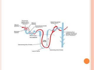
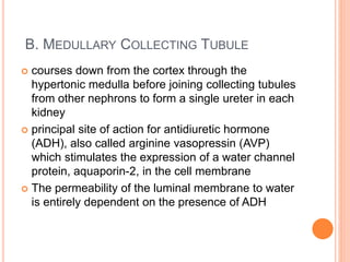
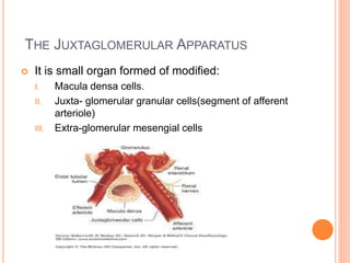
ANATOMY &
PHYSIOLOGY OF
NEPHRON: THE
COLLECTING
TUBULES &
JUXTAGLOMERULAR
APPARATUS Dr.Verdah Sabih
PGT Anesthesia
Holy family Hosp.Rwp
THE COLLECTING TUBULE
 Nephron:basic structural & functional unit of kidney.
 Collecting tubule:final segment of the tubule before
it enters the collecting duct system.
 Because of different embryonic origin(endoderm
rather than mesoderm),not considered a part of
nephron proper
 variably active portion of the Nephron
 last segment to save water for the body
 Each distal convoluted tubule delivers its filtrate to a
collecting duct, most of which begin in the renal
cortex and extend deep into the medulla
 collecting tubule can be divided into:
• cortical
• Medullary
 Together, they normally account for the
reabsorption of 5–7% of the filtered sodium load.
A. CORTICAL COLLECTING TUBULE
 consists of two cell types:
i. principal cells (P cells), :
 which primarily secrete K and participate in aldosterone-
stimulated Na- absorption& water from lumen
 (sites of action of the K-sparing diuretics)
 Reabsorption of Na+ generates a negative luminal voltage, which
provides the driving force for reabsorption of Cl- across the
paracellular pathway
ii. intercalated cells (I cells)
 which are responsible for acid–base regulation.
 Reabsorb K ions and secrete H+ ions into the tubular lumen
 Reabsorption of K+ is mediated by an H+,K+-ATPase located in
the apical cell membrane
 some I cells are capable of secreting bicarbonate ion in response
to large alkaline loads.

lumen
blood
B. MEDULLARY COLLECTING TUBULE
 courses down from the cortex through the
hypertonic medulla before joining collecting tubules
from other nephrons to form a single ureter in each
kidney
 principal site of action for antidiuretic hormone
(ADH), also called arginine vasopressin (AVP)
which stimulates the expression of a water channel
protein, aquaporin-2, in the cell membrane
 The permeability of the luminal membrane to water
is entirely dependent on the presence of ADH
 Dehydration increases ADH secretion whereas
adequate hydration suppresses ADH secretion
 This part of the nephron is responsible for
acidifying urine(secreting H+ against a large
concentration gradient-key role in regulating acid-
base balance)
 Unlike the cortical collecting tubule, the medullary
collecting duct is permeable to urea (special urea
transporters that facilitate urea diffusion across the
luminal and basolateral membranes)
ROLE OF THE COLLECTING TUBULE IN
MAINTAINING A HYPERTONIC MEDULLA
 Differences in permeability to urea in the cortical
and medullary collecting tubules account for up to
half the hypertonicity of the renal medulla
 Cortical collecting tubules are impermeable to urea,
whereas medullary collecting tubules are normally
permeable.
 In the presence of ADH, the innermost part of the
medullary collecting tubules becomes even more
permeable to urea
 when ADH is secreted, water moves out of the
collecting tubules and the urea becomes highly
concentrated
 Urea can then diffuse out deeply into the medullary
interstitium, increasing its tonicity.
THE JUXTAGLOMERULAR APPARATUS
 It is small organ formed of modified:
I. Macula densa cells.
II. Juxta- glomerular granular cells(segment of afferent
arteriole)
III. Extra-glomerular mesengial cells
 Juxtaglomerular cells contain the enzyme renin and
are innervated by the sympathetic nervous system.
 Release of renin depends on β 1 -adrenergic
sympathetic stimulation, changes in afferent
arteriolar wall pressure, and changes in chloride
flow past the macula densa.
 Renin released into the bloodstream catalyzes the
conversion of angiotensinogen, a protein
synthesized by the liver, to angiotensin 1
Nephron
Nephron

Nephron

  • 1.
    ANATOMY & PHYSIOLOGY OF NEPHRON:THE COLLECTING TUBULES & JUXTAGLOMERULAR APPARATUS Dr.Verdah Sabih PGT Anesthesia Holy family Hosp.Rwp
  • 2.
    THE COLLECTING TUBULE Nephron:basic structural & functional unit of kidney.  Collecting tubule:final segment of the tubule before it enters the collecting duct system.  Because of different embryonic origin(endoderm rather than mesoderm),not considered a part of nephron proper  variably active portion of the Nephron  last segment to save water for the body
  • 4.
     Each distalconvoluted tubule delivers its filtrate to a collecting duct, most of which begin in the renal cortex and extend deep into the medulla  collecting tubule can be divided into: • cortical • Medullary  Together, they normally account for the reabsorption of 5–7% of the filtered sodium load.
  • 5.
    A. CORTICAL COLLECTINGTUBULE  consists of two cell types: i. principal cells (P cells), :  which primarily secrete K and participate in aldosterone- stimulated Na- absorption& water from lumen  (sites of action of the K-sparing diuretics)  Reabsorption of Na+ generates a negative luminal voltage, which provides the driving force for reabsorption of Cl- across the paracellular pathway ii. intercalated cells (I cells)  which are responsible for acid–base regulation.  Reabsorb K ions and secrete H+ ions into the tubular lumen  Reabsorption of K+ is mediated by an H+,K+-ATPase located in the apical cell membrane  some I cells are capable of secreting bicarbonate ion in response to large alkaline loads. 
  • 6.
  • 8.
    B. MEDULLARY COLLECTINGTUBULE  courses down from the cortex through the hypertonic medulla before joining collecting tubules from other nephrons to form a single ureter in each kidney  principal site of action for antidiuretic hormone (ADH), also called arginine vasopressin (AVP) which stimulates the expression of a water channel protein, aquaporin-2, in the cell membrane  The permeability of the luminal membrane to water is entirely dependent on the presence of ADH
  • 10.
     Dehydration increasesADH secretion whereas adequate hydration suppresses ADH secretion  This part of the nephron is responsible for acidifying urine(secreting H+ against a large concentration gradient-key role in regulating acid- base balance)  Unlike the cortical collecting tubule, the medullary collecting duct is permeable to urea (special urea transporters that facilitate urea diffusion across the luminal and basolateral membranes)
  • 11.
    ROLE OF THECOLLECTING TUBULE IN MAINTAINING A HYPERTONIC MEDULLA  Differences in permeability to urea in the cortical and medullary collecting tubules account for up to half the hypertonicity of the renal medulla  Cortical collecting tubules are impermeable to urea, whereas medullary collecting tubules are normally permeable.  In the presence of ADH, the innermost part of the medullary collecting tubules becomes even more permeable to urea  when ADH is secreted, water moves out of the collecting tubules and the urea becomes highly concentrated
  • 12.
     Urea canthen diffuse out deeply into the medullary interstitium, increasing its tonicity.
  • 14.
    THE JUXTAGLOMERULAR APPARATUS It is small organ formed of modified: I. Macula densa cells. II. Juxta- glomerular granular cells(segment of afferent arteriole) III. Extra-glomerular mesengial cells
  • 15.
     Juxtaglomerular cellscontain the enzyme renin and are innervated by the sympathetic nervous system.  Release of renin depends on β 1 -adrenergic sympathetic stimulation, changes in afferent arteriolar wall pressure, and changes in chloride flow past the macula densa.  Renin released into the bloodstream catalyzes the conversion of angiotensinogen, a protein synthesized by the liver, to angiotensin 1

Editor's Notes

  • #6 . Increased intracellular [K +] favors K + secretion. Aldosterone enhances Na + –K +- ATPase activity in this part of the nephron by increasing the number of open K + and Na + channels in the luminal membrane.
  • #13 Formation of a concentrated urine when antidiuretic hormone (ADH) levels are high. Note that the fluid leaving the loop of Henle is dilute but becomes concentrated as water is absorbed from the distal tubules and collecting tubules. With high ADH levels, the osmolarity of the urine is about the same as the osmolarity of the renal medullary interstitial fluid in the papilla, which is about 1200 mOsm/L.