Download as PDF, PPTX


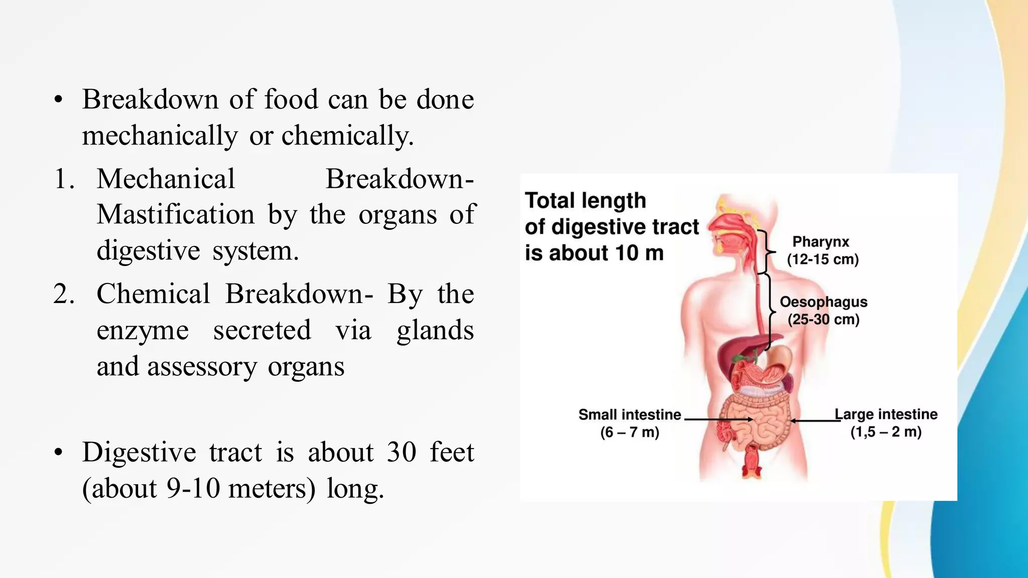
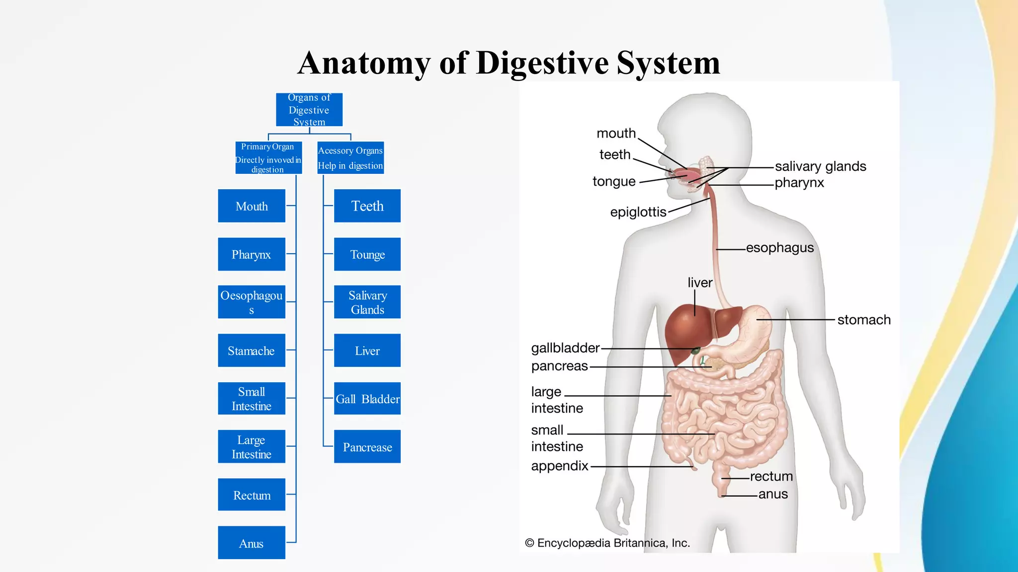


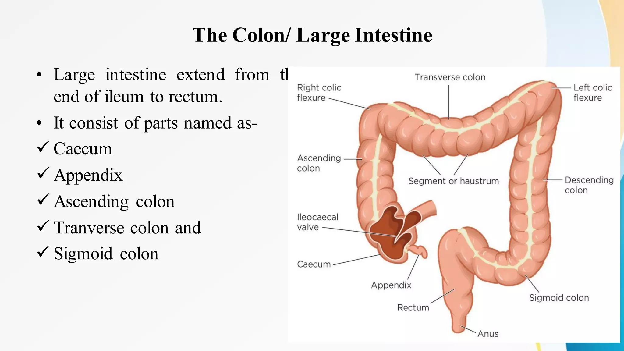


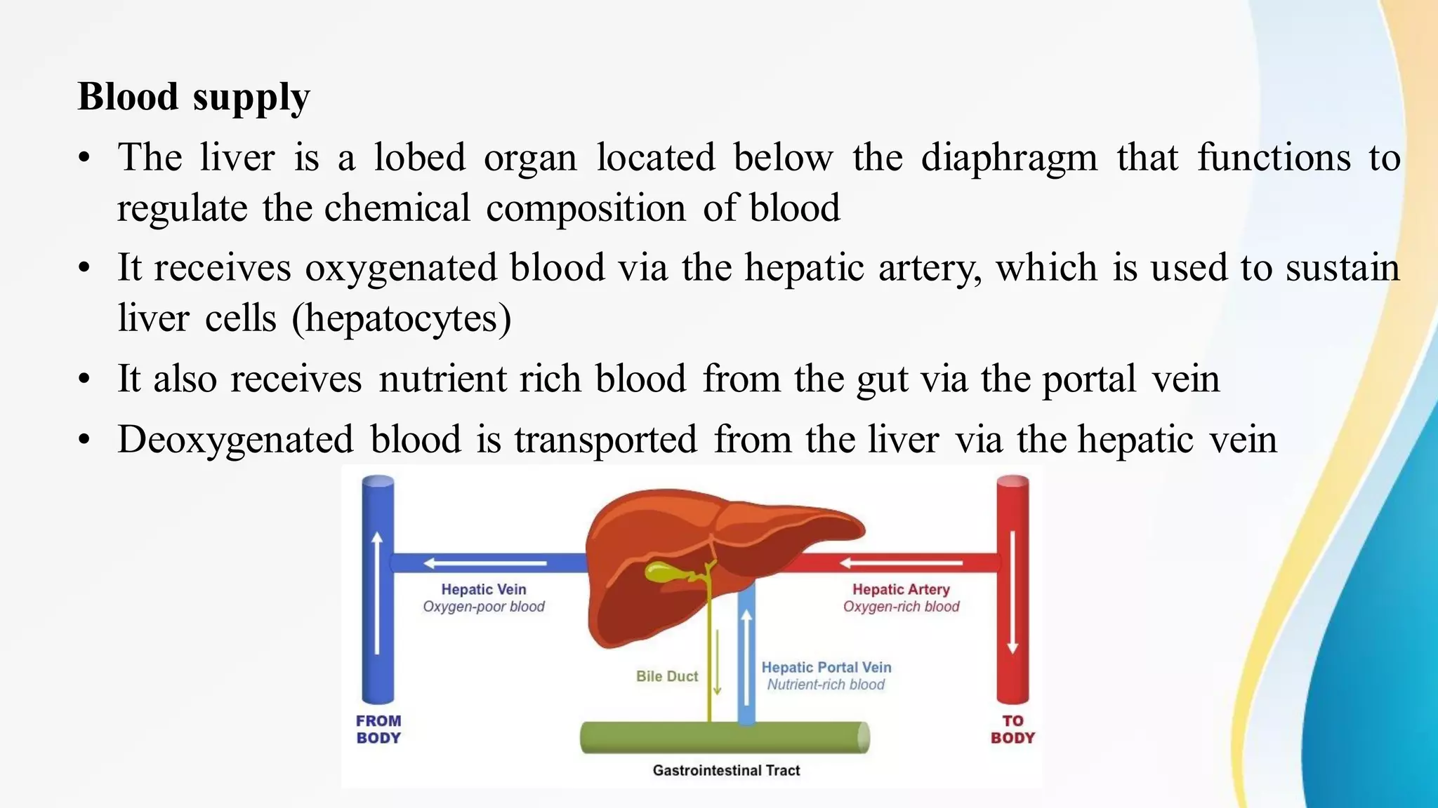


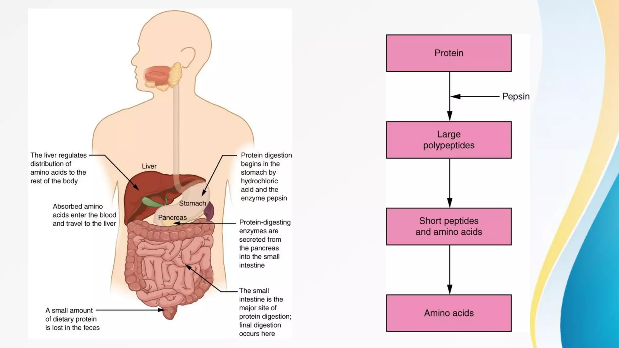


The document summarizes the digestive system, including its organs and their functions. It describes how food travels through the mouth, esophagus, stomach, and intestines. Key digestive organs like the liver, gallbladder and pancreas produce enzymes and bile to break down food into nutrients that can be absorbed. The digestive tract is approximately 30 feet long and works through mechanical and chemical breakdown of food, as well as absorption and elimination of waste.