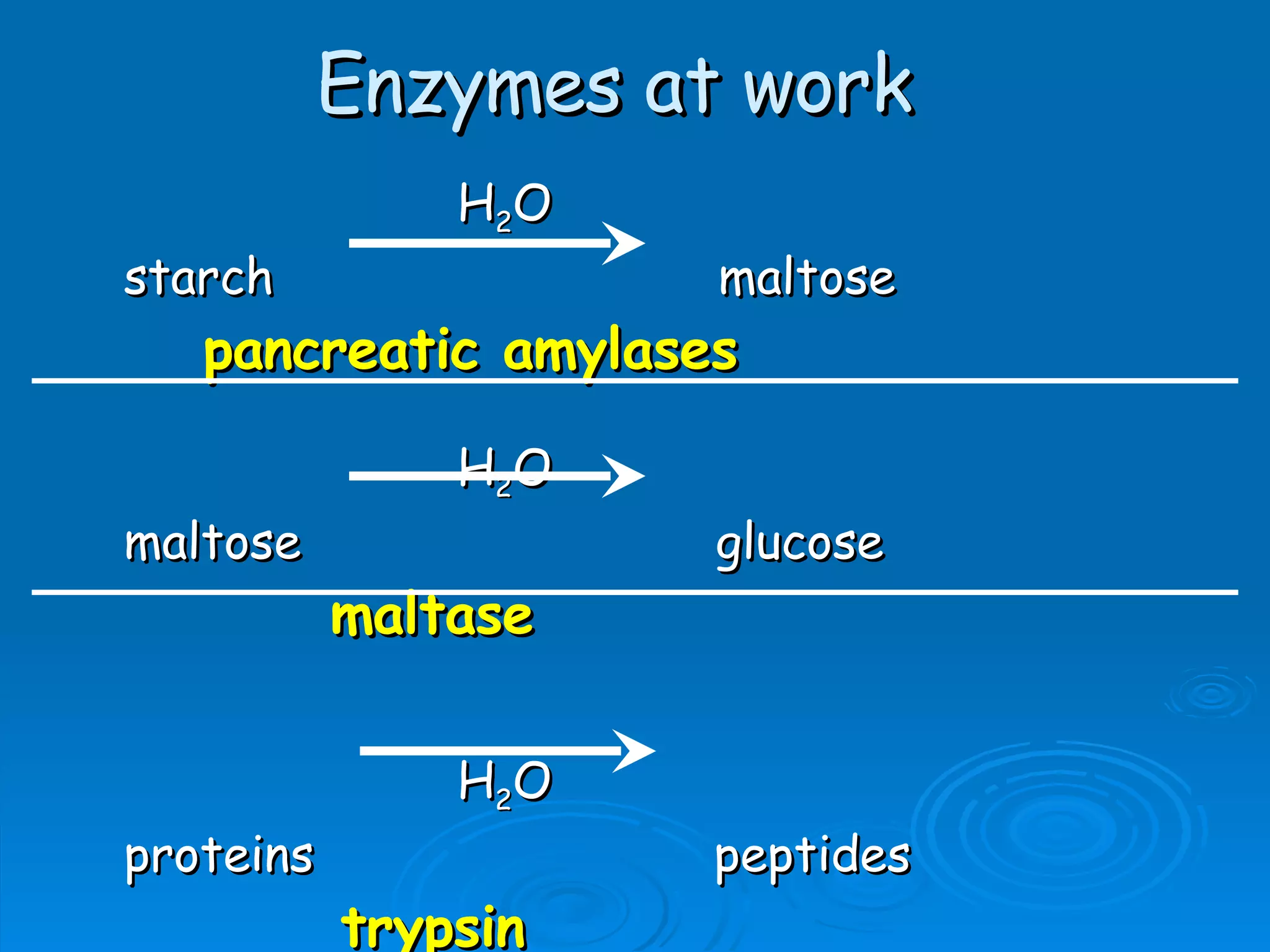
The document outlines the key structures and functions of the human digestive system. It describes the organs involved in digestion, including the mouth, esophagus, stomach, small intestine, pancreas, liver, and large intestine. It also explains the roles of enzymes and chemicals in breaking down carbohydrates, proteins, lipids, and nucleic acids during digestion.


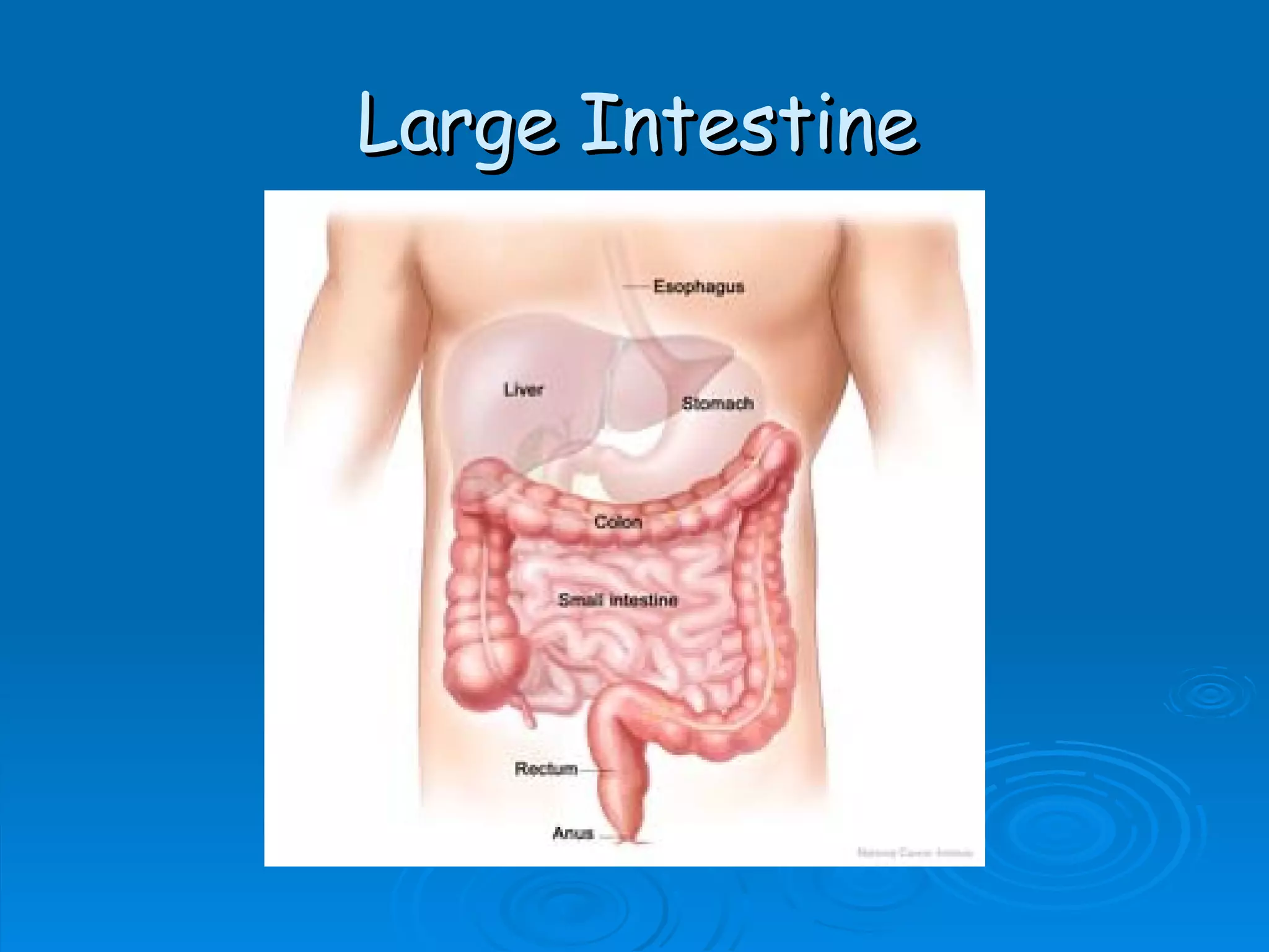
![Large Intestine (colon) Absorption of H 2 O from feces and salts, e.g. high [solute] of the feces will cause watery feces/diarrhea (recall B10, hypertonic). Secretes mucus for lubrication.](https://image.slidesharecdn.com/unit-c1-digestion-1208837072024122-8/75/Digestion-38-2048.jpg)