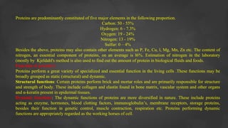
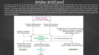
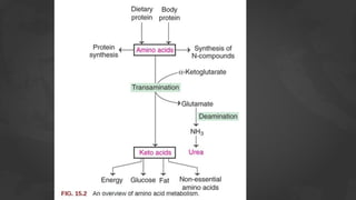
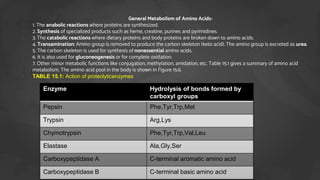
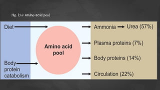
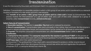
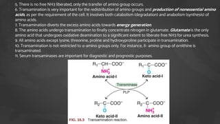
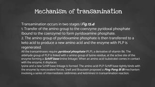
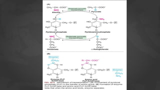
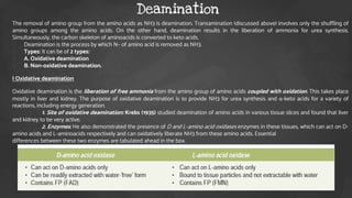
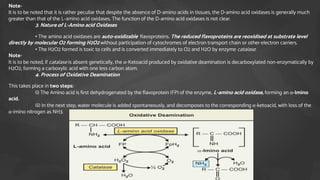
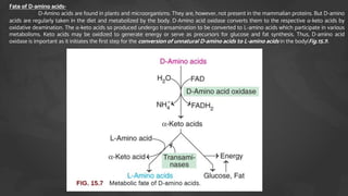
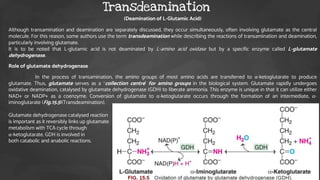
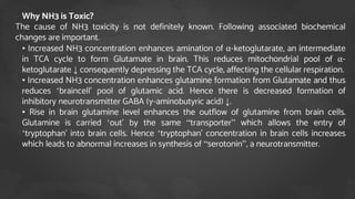
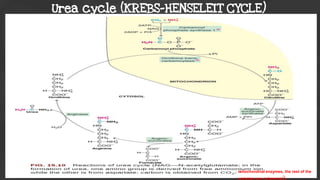
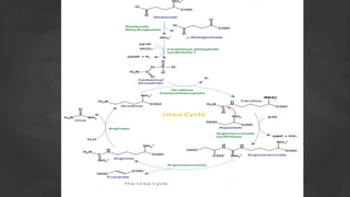
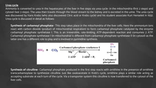
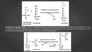
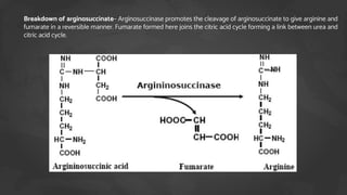
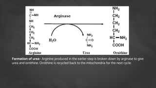
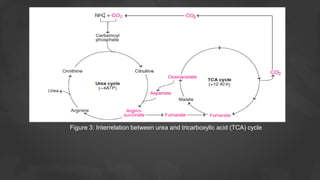
1. The document discusses amino acid and protein metabolism. It covers topics like the amino acid pool, transamination, deamination, the metabolism of ammonia, and the urea cycle.
2. Transamination is the process where the amino group is transferred from one amino acid to a keto acid, catalyzed by transaminase enzymes. Deamination results in the liberation of ammonia for urea synthesis and the conversion of the amino acid carbon skeleton into a keto acid.
3. Glutamate plays a central role as it can accept amino groups via transamination and also undergo oxidative deamination to release ammonia via glutamate dehydrogenase. This links amino acid and urea metabolism to the TCA
2. Here’s what you’ll find in this slides:
1. Introduction to amino acid and protien.
2. Amino acid pool & utilisation of Amino acid.
3. General Metabolism of Amino Acids
a) Transamination
b) Deamination
4. Metabolism of Ammonia
5. Urea cycle & it’s disorders.
Contents of Amino acid metabolism
3. *Proteins are of paramount importance in biological systems. All the major structural and
functional aspects of the body are carried out by protein molecules. All proteins are
polymers of amino acids. Proteins are composed of a number of amino acids linked by
peptide bonds.
*Although about 300 amino acids occur in nature, only 20 of them are seen in human
body. Most of the amino acids (except proline) are alpha amino acid, which means that
the amino group is attached to the same carbon atom to which the carboxyl group is
attached.
1. *Origin of the word 'protein'
2. The term protein is derived from a Greek word proteios meaning holding the first
place. Berzelius (Swedish chemist) suggested the name proteins to the group of
organic compounds that are utmost important to life. Mulder (Dutch chemist) in 1838
used the term proteins for the high molecular weight nitrogen- rich and most abundant
substances present in animals and plants.
Introduction of Amino acid & protein
4. Proteins are predominantly constituted of five major elements in the following proportion.
Carbon: 50 - 55%
Hydrogen: 6 - 7.3%
Oxygen: 19 - 24%
Nitrogen: 13 - 19%
Sulfur: 0 – 4%
Besides the above, proteins may also contain other elements such as P, Fe, Cu, l, Mg, Mn, Zn etc. The content of
nitrogen, an essential component of proteins, on an average is l6%. Estimation of nitrogen in the laboratory
(mostly by Kjeldahl's method is also used to find out the amount of protein in biological fluids and foods.
Function of protein’s
Proteins perform a great variety of specialized and essential function in the living cells .These functions may be
broadly grouped as static (structural) and dynamic.
Structural functions: Certain proteins perform brick and mortar roles and are primarily responsible for structure
and strength of body. These include collagen and elastin found in bone matrix, vascular system and other organs
and α-keratin present in epidermal tissues.
Dynamic functions: The dynamic functions of proteins are more diversified in nature. These include proteins
acting as enzyme, hormones, blood clotting factors, immunoglobulin’s, membrane receptors, storage proteins,
besides their function in genetic control, muscle contraction, respiration etc. Proteins performing dynamic
functions are appropriately regarded as the working horses of cell.
5. The proteins on degradation (proteolysis) release individual amino acids. Amino acids are not just the structural
components of proteins. Each one of the 20 naturally occurring amino acids undergoes its own metabolism and
performs specific functions. Some of the amino acids also serve as precursors for the synthesis of many biologically
important compounds (e.g. melanin, serotonin, creatine etc.). Certain amino acids may directly act as neurotransmitters
(e.g. glycine aspartate, glutamate). Protein metabolism is more appropriately learnt as metabolism of amino acids.
Amino acids-
Amino acids are a group of organic compounds containing two functional groups— amino and carboxyl. The amino
group (—NH2) is basic while the carboxyl group (—COOH) is acidic in nature.
General structure of amino acids-
1. The amino acids are termed as α-amino acids, if both the carboxyl and amino groups are attached to the same
carbon atom, as depicted below
The α-carbon atom binds to a side chain represented by R which is different for each of the 20 amino acids found in
proteins. The amino acids mostly exist in the ionized form in the biological system (shown above).
9. An adult has about 100 g of free amino acids which represent the amino acid pool of the body. The amino acid pool may be an
oversimplification of the facts, since there is no single compartment—rather, several compartments exist. Glutamate and glutamine
together constitute about 50%, and essential amino acids about 10% of the body pool (100 g). The concentration of intracellular amino
acids is always higher than the extracellular amino acids. Amino acids enter the cells against a concentration gradient by active transport.
The amino acid pool of the body is maintained by the sources that contribute (input) and the metabolic pathways that utilize (output) the
amino acids (Fig-15.1).
Amino acid pool
11. The ‘Amino acid pool’ is constantly undergoing depletion because,
1. • Large scale deamination of presumably surplus amino acids take place.
2. • Amino acids and their derivatives viz urea, creatinine are lost in the urine and other excretions.
3. • Amino acids are continually being built up into those proteins, e.g. hair, collagen proteins, which are not part of
dynamic systems.
4. On the other hand, amino acid pool is being always re-established by amino acids, derived from the following:
5. • Reamination of certain non-nitrogenous residues.
6. • Amination of appropriate fragments which are present in the common metabolic pool (and therefore derived from
carbohydrate and fat breakdown).
7. • Amino acids split off from dietary proteins and absorbed from the intestine into the blood.
12. General Metabolism of Amino Acids
The amino acids undergo certain common reactions like transamination
followed by deamination for the liberation of ammonia. The amino group of the
amino acids is utilized for the formation of urea which is an excretory end
product of protein metabolism. The carbon skeleton of the amino acids is first
converted to keto acids (by transamination) which meet one or more of the
following fates.
1. Utilized to generate energy.
2. Used for the synthesis of glucose.
3. Diverted for the formation of fat or ketone bodies.
4. Involved in the production of nonessential amino acids.
A general picture of amino acid metabolism is depicted in Fig-15.2
13.
14. 1. General Metabolism of Amino Acids-
2. 1. The anabolic reactions where proteins are synthesized.
3. 2. Synthesis of specialized products such as heme, creatine, purines and pyrimidines.
4. 3. The catabolic reactions where dietary proteins and body proteins are broken down to amino acids.
5. 4. Transamination: Amino group is removed to produce the carbon skeleton (keto acid). The amino group is excreted as urea.
6. 5. The carbon skeleton is used for synthesis of nonessential amino acids.
7. 6. It is also used for gluconeogenesis or for complete oxidation.
8. 7. Other minor metabolic functions like conjugation, methylation, amidation, etc. Table 15.1 gives a summary of amino acid
metabolism. The amino acid pool in the body is shown in Figure 15.6.
9. TABLE 15.1: Action of proteolyticenzymes
Enzyme Hydrolysis of bonds formed by
carboxyl groups
Pepsin Phe,Tyr,Trp,Met
Trypsin Arg,Lys
Chymotrypsin Phe,Tyr,Trp,Val,Leu
Elastase Ala,Gly,Ser
Carboxypeptidase A C-terminal aromatic amino acid
Carboxypeptidase B C-terminal basic amino acid
16. It was first discovered by Braunstein and Kritzmann (1947). It is a process of combined deamination and amination.
Definition: Transamination is a reversible reaction in which α-NH2 group of one amino acid is transferred to a α-ketoacid
resulting in formation of a new amino acid and a new ketoacid.
1. The transfer of an amino (−NH2) group from an amino acid to a keto acid is known as transamination. This
process involves the interconversion of a pair of amino acids and a pair of keto acids, catalysed by a group of
enzymes called transaminases (recently, aminotransferases).
Salient features of transamination
1. 1. Reversible reaction: The reaction is reversible (Fig-15.3) and is catalysed by enzymes.
2. 2. Site of transamination: Transamination takes place principally in liver, kidney, heart and brain. But the enzyme is
present in almost all mammalian tissues and transamination can be carried out in all tissues to some extent.
3. 3. Enzymes: The enzymes concerned in transamination are called transaminases (better called as amino
transferases).
4. 4. Coenzyme for the reaction: The coenzyme required for the reaction is pyridoxal-P (B6P). In the process of
transamination, the amino acid reacts with enzyme-bound pyridoxal-P to form an enzyme-bound complex of the
Schiff-base type, which then yields ketoacid and
5. pyridoxamine-P. The pyridoxamine-P then reacts with a second ketoacid to produce a similar enzyme bound
Schiffbase complex, which then decomposes forming a second new amino acid and regenerates the pyridoxal
phosphate (PLP) , a coenzyme derived from vitamin B6.
Transamination
17. 1. 5. There is no free NH3 liberated, only the transfer of amino group occurs.
2. 6. Transamination is very important for the redistribution of amino groups and production of nonessential amino
acids, as per the requirement of the cell. It involves both catabolism (degradation) and anabolism (synthesis) of
amino acids.
3. 7. Transamination diverts the excess amino acids towards energy generation.
4. 8. The amino acids undergo transamination to finally concentrate nitrogen in glutamate. Glutamate is the only
amino acid that undergoes oxidative deamination to a significant extent to liberate free NH3 for urea synthesis.
5. 9. All amino acids except lysine, threonine, proline and hydroxyproline participate in transamination.
6. 10. Transamination is not restricted to α-amino groups only. For instance, δ- amino group of ornithine is
transaminated.
7. 11. Serum transaminases are important for diagnostic and prognostic purposes.
18. 1. Transamination occurs in two stages (Fig.15.4)
2. 1. Transfer of the amino group to the coenzyme pyridoxal phosphate
(bound to the coenzyme) to form pyridoxamine phosphate.
3. 2. The amino group of pyridoxamine phosphate is then transferred to a
keto acid to produce a new amino acid and the enzyme with PLP is
regenerated.
4. All the transaminases require pyridoxal phosphate (PLP), a derivative of vitamin B6. The
aldehyde group of PLP is linked with ε-amino group of lysine residue, at the active site of the
enzyme forming a Schiff base (imine linkage). When an amino acid (substrate) comes in contact
with the enzyme, it displaces
5. lysine and a new Schiff base linkage is formed. The amino acid-PLP-Schiff base tightly binds with
the enzyme by noncovalent forces. Snell and Braustein proposed a Ping Pong Bi Bi mechanism
involving a series of intermediates (aldimines and ketimines) in transamination reaction.
Mechanism of transamination
19.
20. The removal of amino group from the amino acids as NH3 is deamination. Transamination (discussed above) involves only the shuffling of
amino groups among the amino acids. On the other hand, deamination results in the liberation of ammonia for urea synthesis.
Simultaneously, the carbon skeleton of aminoacids is converted to keto acids.
1. Deamination is the process by which N– of amino acid is removed as NH3.
2. Types: It can be of 2 types:
3. A. Oxidative deamination
4. B. Non-oxidative deamination.
I Oxidative deamination
Oxidative deamination is the liberation of free ammonia from the amino group of amino acids coupled with oxidation. This takes place
mostly in liver and kidney. The purpose of oxidative deamination is to provide NH3 for urea synthesis and α-keto acids for a variety of
reactions, including energy generation.
1. Site of oxidative deamination: Krebs (1935) studied deamination of amino acids in various tissue slices and found that liver
and kidney to be very active.
2. Enzymes: He also demonstrated the presence of D and L-amino acid oxidases enzymes in these tissues, which can act on D-
amino acids and L-aminoacids respectively and can oxidatively liberate NH3 from these amino acids. Essential
differences between these two enzymes are tabulated ahead in the box.
Deamination
21. Note-
It is to be noted that it is rather peculiar that despite the absence of D-amino acids in tissues, the D-amino acid oxidases is generally much
greater than that of the L-amino acid oxidases. The function of the D-amino acid oxidases is not clear.
3. Nature of L-Amino acid Oxidases
• The amino acid oxidases are auto-oxidizable flavoproteins. The reduced flavoproteins are reoxidised at substrate level
directly by molecular O2 forming H2O2 without participation of cytochromes of electron transport chain or other electron carriers.
• The H2O2 formed is toxic to cells and is converted immediately to O2 and H2O by enzyme catalase.
Note-
It is to be noted, if catalase is absent genetically, the α-Ketoacid produced by oxidative deamination is decarboxylated non-enzymatically by
H2O2, forming a carboxylic acid with one less carbon atom.
4. Process of Oxidative Deamination
This takes place in two steps:
(i) The Amino acid is first dehydrogenated by the flavoprotein (FP) of the enzyme, L-amino acid oxidase, forming an α-Imino
acid.
(ii) In the next step, water molecule is added spontaneously, and decomposes to the corresponding α-ketoacid, with loss of the
α-imino nitrogen as NH3.
22. Fate of D-amino acids-
D-Amino acids are found in plants and microorganisms. They are, however, not present in the mammalian proteins. But D-amino
acids are regularly taken in the diet and metabolized by the body. D-Amino acid oxidase converts them to the respective α-keto acids by
oxidative deamination. The α-keto acids so produced undergo transamination to be converted to L-amino acids which participate in various
metabolisms. Keto acids may be oxidized to generate energy or serve as precursors for glucose and fat synthesis. Thus, D-amino acid
oxidase is important as it initiates the first step for the conversion of unnatural D-amino acids to L-amino acids in the body(Fig.15.7).
23. II Non-oxidative deamination-
Some of the amino acids can be deaminated to liberate NH3 without undergoing oxidation
(a) Amino acid dehydrases : Serine, threonine and homoserine are the hydroxy amino acids. They undergo non-oxidative
deamination catalysed by PLPdependent dehydrases (dehydratases).
(b) Amino acid desulfhydrases : The sulfur amino acids, namely cysteine and homocysteine, undergo deamination coupled
with desulfhydration to give keto acids.
(c) Deamination of histidine : The enzyme histidase acts on histidine to liberate NH3 by a non-oxidative deamination process.
24. (Deamination of L-Glutamic Acid)
Although transamination and deamination are separately discussed, they occur simultaneously, often involving glutamate as the central
molecule. For this reason, some authors use the term transdeamination while describing the reactions of transamination and deamination,
particularly involving glutamate.
It is to be noted that L-glutamic acid is not deaminated by L-amino acid oxidase but by a specific enzyme called L-glutamate
dehydrogenase.
Role of glutamate dehydrogenase
In the process of transamination, the amino groups of most amino acids are transferred to α-ketoglutarate to produce
glutamate. Thus, glutamate serves as a ‘collection centre’ for amino groups in the biological system. Glutamate rapidly undergoes
oxidative deamination, catalysed by glutamate dehydrogenase (GDH) to liberate ammonia. This enzyme is unique in that it can utilize either
NAD+ or NADP+ as a coenzyme. Conversion of glutamate to α-ketoglutarate occurs through the formation of an intermediate, α-
iminoglutarate (Fig.15.5)(Transdeamination).
Glutamate dehydrogenase catalysed reaction
is important as it reversibly links up glutamate
metabolism with TCA cycle through
α-ketoglutarate. GDH is involved in
both catabolic and anabolic reactions.
Transdeamination
25. Ammonia is constantly being liberated in the metabolism of amino acids (mostly) and other nitrogenous compounds.
At the physiological pH, ammonia exists as ammonium (NH4+) ion.
I Formation of ammonia
The production of NH3 occurs from the amino acids (transamination and deamination), biogenic amines, amino group
of purines and pyrimidines and by the action of intestinal bacteria (urease) on urea.
II Transport and storage of NH3
Despite a regular and constant production of NH3 from various tissues, its concentration in the circulation is
surprisingly low (normal plasma 10–20mg/dl). This is mostly because the body has an efficient mechanism for NH3
transport and its immediate utilization for urea synthesis. The transport of ammonia between various tissues and the
liver mostly occurs in the form of glutamine or alanine and not as free ammonia.
Notes- Ammonia store house is Glutamine which is present at the highest concentration 8mg/dl in adult blood
it is freely diffusible in tissues hence ammonia is easily transported.
III Functions of ammonia
Ammonia is not just a waste product of nitrogen metabolism. It is involved (directly or via glutamine) for the synthesis
of many compounds in the body. These include nonessential amino acids, purines, pyrimidines, amino sugars,
asparagine etc. Ammonium ions (NH4+) are very important to maintain acid-base balance of the body.
IV Toxicity of ammonia
Even a marginal elevation in the blood ammonia concentration is harmful to the brain. Ammonia, when it
accumulates in the body, results in slurring of speech and blurring of the vision and causes tremors. It may lead to
coma and, finally, death, if not corrected.
Metabolism of Ammonia
26. Why NH3 is Toxic?
The cause of NH3 toxicity is not definitely known. Following associated biochemical
changes are important.
• • Increased NH3 concentration enhances amination of α-ketoglutarate, an intermediate
in TCA cycle to form Glutamate in brain. This reduces mitochondrial pool of α-
ketoglutarate ↓ consequently depressing the TCA cycle, affecting the cellular respiration.
• • Increased NH3 concentration enhances glutamine formation from Glutamate and thus
reduces ‘braincell’ pool of glutamic acid. Hence there is decreased formation of
inhibitory neurotransmitter GABA (γ-aminobutyric acid) ↓.
• • Rise in brain glutamine level enhances the outflow of glutamine from brain cells.
Glutamine is carried ‘out’ by the same “transporter” which allows the entry of
‘tryptophan’ into brain cells. Hence ‘tryptophan’ concentration in brain cells increases
which leads to abnormal increases in synthesis of “serotonin”, a neurotransmitter.
29. Urea cycle
Ammonia is converted to urea in the hepatocytes of the liver in five steps via urea cycle- in the mitochondria (first 2 steps) and
cytosol (last 3 steps). The urea then travels through the blood stream to the kidney and is excreted in the urine. The urea cycle
was discovered by Hans Krebs (who also discovered Citric acid or Krebs cycle) and his student associate Kurt Henseleit in 1932.
Urea cycle is discussed in detail as follows:
1. Synthesis of carbamoyl phosphate– This step takes place in the mitochondria of the liver cells. Here the ammonium ions
react with carbon dioxide (product of mitochondrial respiration) to form carbamoyl phosphate catalyzed by the enzyme
carbamoyl phosphate synthetase I. This is an irreversible, rate-limiting, ATP-dependent reaction and consumes 2 ATP.
Carbamoyl phosphate synthetase I (in mitochondria) is different from carbamoyl phosphate synthetase II (in cytosol) as the
latter one has a different role to play and is involved in pyrimidine synthesis.
2. Synthesis of citrulline– Carbamoyl phosphate produced in the first step reacts with ornithine in the presence of ornithine
transcarbamoylase to synthesize citrulline. Just like oxaloacetate in Kreb’s cycle, ornithine plays a similar role acting as
accepting substrate at each turn of the cycle. Via a transporter system this citrulline is now transferred to the cytosol of the
liver cells.
30. Formation of arginosuccinate– In this ATP dependent step, the carbonyl carbon of citrulline is attacked by the lone pair of the amine
in aspartate to produce arginosuccinate in presence of arginiosuccinate synthetase. In this step, the second nitrogen of urea is
incorporated by condensation. ATP is broken down into AMP and pyrophosphate.
31. Breakdown of arginosuccinate– Arginosuccinase promotes the cleavage of arginosuccinate to give arginine and
fumarate in a reversible manner. Fumarate formed here joins the citric acid cycle forming a link between urea and
citric acid cycle.
32. 1. Formation of urea– Arginine produced in the earlier step is broken down by arginase to give
urea and ornithine. Ornithine is recycled back to the mitochondria for the next cycle.
34. A) The Mitochondrial Stage
The first two steps of the urea cycle occur in the mitochondria of the cell.
• Step 1: The enzyme carbamoyl phosphate synthetase (CPS) takes ammonia and bicarbonate, and forms
carbamoyl phosphate with the use of ATP. This is the step in the cycle which determines how fast the cycle
progresses.
• Step 2: Ornithine transcarbamoylase (OTC) then condenses carbamoyl phosphate and ornithine, which forms
citrulline.
This citrulline is then moved out of the mitochondria into the cytosol of the cell by the transporter.
B) The Cytosolic Stage
• Step 3: Argininosuccinate synthetase (AS) takes the citrulline formed in the mitochondrial stage, and condenses
it with aspartate to form argininosuccinate.
• Step 4: Argininosuccinate is then broken into arginine and fumarate by argininosuccinase.
• Step 5: Arginine is then further broken down into urea and ornithine by arginase.
Arginine can also be acquired from the diet, and this can also be taken in by the liver cells and broken down into urea
and ornithine by arginase.
The ornithine is then transported into the mitochondria by ornithine translocase. There, it is used by OTC again, to
form citrulline.
The citrulline is then processed to form urea and ornithine again, and the cycle continues.
During the cycle, urea is the only new product which is formed, while all other molecules used in the cycle are
recycled.
35. 1. Regulation of urea cycle
The first reaction catalysed by carbamoyl phosphate synthase I (CPS I) is rate-
limiting reaction or committed step in urea synthesis. CPS I is allosterically activated by
N-acetylglutamate (NAG). The rate of urea synthesis in liver is correlated with the
concentration of N-acetylglutamate. High concentrations of arginine increase NAG.
The consumption of a protein-rich meal increases the level of NAG in liver, leading to
enhanced urea synthesis. Carbamoyl phosphate synthase I and glutamate
dehydrogenase are localized in the mitochondria. They coordinate with each other in
the formation of NH3, and its utilization for the synthesis of carbamoyl phosphate. The
remaining four enzymes of urea cycle are mostly controlled by the concentration of
their respective substrates.
*Bioenergetic-The urea cycle is irreversible and consumes 4 ATP. Two ATP are utilized
for the synthesis of carbamoyl phosphate. One ATP is converted to AMP and PPi to
produce arginosuccinate which equals to 2 ATP. Hence 4 ATP are actually consumed.
36. Urea cycle is linked with TCA cycle in three different ways (Figure 3). This is regarded as
bicyclic integration between the two cycles.
• 1. The production of fumarate in urea cycle is the most important integrating point
with TCA cycle. Fumarate is converted to malate and then to oxaloacetate in TCA
cycle. Oxaloacetate undergoes transamination to produce aspartate which enters urea
cycle. Here, it combines with citrulline to produce arginosuccinate. Oxaloacetate is an
important metabolite which can combine with acetyl CoA to form citrate and get
finally oxidized. Oxaloacetate can also serve as a precursor for the synthesis of
glucose (gluconeogenesis).
• 2. ATP (12) are generated in the TCA cycle while ATP (4) are utilized for urea synthesis.
• 3. Citric acid cycle is an important metabolic pathway for the complete oxidation of
various metabolites to CO2 and H2O. The CO2 liberated in TCA cycle (in the
mitochondria) can be utilized in urea cycle.
Integration between urea cycle and TCA cycle
38. • Metabolic defects associated with each of the five enzymes of urea cycle have been
• reported are given below. All the disorders invariably lead to a build-up in blood
ammonia (hyperammonemia), leading to toxicity. Other metabolites of urea cycle
also accumulate which, however, depends on the specific enzyme defect. The clinical
symptoms associated with defect in urea cycle enzymes include vomiting, lethargy,
irritability, anoxia and mental retardation.
Metabolic disorders of urea cycle