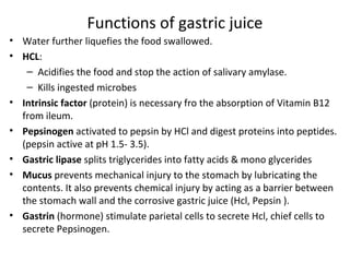
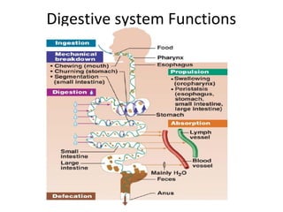
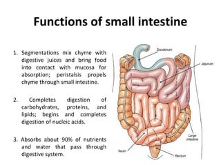
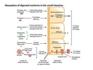
The digestive system
The digestive system performs six basic activities:
1. Ingestion. This process involves taking foods and liquids into the mouth (eating).
2. Secretion. Each day, cells within the walls of the GI tract and accessory digestive organs
secrete a total of about 7 liters of water, acid, buffers, and enzymes into the lumen
(interior space) of the tract.
3. Mixing and propulsion. Alternating contractions and relaxations of smooth muscle in
the walls of the GI tract mix food and secretions and propel them toward the anus. This
capability of the GI tract to mix and move material along its length is called motility.
4. Digestion. Mechanical and chemical processes break down ingested food into small
molecules. In mechanical digestion the teeth cut and grind food before it is swallowed,
and then smooth muscles of the stomach and small intestine churn the food. As a result,
food molecules become dissolved and thoroughly mixed with digestive enzymes.
In chemical digestion the large carbohydrate, lipid, protein, and nucleic acid molecules
in food are split into smaller molecules by hydrolysis. Digestive enzymes produced by the
salivary glands, tongue, stomach, pancreas, and small intestine catalyze these catabolic
reactions. A few substances in food can be absorbed without chemical digestion. These
include vitamins, ions, cholesterol, and water.
5. Absorption. The entrance of ingested and secreted fluids, ions, and the products of
digestion into the epithelial cells lining the lumen of the GI tract is called absorption. The
absorbed substances pass into blood or lymph and circulate to cells throughout the body.
6. Defecation. Wastes, indigestible substances, bacteria, cells sloughed from the lining of
the GI tract, and digested materials that were not absorbed in their journey through the
digestive tract leave the body through the anus in a process called defecation. The
eliminated material is termed feces or stool.
Digestive system Functions
Peritoneum
The peritoneum is the largest serous membrane of the body; it lines the wall of the
abdominal cavity and covers some abdominal organs. It helps to hold the organs in
place; and stores fat.
The peritoneum is divided into parietal peritoneum, which lines the wall of the
abdominopelvic cavity, and visceral peritoneum, which covers the organs in the cavity
(Figure d). The space between the parietal and visceral portions of the peritoneum is
called peritoneal cavity containing lubricating fluid.
some organs lie on the posterior abdominal wall and are covered by peritoneum only on
their anterior surfaces; they are not in the peritoneal cavity. Such organs, including the
kidneys, ascending and descending colons of the large intestine, duodenum of the small
intestine, and pancreas, are said to be retroperitoneal (retro- behind).
Unlike the pericardium and pleurae, which smoothly cover the heart and lungs, the
peritoneum contains large folds that weave between the viscera. The folds bind the
organs to one another and to the walls of the abdominal cavity. They also contain blood
vessels, lymphatic vessels, and nerves that supply the abdominal organs.
There are five major peritoneal folds: the greater omentum, falciform ligament, lesser
omentum, mesentery, and mesocolon
LAYERS OF THE GI TRACT
The mucosa, or inner lining of the GI tract, is a mucous membrane.
It is composed of (1) a layer of epithelium in direct contact with the contents of the GI
tract, (2) a layer of connective tissue called the lamina propria, and (3) a thin layer of
smooth muscle (muscularis mucosae).
The submucosa consists of areolar connective tissue that binds the mucosa to the
muscularis. It contains many blood and lymphatic vessels that receive absorbed food
molecules. molecules. Also the submucosa consists of an extensive network of neurons
known as the submucosal plexus.
The muscularis externa is responsible for segmentation and peristalsis. This tunic
typically has an inner circular layer and an outer longitudinal layer of smooth muscle
cells. In several places along the tract, the circular layer thickens to form sphincters that
act as valves to prevent backflow and control food passage.
Contains the myenteric plexus of Auerbach, the other major intrinsic nerve plexus.
Located between the two layers of smooth muscle, controls motility of the G.I. tract.
The serosa is the protective outermost layer. The serosa is a serous membrane
composed of areolar connective tissue and simple squamous epithelium. The serosa is
also called the visceral peritoneum because it forms a portion of the peritoneum. The
esophagus does not have serosa; instead only a single layer of areolar connective tissue
called the adventitia forms the superficial layer of this organ.
The enteric nervous system coordinates digestion, secretion,
and motility to optimize nutrient absorption.
Its activity is modified by information from the CNS and ANS
Enteric Nervous System
The gastrointestinal
tract is regulated by an
intrinsic set of nerves
known as the enteric
nervous system and by
an extrinsic set of
nerves that are part of
the autonomic
nervous system.
Enteric Nervous System
The neurons of the ENS are arranged into two plexuses: the myenteric
plexus and submucosal plexus .
The myenteric plexus or plexus of Auerbach , is located between the
longitudinal and circular smooth muscle layers of the muscularis.
The submucosal plexus, or plexus of Meissner, is found within the
submucosa.
The plexuses of the ENS consist of motor neurons, interneurons, and
sensory neurons . Because the motor neurons of the myenteric plexus
supply the longitudinal and circular smooth muscle layers of the
muscularis, this plexus mostly controls GI tract motility (movement),
The motor neurons of the submucosal plexus supply the secretory cells
of the mucosal epithelium, controlling the secretions of the organs of
the GI tract. The interneurons of the ENS interconnect the neurons of
the myenteric and submucosal plexuses. The sensory neurons of the
ENS supply the mucosal epithelium.
Even though the neurons of the ENS can function independently, they
are subject to regulated by the neurons of autonomic nervous system.
The vagus (X) nerves supply parasympathetic fibers to most parts of the
GI tract. The parasympathetic nerves that supply the GI tract form neural
connections with the ENS. Parasympathetic preganglionic neurons of the
vagus nerves synapse with neurons located in the myenteric and
submucosal plexuses. Some of the parasympathetic postganglionic
neurons in turn synapse with neurons in the ENS; others directly
innervate smooth muscle and glands within the wall of the GI tract.
Stimulation of the parasympathetic nerves causes an increase in GI
secretion and motility by increasing the activity of ENS neurons.
Sympathetic postganglionic neurons synapse with neurons located in the
myenteric plexus and the submucosal plexus. In general, the sympathetic
nerves that supply the GI tract cause a decrease in GI secretion and
motility by inhibiting the neurons of the ENS. Emotions such as anger,
fear, and anxiety may slow digestion because they stimulate the
sympathetic nerves that supply the GI tract.
Autonomic Nervous System
The Human Digestive System
• Teeth mechanically break
down food into small pieces.
• The tongue mix food with saliva
and moves the food around
until it forms a ball called a
bolus.
• The bolus is passed to the
pharynx (throat) and the
epiglottis is a flap like structure
at the back off the throat that
closes over the trachea, makes
sure the bolus passes into the
esophagus and not down the
windpipe!
The Human Digestive System
• The bolus passes down the
esophagus by peristalsis.
• Peristalsis is a wave of
muscular contractions that
push the bolus down
towards the stomach.
The Human Digestive System
• To enter the stomach, the bolus
must pass through the lower
esophageal sphincter, a tight
muscle that keeps stomach acid
out of the esophagus.
• The stomach has folds called
rugae and is a big muscular
pouch which churns the bolus
(Physical Digestion) and mixes it
with gastric juice (mixture of
stomach acid, mucus and enzymes).
• The digested bolus is now called
chyme and it leaves the stomach
by passing through the pyloric
sphincter.
Actions of Stomach
Composition of Gastric juice
About 2 liters of gastric juice secreted daily by secretory glands in the
mucosa of stomach consisting of
Functions of gastric juice
• Water further liquefies the food swallowed.
• HCL:
– Acidifies the food and stop the action of salivary amylase.
– Kills ingested microbes
• Intrinsic factor (protein) is necessary fro the absorption of Vitamin B12
from ileum.
• Pepsinogen activated to pepsin by HCl and digest proteins into peptides.
(pepsin active at pH 1.5- 3.5).
• Gastric lipase splits triglycerides into fatty acids & mono glycerides
• Mucus prevents mechanical injury to the stomach by lubricating the
contents. It also prevents chemical injury by acting as a barrier between
the stomach wall and the corrosive gastric juice (Hcl, Pepsin ).
• Gastrin (hormone) stimulate parietal cells to secrete Hcl, chief cells to
secrete Pepsinogen.
There is always a small quantity of gastric juice present in the stomach, even when it
contains no food. This is known as fasting juice. Secretion reaches its maximum level
about 1 hour after a meal then declines to the fasting level after 4 hours.
There are three phases of secretion of gastric juice
1. Cephalic phase: this flow of juice occurs before food reaches the stomach and is due
to reflex stimulation of the vagus (parasympathetic) nerves initiated by sight, smell or
taste of food. When the vagus nerves have been cut(vagotomy) this phase of gastric
secretion stops. Sympathetic stimulation also inhibits gastric activity.
eg: during emotional states
2.Gastric phase: the presence of food in the pyloric antrum and duodenum cause
enteroendocrine cells to secrete gastrin hormone which passes directly into the
circulating blood. Gastrin , circulating in the blood which supplies the stomach,
stimulates the gastric glands to produce more gastric juice. In this way secretion of
digestive juice is continued After completion of a meal and the end of the cephalic
phase. Gastrin secretion suppressed when the pH in the pyloric antrum falls to about
1.5.
3. Intestinal Phase: when the partially digested contents of the stomach reach the small
intestine, two hormones, secretin and cholecystokinin are produced by endocrine cells in
the intestinal mucosa. These hormones are passes directly into the circulating blood and
they slow down the secretion of gastric juice and reduce gastric motility. By slowing the
emptying rate of the stomach, the chyme in the duodenum becomes more throughly
mixed with bile and pancreatic juice. This phase of gastric sceretion is most marked
following a meal with a high fat content.
Small intestine
• Small intestine is the part of gastrointestinal (GI) tract, extending between the
pyloric sphincter of stomach and ileocecal sphincter, which opens into large
intestine.
• It is called small intestine because of its small diameter, compared to that of
the large intestine. But it is longer than large intestine. Its length is about
6 meter.
• Important function of small intestine is absorption. In the small intestine the
chemical digestion of food is completed and Maximum absorption of digested
food products takes place in small intestine.
• Small intestine consists of three portions:
1. Proximal part known as duodenum
2. Middle part known as jejunum
3. Distal part known as ileum.
Small intestine
• The shortest section, the duodenum, resembles the shape of a “C” and receives
food through the pyloric sphincter. This is about 25 cm long and curves around
the head of the pancreas. Secretions from the gall bladder and pancreas merge in
a common structure – the hepatopancreatic ampulla – and enter the duodenum
at the duodenal papilla. The duodenal papilla is guarded by a ring of smooth
muscle, the hepatopancreatic sphincter (of Oddi).
• The next section, the jejunum, is about 3 feet long and has a thicker layer of
smooth muscle. It is named jejunum because on death it is found to be empty.
• The ileum (ileum intestine), the last section of the small intestine, is about 6 feet
long and ends at the ileocecal sphincter (valve), where it joins the large intestine.
which controls the flow of material from the ileum to the caecum, the first part of
the large intestine, and prevents backflow.
INTESTINAL VILLI
• Wall of the small intestine has all the four layers as in stomach :
1. Mucus layer
2. Submucus layer
3. Muscular layer
4. Serous or fibrous layer.
• Mucous membrane of small intestine is covered by minute projections
called villi. The height of villi is about 1 mm and the diameter is less than
1 mm. Villi are lined by columnar cells, which are called enterocytes. Each
enterocyte gives rise to hair-like projections called microvilli. Villi and
microvilli increase Small Intestine the surface area of mucous membrane
by many folds. Within each villus, Goblet cells that secrete mucus are
interspersed between the enterocytes. These epithelial cells enclose a
network of blood capilliaries and a central lymph capillary called lacteal
because absorbed fat gives the lymph a milky appearance.
The intestinal glands are simple tubular glands situated below the surface
between the villi.
Secretions from small intestine
• Digestive Enzymes of small intestine released by enterocytes of the villi. act
on the partially digested food and convert them into final digestive
products.
• Proteolytic Enzymes peptidases convert peptides into amino acids.
• Amylolytic Enzymes Lactase, sucrase and maltase convert the disaccharides
(lactose, sucrose and maltose) into two molecules of monosaccharides.
Dextrinase converts dextrin, maltose and maltriose into glucose.
• Lipolytic Enzyme Intestinal lipase acts on triglycerides and converts them
into fatty acids.
• Mucus protects the intestinal wall from the acid chyme, which enters the
intestine from stomach; thereby it prevents the intestinal ulcer.
• Defensins secreted by paneth cells of intestinal glands are the
antimicrobial peptides.
• Enterokinase present in intestinal juice activates trypsinogen into trypsin„
• Intrinsic factor plays an important role in erythropoiesis. It is necessary for
the absorption of vitamin B12.
Functions of small intestine
1. Segmentations mix chyme with
digestive juices and bring food
into contact with mucosa for
absorption; peristalsis propels
chyme through small intestine.
2. Completes digestion of
carbohydrates, proteins, and
lipids; begins and completes
digestion of nucleic acids.
3. Absorbs about 90% of nutrients
and water that pass through
digestive system.
Mechanical Digestion in the Small Intestine
The two types of movements of the small intestine—segmentations and a type of
peristalsis called migrating motility complexes—are governed mainly by the
myenteric plexus.
Segmentations are localized, mixing contractions that occur in portions of
intestine distended by a large volume of chyme.
Segmentations mix chyme with the digestive juices and bring the particles of food
into contact with the mucosa for absorption; they do not push the intestinal
contents along the tract.
A segmentation starts with the contractions of circular muscle fibers in a portion
of the small intestine, an action that constricts the intestine into segments. Next,
muscle fibers that encircle the middle of each segment also contract, dividing
each segment again. Finally, the fibers that first contracted relax, and each small
segment unites with an adjoining small segment so that large segments are
formed again. As this sequence of events repeats, the chyme sloshes back and
forth. Segmentations occur most rapidly in the duodenum, about 12 times per
minute, and progressively slow to about 8 times per minute in the ileum. This
movement is similar to alternately squeezing the middle and then the ends of
a capped tube of toothpaste.
Peristalsis in the Small Intestine
After most of a meal has been absorbed, which lessens distension of the wall of
the small intestine, segmentation stops and peristalsis begins. The type of
peristalsis that occurs in the small intestine, termed a migrating motility
complex (MMC), begins in the lower portion of the stomach and pushes chyme
forward along a short stretch of small intestine before dying out.
They move toward the anus at a velocity of 0.5 to 2.0 cm/sec, faster in the
proximal intestine and slower in the terminal intestine. They normally are very
weak and usually die out after traveling only 3 to 5 centimeters, very rarely
farther than 10 centimeters.
The MMC slowly migrates down the small intestine, reaching the end of the
ileum in 90–120 minutes. Then another MMC begins in the stomach.
Altogether, chyme remains in the small intestine for 3–5 hours.
Chemical Digestion in the Small Intestine
• The chyme entering the small intestine contains partially digested
carbohydrates, proteins, and lipids. The completion of the digestion of
carbohydrates, proteins, and lipids is a collective effort of pancreatic juice,
bile, and intestinal juice in the small intestine.
• Digestion of Carbohydrates
Chemical Digestion in the Small Intestine
Digestion of Proteins
Digestion of Lipids
Pancreatic juice contains two nucleases: ribonuclease, which digests RNA, and
deoxyribonuclease, which digests DNA. The nucleotides that result from the action of
the two nucleases are further digested by brush-border enzymes called nucleosidases
and phosphatases into pentoses, phosphates, and nitrogenous bases. These products
are absorbed via active transport.
Digestion of Nucleic
cids
Absorption of digested nutrients in the small intestine.
Large Intestine
Functions:
• Reabsorb water and compact material
into feces, absorb vitamins produced by
bacteria
• Store fecal matter prior to defecation
• Other than digestion of enteric bacteria,
no further digestion takes place
• Vitamins, water, and electrolytes are
reclaimed
Chemical Digestion in the Large Intestine
The final stage of digestion occurs in the colon through the activity of bacteria. Mucus is
secreted by the glands of the large intestine, but no enzymes are secreted. Chyme is prepared
for elimination by the action of bacteria, which ferment any remaining carbohydrates and
release hydrogen, carbon dioxide, and methane gases. Bacteria also convert any remaining
proteins to amino acids and break down the amino acids into simpler substances: indole,
hydrogen sulfide, and fatty acids. Some of the indole is eliminated in the feces and contributes
to their odor; the rest is absorbed and transported to the liver, where these compounds are
converted to less toxic compounds and excreted in the urine. Bacteria also decompose bilirubin
to simpler pigments, including stercobilin, which gives feces their brown color.
Absorption and Feces Formation in the Large Intestine
Chyme has remained in the large intestine 3–10 hours; it has become solid or semisolid because
of water absorption and is now called feces. Chemically, feces consist of water, inorganic salts,
and sloughed-off epithelial cells from the mucosa of the gastrointestinal tract, bacteria, and
products of bacterial decomposition, unabsorbed digested materials, and indigestible parts of
food. Normally, 0.5–1 liter of water that enters the large intestine, all but about 100–200 mL is
normally absorbed via osmosis. The large intestine also absorbs ions, including sodium and
chloride, and some vitamins.
Salivary glands
Salivary glands produce saliva, which dissolves food chemicals, moistens
food, and contains chemicals that begin the breakdown of starches.
Intrinsic - buccal
Extrinsic - parotid, submandibular, sublingual
parotid - only serous cells (no mucin)
submandibular and buccal - about equal amounts serous and mucous cells
sublingual - only mucous cells
Salivia contains:
1. serous fluid
2. mucin
3. salivary amylase
4. lingual lipase
5. lysozyme
6. IgA
7. defensins
8. some antimicrobial cyanide compound
The digestive system

The digestive system

  • 1.
  • 3.
    The digestive systemperforms six basic activities: 1. Ingestion. This process involves taking foods and liquids into the mouth (eating). 2. Secretion. Each day, cells within the walls of the GI tract and accessory digestive organs secrete a total of about 7 liters of water, acid, buffers, and enzymes into the lumen (interior space) of the tract. 3. Mixing and propulsion. Alternating contractions and relaxations of smooth muscle in the walls of the GI tract mix food and secretions and propel them toward the anus. This capability of the GI tract to mix and move material along its length is called motility. 4. Digestion. Mechanical and chemical processes break down ingested food into small molecules. In mechanical digestion the teeth cut and grind food before it is swallowed, and then smooth muscles of the stomach and small intestine churn the food. As a result, food molecules become dissolved and thoroughly mixed with digestive enzymes. In chemical digestion the large carbohydrate, lipid, protein, and nucleic acid molecules in food are split into smaller molecules by hydrolysis. Digestive enzymes produced by the salivary glands, tongue, stomach, pancreas, and small intestine catalyze these catabolic reactions. A few substances in food can be absorbed without chemical digestion. These include vitamins, ions, cholesterol, and water. 5. Absorption. The entrance of ingested and secreted fluids, ions, and the products of digestion into the epithelial cells lining the lumen of the GI tract is called absorption. The absorbed substances pass into blood or lymph and circulate to cells throughout the body. 6. Defecation. Wastes, indigestible substances, bacteria, cells sloughed from the lining of the GI tract, and digested materials that were not absorbed in their journey through the digestive tract leave the body through the anus in a process called defecation. The eliminated material is termed feces or stool.
  • 4.
  • 5.
    Peritoneum The peritoneum isthe largest serous membrane of the body; it lines the wall of the abdominal cavity and covers some abdominal organs. It helps to hold the organs in place; and stores fat. The peritoneum is divided into parietal peritoneum, which lines the wall of the abdominopelvic cavity, and visceral peritoneum, which covers the organs in the cavity (Figure d). The space between the parietal and visceral portions of the peritoneum is called peritoneal cavity containing lubricating fluid. some organs lie on the posterior abdominal wall and are covered by peritoneum only on their anterior surfaces; they are not in the peritoneal cavity. Such organs, including the kidneys, ascending and descending colons of the large intestine, duodenum of the small intestine, and pancreas, are said to be retroperitoneal (retro- behind). Unlike the pericardium and pleurae, which smoothly cover the heart and lungs, the peritoneum contains large folds that weave between the viscera. The folds bind the organs to one another and to the walls of the abdominal cavity. They also contain blood vessels, lymphatic vessels, and nerves that supply the abdominal organs. There are five major peritoneal folds: the greater omentum, falciform ligament, lesser omentum, mesentery, and mesocolon
  • 7.
    LAYERS OF THEGI TRACT
  • 8.
    LAYERS OF THEGI TRACT
  • 9.
    The mucosa, orinner lining of the GI tract, is a mucous membrane. It is composed of (1) a layer of epithelium in direct contact with the contents of the GI tract, (2) a layer of connective tissue called the lamina propria, and (3) a thin layer of smooth muscle (muscularis mucosae). The submucosa consists of areolar connective tissue that binds the mucosa to the muscularis. It contains many blood and lymphatic vessels that receive absorbed food molecules. molecules. Also the submucosa consists of an extensive network of neurons known as the submucosal plexus. The muscularis externa is responsible for segmentation and peristalsis. This tunic typically has an inner circular layer and an outer longitudinal layer of smooth muscle cells. In several places along the tract, the circular layer thickens to form sphincters that act as valves to prevent backflow and control food passage. Contains the myenteric plexus of Auerbach, the other major intrinsic nerve plexus. Located between the two layers of smooth muscle, controls motility of the G.I. tract. The serosa is the protective outermost layer. The serosa is a serous membrane composed of areolar connective tissue and simple squamous epithelium. The serosa is also called the visceral peritoneum because it forms a portion of the peritoneum. The esophagus does not have serosa; instead only a single layer of areolar connective tissue called the adventitia forms the superficial layer of this organ.
  • 10.
    The enteric nervoussystem coordinates digestion, secretion, and motility to optimize nutrient absorption. Its activity is modified by information from the CNS and ANS Enteric Nervous System The gastrointestinal tract is regulated by an intrinsic set of nerves known as the enteric nervous system and by an extrinsic set of nerves that are part of the autonomic nervous system.
  • 11.
    Enteric Nervous System Theneurons of the ENS are arranged into two plexuses: the myenteric plexus and submucosal plexus . The myenteric plexus or plexus of Auerbach , is located between the longitudinal and circular smooth muscle layers of the muscularis. The submucosal plexus, or plexus of Meissner, is found within the submucosa. The plexuses of the ENS consist of motor neurons, interneurons, and sensory neurons . Because the motor neurons of the myenteric plexus supply the longitudinal and circular smooth muscle layers of the muscularis, this plexus mostly controls GI tract motility (movement), The motor neurons of the submucosal plexus supply the secretory cells of the mucosal epithelium, controlling the secretions of the organs of the GI tract. The interneurons of the ENS interconnect the neurons of the myenteric and submucosal plexuses. The sensory neurons of the ENS supply the mucosal epithelium.
  • 12.
    Even though theneurons of the ENS can function independently, they are subject to regulated by the neurons of autonomic nervous system. The vagus (X) nerves supply parasympathetic fibers to most parts of the GI tract. The parasympathetic nerves that supply the GI tract form neural connections with the ENS. Parasympathetic preganglionic neurons of the vagus nerves synapse with neurons located in the myenteric and submucosal plexuses. Some of the parasympathetic postganglionic neurons in turn synapse with neurons in the ENS; others directly innervate smooth muscle and glands within the wall of the GI tract. Stimulation of the parasympathetic nerves causes an increase in GI secretion and motility by increasing the activity of ENS neurons. Sympathetic postganglionic neurons synapse with neurons located in the myenteric plexus and the submucosal plexus. In general, the sympathetic nerves that supply the GI tract cause a decrease in GI secretion and motility by inhibiting the neurons of the ENS. Emotions such as anger, fear, and anxiety may slow digestion because they stimulate the sympathetic nerves that supply the GI tract. Autonomic Nervous System
  • 13.
    The Human DigestiveSystem • Teeth mechanically break down food into small pieces. • The tongue mix food with saliva and moves the food around until it forms a ball called a bolus. • The bolus is passed to the pharynx (throat) and the epiglottis is a flap like structure at the back off the throat that closes over the trachea, makes sure the bolus passes into the esophagus and not down the windpipe!
  • 14.
    The Human DigestiveSystem • The bolus passes down the esophagus by peristalsis. • Peristalsis is a wave of muscular contractions that push the bolus down towards the stomach.
  • 16.
    The Human DigestiveSystem • To enter the stomach, the bolus must pass through the lower esophageal sphincter, a tight muscle that keeps stomach acid out of the esophagus. • The stomach has folds called rugae and is a big muscular pouch which churns the bolus (Physical Digestion) and mixes it with gastric juice (mixture of stomach acid, mucus and enzymes). • The digested bolus is now called chyme and it leaves the stomach by passing through the pyloric sphincter.
  • 17.
  • 18.
    Composition of Gastricjuice About 2 liters of gastric juice secreted daily by secretory glands in the mucosa of stomach consisting of
  • 19.
    Functions of gastricjuice • Water further liquefies the food swallowed. • HCL: – Acidifies the food and stop the action of salivary amylase. – Kills ingested microbes • Intrinsic factor (protein) is necessary fro the absorption of Vitamin B12 from ileum. • Pepsinogen activated to pepsin by HCl and digest proteins into peptides. (pepsin active at pH 1.5- 3.5). • Gastric lipase splits triglycerides into fatty acids & mono glycerides • Mucus prevents mechanical injury to the stomach by lubricating the contents. It also prevents chemical injury by acting as a barrier between the stomach wall and the corrosive gastric juice (Hcl, Pepsin ). • Gastrin (hormone) stimulate parietal cells to secrete Hcl, chief cells to secrete Pepsinogen.
  • 20.
    There is alwaysa small quantity of gastric juice present in the stomach, even when it contains no food. This is known as fasting juice. Secretion reaches its maximum level about 1 hour after a meal then declines to the fasting level after 4 hours. There are three phases of secretion of gastric juice 1. Cephalic phase: this flow of juice occurs before food reaches the stomach and is due to reflex stimulation of the vagus (parasympathetic) nerves initiated by sight, smell or taste of food. When the vagus nerves have been cut(vagotomy) this phase of gastric secretion stops. Sympathetic stimulation also inhibits gastric activity. eg: during emotional states 2.Gastric phase: the presence of food in the pyloric antrum and duodenum cause enteroendocrine cells to secrete gastrin hormone which passes directly into the circulating blood. Gastrin , circulating in the blood which supplies the stomach, stimulates the gastric glands to produce more gastric juice. In this way secretion of digestive juice is continued After completion of a meal and the end of the cephalic phase. Gastrin secretion suppressed when the pH in the pyloric antrum falls to about 1.5. 3. Intestinal Phase: when the partially digested contents of the stomach reach the small intestine, two hormones, secretin and cholecystokinin are produced by endocrine cells in the intestinal mucosa. These hormones are passes directly into the circulating blood and they slow down the secretion of gastric juice and reduce gastric motility. By slowing the emptying rate of the stomach, the chyme in the duodenum becomes more throughly mixed with bile and pancreatic juice. This phase of gastric sceretion is most marked following a meal with a high fat content.
  • 23.
    Small intestine • Smallintestine is the part of gastrointestinal (GI) tract, extending between the pyloric sphincter of stomach and ileocecal sphincter, which opens into large intestine. • It is called small intestine because of its small diameter, compared to that of the large intestine. But it is longer than large intestine. Its length is about 6 meter. • Important function of small intestine is absorption. In the small intestine the chemical digestion of food is completed and Maximum absorption of digested food products takes place in small intestine. • Small intestine consists of three portions: 1. Proximal part known as duodenum 2. Middle part known as jejunum 3. Distal part known as ileum.
  • 24.
    Small intestine • Theshortest section, the duodenum, resembles the shape of a “C” and receives food through the pyloric sphincter. This is about 25 cm long and curves around the head of the pancreas. Secretions from the gall bladder and pancreas merge in a common structure – the hepatopancreatic ampulla – and enter the duodenum at the duodenal papilla. The duodenal papilla is guarded by a ring of smooth muscle, the hepatopancreatic sphincter (of Oddi). • The next section, the jejunum, is about 3 feet long and has a thicker layer of smooth muscle. It is named jejunum because on death it is found to be empty. • The ileum (ileum intestine), the last section of the small intestine, is about 6 feet long and ends at the ileocecal sphincter (valve), where it joins the large intestine. which controls the flow of material from the ileum to the caecum, the first part of the large intestine, and prevents backflow.
  • 25.
    INTESTINAL VILLI • Wallof the small intestine has all the four layers as in stomach : 1. Mucus layer 2. Submucus layer 3. Muscular layer 4. Serous or fibrous layer. • Mucous membrane of small intestine is covered by minute projections called villi. The height of villi is about 1 mm and the diameter is less than 1 mm. Villi are lined by columnar cells, which are called enterocytes. Each enterocyte gives rise to hair-like projections called microvilli. Villi and microvilli increase Small Intestine the surface area of mucous membrane by many folds. Within each villus, Goblet cells that secrete mucus are interspersed between the enterocytes. These epithelial cells enclose a network of blood capilliaries and a central lymph capillary called lacteal because absorbed fat gives the lymph a milky appearance.
  • 26.
    The intestinal glandsare simple tubular glands situated below the surface between the villi.
  • 27.
    Secretions from smallintestine • Digestive Enzymes of small intestine released by enterocytes of the villi. act on the partially digested food and convert them into final digestive products. • Proteolytic Enzymes peptidases convert peptides into amino acids. • Amylolytic Enzymes Lactase, sucrase and maltase convert the disaccharides (lactose, sucrose and maltose) into two molecules of monosaccharides. Dextrinase converts dextrin, maltose and maltriose into glucose. • Lipolytic Enzyme Intestinal lipase acts on triglycerides and converts them into fatty acids. • Mucus protects the intestinal wall from the acid chyme, which enters the intestine from stomach; thereby it prevents the intestinal ulcer. • Defensins secreted by paneth cells of intestinal glands are the antimicrobial peptides. • Enterokinase present in intestinal juice activates trypsinogen into trypsin„ • Intrinsic factor plays an important role in erythropoiesis. It is necessary for the absorption of vitamin B12.
  • 28.
    Functions of smallintestine 1. Segmentations mix chyme with digestive juices and bring food into contact with mucosa for absorption; peristalsis propels chyme through small intestine. 2. Completes digestion of carbohydrates, proteins, and lipids; begins and completes digestion of nucleic acids. 3. Absorbs about 90% of nutrients and water that pass through digestive system.
  • 29.
    Mechanical Digestion inthe Small Intestine The two types of movements of the small intestine—segmentations and a type of peristalsis called migrating motility complexes—are governed mainly by the myenteric plexus. Segmentations are localized, mixing contractions that occur in portions of intestine distended by a large volume of chyme. Segmentations mix chyme with the digestive juices and bring the particles of food into contact with the mucosa for absorption; they do not push the intestinal contents along the tract. A segmentation starts with the contractions of circular muscle fibers in a portion of the small intestine, an action that constricts the intestine into segments. Next, muscle fibers that encircle the middle of each segment also contract, dividing each segment again. Finally, the fibers that first contracted relax, and each small segment unites with an adjoining small segment so that large segments are formed again. As this sequence of events repeats, the chyme sloshes back and forth. Segmentations occur most rapidly in the duodenum, about 12 times per minute, and progressively slow to about 8 times per minute in the ileum. This movement is similar to alternately squeezing the middle and then the ends of a capped tube of toothpaste.
  • 30.
    Peristalsis in theSmall Intestine After most of a meal has been absorbed, which lessens distension of the wall of the small intestine, segmentation stops and peristalsis begins. The type of peristalsis that occurs in the small intestine, termed a migrating motility complex (MMC), begins in the lower portion of the stomach and pushes chyme forward along a short stretch of small intestine before dying out. They move toward the anus at a velocity of 0.5 to 2.0 cm/sec, faster in the proximal intestine and slower in the terminal intestine. They normally are very weak and usually die out after traveling only 3 to 5 centimeters, very rarely farther than 10 centimeters. The MMC slowly migrates down the small intestine, reaching the end of the ileum in 90–120 minutes. Then another MMC begins in the stomach. Altogether, chyme remains in the small intestine for 3–5 hours.
  • 31.
    Chemical Digestion inthe Small Intestine • The chyme entering the small intestine contains partially digested carbohydrates, proteins, and lipids. The completion of the digestion of carbohydrates, proteins, and lipids is a collective effort of pancreatic juice, bile, and intestinal juice in the small intestine. • Digestion of Carbohydrates
  • 32.
    Chemical Digestion inthe Small Intestine Digestion of Proteins Digestion of Lipids Pancreatic juice contains two nucleases: ribonuclease, which digests RNA, and deoxyribonuclease, which digests DNA. The nucleotides that result from the action of the two nucleases are further digested by brush-border enzymes called nucleosidases and phosphatases into pentoses, phosphates, and nitrogenous bases. These products are absorbed via active transport. Digestion of Nucleic cids
  • 33.
    Absorption of digestednutrients in the small intestine.
  • 34.
    Large Intestine Functions: • Reabsorbwater and compact material into feces, absorb vitamins produced by bacteria • Store fecal matter prior to defecation • Other than digestion of enteric bacteria, no further digestion takes place • Vitamins, water, and electrolytes are reclaimed
  • 36.
    Chemical Digestion inthe Large Intestine The final stage of digestion occurs in the colon through the activity of bacteria. Mucus is secreted by the glands of the large intestine, but no enzymes are secreted. Chyme is prepared for elimination by the action of bacteria, which ferment any remaining carbohydrates and release hydrogen, carbon dioxide, and methane gases. Bacteria also convert any remaining proteins to amino acids and break down the amino acids into simpler substances: indole, hydrogen sulfide, and fatty acids. Some of the indole is eliminated in the feces and contributes to their odor; the rest is absorbed and transported to the liver, where these compounds are converted to less toxic compounds and excreted in the urine. Bacteria also decompose bilirubin to simpler pigments, including stercobilin, which gives feces their brown color. Absorption and Feces Formation in the Large Intestine Chyme has remained in the large intestine 3–10 hours; it has become solid or semisolid because of water absorption and is now called feces. Chemically, feces consist of water, inorganic salts, and sloughed-off epithelial cells from the mucosa of the gastrointestinal tract, bacteria, and products of bacterial decomposition, unabsorbed digested materials, and indigestible parts of food. Normally, 0.5–1 liter of water that enters the large intestine, all but about 100–200 mL is normally absorbed via osmosis. The large intestine also absorbs ions, including sodium and chloride, and some vitamins.
  • 37.
    Salivary glands Salivary glandsproduce saliva, which dissolves food chemicals, moistens food, and contains chemicals that begin the breakdown of starches. Intrinsic - buccal Extrinsic - parotid, submandibular, sublingual parotid - only serous cells (no mucin) submandibular and buccal - about equal amounts serous and mucous cells sublingual - only mucous cells Salivia contains: 1. serous fluid 2. mucin 3. salivary amylase 4. lingual lipase 5. lysozyme 6. IgA 7. defensins 8. some antimicrobial cyanide compound