Digestive System
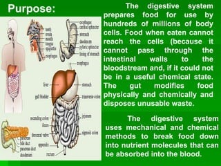
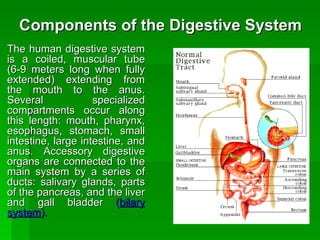
The digestive system prepares food for use by hundreds of millions of body cells. Food when eaten cannot reach the cells (because it cannot pass through the intestinal walls to the bloodstream and, if it could not be in a useful chemical state. The gut modifies food physically and chemically and disposes unusable waste.   Purpose: The digestive system uses mechanical and chemical methods to break food down into nutrient molecules that can be absorbed into the blood.
Types of Digestive System in Animals There are two types of plans and locations of digestion : Sac-like plans  are found in many invertebrates, who have a single opening for food intake and the discharge of wastes.   Coelenteronic:  incomplete digestive system, having a single opening that serves as the mouth and the anus. OR Vertebrates use the more efficient  tube-within-a-tube-plan  with food entering through one opening (the mouth) and wastes leaving through another (the anus). OR Enteronic:  complete type of digestive system, having an anterior opening called mouth and posterior opening called anus
Types of Digestion Intracellular Digestion:  food is taken into cells by phagocytosis with digestive enzymes being secreted into the phagocytic vesicles occurs in sponges, coelenterates and most protozoans. Extracellular Digestion:  occurs in the lumen (opening) of the digestive system, with the nutrient molecules being transferred to the blood or body fluid; occurs in chordates, annelids, and crustaceans.
Digestion  is the process of breaking down food into molecules small enough for the body to absorb. Proteins, carbohydrates, and fat in our diets must be broken down and later, reassembled in forms useful to our body. herbivores:  organisms which feed on plants carnivores:  organisms which feed on flesh of animals omnivores:  organisms which feed on both plants and animals
Stages in the Digestive Process   movement:  propels food through the digestive system  secretion : release of digestive juices in response to a specific stimulus  digestion : breakdown of food into molecular components small enough to cross the plasma membrane  absorption : passage of the molecules into the body's interior and their passage throughout the body  elimination:  removal of undigested
Components of the Digestive System  The human digestive system is a coiled, muscular tube (6-9 meters long when fully extended) extending from the mouth to the anus. Several specialized compartments occur along this length: mouth, pharynx, esophagus, stomach, small intestine, large intestine, and anus. Accessory digestive organs are connected to the main system by a series of ducts: salivary glands, parts of the pancreas, and the liver and gall bladder  ( bilary system ).
The Anatomy and Physiology of Digestion Food enters the digestive system via the mouth or oral cavity, mucous membrane lined. The lips (labia) protect its outer opening, cheeks form lateral walls, hard palate and soft palate form anterior / posterior roof. The floor of the mouth is the muscular tongue. Food is first processed (bitten off) by teeth, especially the anterior incisors. Suitably sized portions then retained in closed mouth and chewed or masticated (especially by cheek teeth, premolars, molars) aided by saliva Ducted salivary glands open at various points into mouth. This process involves teeth (muscles of mastication move jaws) and tongue (extrinsic and intrinsic muscles  Mouth and Teeth
Structure of a tooth Enamel:  material covering the crown which is the hardest substance in the body Crown:  portion above the gum Dentine:  bulk of the teeth Root:  portion embedded in a socket of jawbone Neck:  slightly constricted region between the root and the crown and is covered by the gum or gingiva Periodontal membrane:  covers the roof and hold the tooth firmly in the socket Pulp:  consists of connective tissue containing a dense network of capillaries, nerves, and lymph vessels Gingiva 6 Pulp cavity 11 Neck 5 Apex/ical foramen 10 Gingival sulcus 4 Alveolar bone 9 Dentine 3 Root 8 Crown 2 Periodontal 7 Enamel 1 Name Number Name Number
Swallowing Swallowing moves food from the mouth through the pharynx into the esophagus and then to the stomach. Step 1:  A mass of chewed, moistened food, a bolus, is moved to the back of the moth by the tongue. In the pharynx, the bolus triggers an involuntary swallowing reflex that prevents food from entering the lungs, and directs the bolus into the esophagus.  Step 2:  Muscles in the esophagus propel the bolus by waves of involuntary muscular contractions  (peristalsis)  of smooth muscle lining the esophagus.  Step 3:  The bolus passes through the gastroesophogeal sphincter, into the stomach. Heartburn results from irritation of the esophagus by gastric juices that leak through this sphincter.
Structure of the throat and the mechanics of swallowing.
Peristalsis and the movement of food from the mouth to the stomach.  The muscles in the esophagus squeeze the food downward with a wavelike motion, a process referred to as  peristalsis .
Esophagus The esophagus (about 10") is the first part of the digestive tract proper and shares its distinctive structure.  Basic tissue layers of the gut are: mucosa.  Innermost, moist lining membrane. Epithelium (friction resistant stratified squamous in esophagus, simple beyond) plus a little connective tissue and smooth muscle.  submucosa.  Soft connective tissue layer, blood vessels, nerves, lymphatics  muscularis externa.  Typically circular inner layer, longitudinal outer layer of smooth muscle  serosal fluid producing single layer.
Stomach The stomach is a J-shaped, expandable sack, normally on the left side of the upper abdomen. Several muscle layers surround the stomach, serving to churn food. The stomach can expand to hold about 2 L of food (= 1/2 gal). The stomach contains  hydrochloric acid  (HCl ) strong enough to dissolve metal (pH about 1.5 to 3, usually around 2), which kills bacteria and helps denature the proteins in our food, making them more vulnerable to attack by pepsin. The stomach secretes mucus to protect itself from being digested by its own acid and enzymes. The stomach also manufactures  pepsin , an enzyme to digest protein.
 
Summary The digestive process begins with the senses. When we see and smell food, especially when we are hungry, our mouths begin to "water." Saliva is secreted into the mouth from salivary glands located in the cheeks and jaw. Saliva contains the first enzymes that will act upon our food.  The first step of digestion is  mastication ,  the chewing process. The food particles must be broken down into very tiny pieces and mixed with saliva in order for the enzymes to do their job. Many digestive disturbances could be avoided with proper chewing. In our fast-paced society most people eat too fast and swallow their food without proper mastication. When chewed properly food will be in an almost liquid form when swallowed. From the mouth, the food moves to the stomach by way of the esophagus, a muscular tube which passes through a hole in the diaphragm to enter the abdominal area. The muscles in the esophagus squeeze the food downward with a wavelike motion, a process referred to as  peristalsis . Because of this muscular action, food can be swallowed in zero gravity, or even when the body is upside down.
Whereas the pH (acidity) in the mouth and esophagus is very basic (high pH), the pH in the stomach is very acidic (low pH). This low or acid pH is the result of the secretion of hydrochloric acid (HCL), and is necessary for the action of the enzymes in the stomach that are responsible for breaking down protein. The stomach is protected from this acid by a mucus lining. The stomach acid is kept out of the esophagus by the action of the  cardiac valve , a sphincter muscle which opens when food passes through, but which immediately closes to prevent the contents of the stomach from backing up into the esophagus.  Another sphincter valve, the  pyloric sphincter , keeps the food in the stomach until it has been properly churned and mixed. Once the enzymes that require an acid environment have had time to do their job, the pyloric sphincter opens and the food passes into the small intestines where most of the absorption of nutrients takes place.
 

Digestive features

  • 1.
  • 2.
    The digestive systemprepares food for use by hundreds of millions of body cells. Food when eaten cannot reach the cells (because it cannot pass through the intestinal walls to the bloodstream and, if it could not be in a useful chemical state. The gut modifies food physically and chemically and disposes unusable waste. Purpose: The digestive system uses mechanical and chemical methods to break food down into nutrient molecules that can be absorbed into the blood.
  • 3.
    Types of DigestiveSystem in Animals There are two types of plans and locations of digestion : Sac-like plans are found in many invertebrates, who have a single opening for food intake and the discharge of wastes. Coelenteronic: incomplete digestive system, having a single opening that serves as the mouth and the anus. OR Vertebrates use the more efficient tube-within-a-tube-plan with food entering through one opening (the mouth) and wastes leaving through another (the anus). OR Enteronic: complete type of digestive system, having an anterior opening called mouth and posterior opening called anus
  • 4.
    Types of DigestionIntracellular Digestion: food is taken into cells by phagocytosis with digestive enzymes being secreted into the phagocytic vesicles occurs in sponges, coelenterates and most protozoans. Extracellular Digestion: occurs in the lumen (opening) of the digestive system, with the nutrient molecules being transferred to the blood or body fluid; occurs in chordates, annelids, and crustaceans.
  • 5.
    Digestion isthe process of breaking down food into molecules small enough for the body to absorb. Proteins, carbohydrates, and fat in our diets must be broken down and later, reassembled in forms useful to our body. herbivores: organisms which feed on plants carnivores: organisms which feed on flesh of animals omnivores: organisms which feed on both plants and animals
  • 6.
    Stages in theDigestive Process movement: propels food through the digestive system secretion : release of digestive juices in response to a specific stimulus digestion : breakdown of food into molecular components small enough to cross the plasma membrane absorption : passage of the molecules into the body's interior and their passage throughout the body elimination: removal of undigested
  • 7.
    Components of theDigestive System The human digestive system is a coiled, muscular tube (6-9 meters long when fully extended) extending from the mouth to the anus. Several specialized compartments occur along this length: mouth, pharynx, esophagus, stomach, small intestine, large intestine, and anus. Accessory digestive organs are connected to the main system by a series of ducts: salivary glands, parts of the pancreas, and the liver and gall bladder ( bilary system ).
  • 8.
    The Anatomy andPhysiology of Digestion Food enters the digestive system via the mouth or oral cavity, mucous membrane lined. The lips (labia) protect its outer opening, cheeks form lateral walls, hard palate and soft palate form anterior / posterior roof. The floor of the mouth is the muscular tongue. Food is first processed (bitten off) by teeth, especially the anterior incisors. Suitably sized portions then retained in closed mouth and chewed or masticated (especially by cheek teeth, premolars, molars) aided by saliva Ducted salivary glands open at various points into mouth. This process involves teeth (muscles of mastication move jaws) and tongue (extrinsic and intrinsic muscles Mouth and Teeth
  • 9.
    Structure of atooth Enamel: material covering the crown which is the hardest substance in the body Crown: portion above the gum Dentine: bulk of the teeth Root: portion embedded in a socket of jawbone Neck: slightly constricted region between the root and the crown and is covered by the gum or gingiva Periodontal membrane: covers the roof and hold the tooth firmly in the socket Pulp: consists of connective tissue containing a dense network of capillaries, nerves, and lymph vessels Gingiva 6 Pulp cavity 11 Neck 5 Apex/ical foramen 10 Gingival sulcus 4 Alveolar bone 9 Dentine 3 Root 8 Crown 2 Periodontal 7 Enamel 1 Name Number Name Number
  • 10.
    Swallowing Swallowing movesfood from the mouth through the pharynx into the esophagus and then to the stomach. Step 1: A mass of chewed, moistened food, a bolus, is moved to the back of the moth by the tongue. In the pharynx, the bolus triggers an involuntary swallowing reflex that prevents food from entering the lungs, and directs the bolus into the esophagus. Step 2: Muscles in the esophagus propel the bolus by waves of involuntary muscular contractions (peristalsis) of smooth muscle lining the esophagus. Step 3: The bolus passes through the gastroesophogeal sphincter, into the stomach. Heartburn results from irritation of the esophagus by gastric juices that leak through this sphincter.
  • 11.
    Structure of thethroat and the mechanics of swallowing.
  • 12.
    Peristalsis and themovement of food from the mouth to the stomach. The muscles in the esophagus squeeze the food downward with a wavelike motion, a process referred to as peristalsis .
  • 13.
    Esophagus The esophagus(about 10") is the first part of the digestive tract proper and shares its distinctive structure. Basic tissue layers of the gut are: mucosa. Innermost, moist lining membrane. Epithelium (friction resistant stratified squamous in esophagus, simple beyond) plus a little connective tissue and smooth muscle. submucosa. Soft connective tissue layer, blood vessels, nerves, lymphatics muscularis externa. Typically circular inner layer, longitudinal outer layer of smooth muscle serosal fluid producing single layer.
  • 14.
    Stomach The stomachis a J-shaped, expandable sack, normally on the left side of the upper abdomen. Several muscle layers surround the stomach, serving to churn food. The stomach can expand to hold about 2 L of food (= 1/2 gal). The stomach contains hydrochloric acid (HCl ) strong enough to dissolve metal (pH about 1.5 to 3, usually around 2), which kills bacteria and helps denature the proteins in our food, making them more vulnerable to attack by pepsin. The stomach secretes mucus to protect itself from being digested by its own acid and enzymes. The stomach also manufactures pepsin , an enzyme to digest protein.
  • 15.
  • 16.
    Summary The digestiveprocess begins with the senses. When we see and smell food, especially when we are hungry, our mouths begin to "water." Saliva is secreted into the mouth from salivary glands located in the cheeks and jaw. Saliva contains the first enzymes that will act upon our food. The first step of digestion is mastication , the chewing process. The food particles must be broken down into very tiny pieces and mixed with saliva in order for the enzymes to do their job. Many digestive disturbances could be avoided with proper chewing. In our fast-paced society most people eat too fast and swallow their food without proper mastication. When chewed properly food will be in an almost liquid form when swallowed. From the mouth, the food moves to the stomach by way of the esophagus, a muscular tube which passes through a hole in the diaphragm to enter the abdominal area. The muscles in the esophagus squeeze the food downward with a wavelike motion, a process referred to as peristalsis . Because of this muscular action, food can be swallowed in zero gravity, or even when the body is upside down.
  • 17.
    Whereas the pH(acidity) in the mouth and esophagus is very basic (high pH), the pH in the stomach is very acidic (low pH). This low or acid pH is the result of the secretion of hydrochloric acid (HCL), and is necessary for the action of the enzymes in the stomach that are responsible for breaking down protein. The stomach is protected from this acid by a mucus lining. The stomach acid is kept out of the esophagus by the action of the cardiac valve , a sphincter muscle which opens when food passes through, but which immediately closes to prevent the contents of the stomach from backing up into the esophagus. Another sphincter valve, the pyloric sphincter , keeps the food in the stomach until it has been properly churned and mixed. Once the enzymes that require an acid environment have had time to do their job, the pyloric sphincter opens and the food passes into the small intestines where most of the absorption of nutrients takes place.
  • 18.