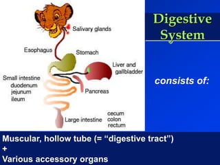
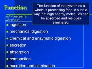
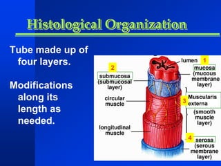
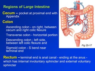
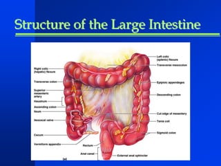
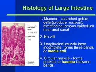
Digestive System 
Objectives: 
Discuss the general functions and anatomy 
of the digestive tract 
Describe the individual organs of the 
system, including a discussion of the gross 
and microscopic anatomy.
Digestive 
System 
consists of: 
Muscular, hollow tube (= “digestive tract”) 
+ 
Various accessory organs
Function 
Individual parts 
function in: 
 ingestion 
The function of the system as a 
whole is processing food in such a 
way that high energy molecules can 
be absorbed and residues 
 mechanical digestion 
eliminated. 
 chemical and enzymatic digestion 
 secretion 
 absorption 
 compaction 
 excretion and elimination
Muscularis 
externa 
Histological Organization 
Tube made up of 
four layers. 
Modifications 
along its 
length as 
needed. 
1 
2 
3 
4
The 4 Layers of the Gut 
1) Mucosa 
Epithelial cells – protection, absorption 
2) Submucosa – made up of loose connective tissue 
contains submucosal plexus and blood vessels 
3) Muscularis externa – smooth muscle, usually two layers - 
outer layer: longitudinal 
inner layer: circular 
4) Serosa 
outer layer, functions in protection
Ingestion 
 Ingestion is the first 
step in the process 
of digestion. 
 Ingestion means 
that food is taken 
into the mouth, 
chewed, and 
swallowed.
Oral Cavity 
 Also called the mouth, the oral 
cavity is the place where 
ingestion happens. 
 Hard and soft palates - form 
roof of mouth 
 Tongue - skeletal muscle 
 Salivary glands - three pairs 
 Teeth
Oral Cavity 
Digestion starts here by two 
means: 
 Mechanical – teeth tear, 
shred, and grind food. 
 Chemical – enzymes in 
saliva break down food 
molecules such as 
proteins and complex 
sugars.
Structure of Teeth 
Crown - exposed surface of tooth 
Neck - boundary between root and crown 
Enamel - outer surface 
Dentin – bone-like, but noncellular 
Pulp cavity - hollow with blood vessels and 
nerves 
Root canal - canal length of root
Types and Numbers of Teeth 
Dental succession: 
Deciduous (baby, primary) teeth - 20, replaced by 
Permanent teeth - 32 teeth
Types and Numbers of Teeth 
 All teeth are formed 
before milk teeth or 
primary teeth are lost. 
 It is the action of adult 
teeth moving into 
place that causes 
primary teeth to fall 
out.
Three pairs of Salivary Glands 
Salivary glands help with: 
• digestion 
• lubrication (swallowing) 
• moistening (tasting) 
They secrete enzyme-filled 
saliva to help break down 
food
Ingestion 
 The tongue is a 
muscle that pushes 
your food toward 
teeth to be 
mechanically 
processed and 
shapes your food into 
small rounded pieces 
to be swallowed.
Swallowing 
As you swallow, your 
tongue pushes the food to 
the back of the mouth. 
Since both air and food go 
through the pharynx, a flap 
of tissue called the 
epiglottis covers the larynx 
to prevent food from 
travelling into the lungs
Lesser curvature 
Greater curvature 
Cardia - end under the heart 
Fundus - bulge above the esophageal opening 
Body - largest region 
Pylorus - J curve, inferior end, terminates in 
Cardiac and Pyloric sphincters (importance?) 
Rugae – highly extendable interior folds 
Figs 25-10/11 
Gross Anatomy of the Stomach
Histology of Stomach 
Type of epithelium lining stomach? 
Fig 25.13 
Gastric pits – shallow pits, external half rapidly reproduces 
for replacement 
Gastric glands – deep in lamina propria, 3 types of cells 
1. Parietal cells (produce HCl and intrinsic factor) 
2. Chief cells (produce pepsinogen) 
3. Enteroendocrine cells – G cells (several hormones 
including gastrin which stimulates both parietal and chief cells)
Regions of Small Intestine 
SI is longest part of dig. tube 
 Duodenum (short, 12 inches) 
– fixed shape & position 
 Jejunum (2.5 m long) 
– Most of digestion 
 Ileum (longest at 3.5 m) 
– Most of absorption, ends in 
 Ileocecal valve – slit valve into large intestine (colon)
Structure of Small Intestinal Wall Fig 25.15 
Plicae circulares – circular pleats around the interior of the 
small intestine. Slows movement of food, increases surface 
area of intestine. 
Villi – minute finger-like projections, contain capillaries & 
lacteals. Further increase surface area. 
Microvilli – sub-microscopic size, projections on single cells. 
Aid in absorption of nutrients. 
Intestinal glands (crypts) 
- intestinal juice production 
- Cell regeneration
Histology of Small Intestinal Wall
Histology of S.I. Wall (cont)
Regions of Large Intestine 
Cecum – pocket at proximal end with 
Appendix 
Colon 
Ascending colon - on right, between 
cecum and right colic flexure 
Transverse colon - horizontal portion 
Descending colon - left side, 
between left colic flexure and 
Sigmoid colon - S bend near 
terminal end 
Fig 25-17 
Rectum – terminal end is anal canal - ending at the anus - 
which has internal involuntary sphincter and external voluntary 
sphincter
Structure of the Large Intestine
Histology of Large Intestine 
1. Mucosa - abundant goblet 
cells (produce mucous), 
stratified squamous epithelium 
near anal canal 
2. No villi 
3. Longitudinal muscle layer 
incomplete, forms three bands 
or taenia coli 
4. Circular muscle - forms 
pockets or haustra between 
bands

Digestive Tract

  • 1.
    Digestive System Objectives: Discuss the general functions and anatomy of the digestive tract Describe the individual organs of the system, including a discussion of the gross and microscopic anatomy.
  • 2.
    Digestive System consistsof: Muscular, hollow tube (= “digestive tract”) + Various accessory organs
  • 3.
    Function Individual parts function in:  ingestion The function of the system as a whole is processing food in such a way that high energy molecules can be absorbed and residues  mechanical digestion eliminated.  chemical and enzymatic digestion  secretion  absorption  compaction  excretion and elimination
  • 4.
    Muscularis externa HistologicalOrganization Tube made up of four layers. Modifications along its length as needed. 1 2 3 4
  • 5.
    The 4 Layersof the Gut 1) Mucosa Epithelial cells – protection, absorption 2) Submucosa – made up of loose connective tissue contains submucosal plexus and blood vessels 3) Muscularis externa – smooth muscle, usually two layers - outer layer: longitudinal inner layer: circular 4) Serosa outer layer, functions in protection
  • 6.
    Ingestion  Ingestionis the first step in the process of digestion.  Ingestion means that food is taken into the mouth, chewed, and swallowed.
  • 7.
    Oral Cavity Also called the mouth, the oral cavity is the place where ingestion happens.  Hard and soft palates - form roof of mouth  Tongue - skeletal muscle  Salivary glands - three pairs  Teeth
  • 8.
    Oral Cavity Digestionstarts here by two means:  Mechanical – teeth tear, shred, and grind food.  Chemical – enzymes in saliva break down food molecules such as proteins and complex sugars.
  • 9.
    Structure of Teeth Crown - exposed surface of tooth Neck - boundary between root and crown Enamel - outer surface Dentin – bone-like, but noncellular Pulp cavity - hollow with blood vessels and nerves Root canal - canal length of root
  • 10.
    Types and Numbersof Teeth Dental succession: Deciduous (baby, primary) teeth - 20, replaced by Permanent teeth - 32 teeth
  • 11.
    Types and Numbersof Teeth  All teeth are formed before milk teeth or primary teeth are lost.  It is the action of adult teeth moving into place that causes primary teeth to fall out.
  • 12.
    Three pairs ofSalivary Glands Salivary glands help with: • digestion • lubrication (swallowing) • moistening (tasting) They secrete enzyme-filled saliva to help break down food
  • 13.
    Ingestion  Thetongue is a muscle that pushes your food toward teeth to be mechanically processed and shapes your food into small rounded pieces to be swallowed.
  • 14.
    Swallowing As youswallow, your tongue pushes the food to the back of the mouth. Since both air and food go through the pharynx, a flap of tissue called the epiglottis covers the larynx to prevent food from travelling into the lungs
  • 15.
    Lesser curvature Greatercurvature Cardia - end under the heart Fundus - bulge above the esophageal opening Body - largest region Pylorus - J curve, inferior end, terminates in Cardiac and Pyloric sphincters (importance?) Rugae – highly extendable interior folds Figs 25-10/11 Gross Anatomy of the Stomach
  • 16.
    Histology of Stomach Type of epithelium lining stomach? Fig 25.13 Gastric pits – shallow pits, external half rapidly reproduces for replacement Gastric glands – deep in lamina propria, 3 types of cells 1. Parietal cells (produce HCl and intrinsic factor) 2. Chief cells (produce pepsinogen) 3. Enteroendocrine cells – G cells (several hormones including gastrin which stimulates both parietal and chief cells)
  • 17.
    Regions of SmallIntestine SI is longest part of dig. tube  Duodenum (short, 12 inches) – fixed shape & position  Jejunum (2.5 m long) – Most of digestion  Ileum (longest at 3.5 m) – Most of absorption, ends in  Ileocecal valve – slit valve into large intestine (colon)
  • 18.
    Structure of SmallIntestinal Wall Fig 25.15 Plicae circulares – circular pleats around the interior of the small intestine. Slows movement of food, increases surface area of intestine. Villi – minute finger-like projections, contain capillaries & lacteals. Further increase surface area. Microvilli – sub-microscopic size, projections on single cells. Aid in absorption of nutrients. Intestinal glands (crypts) - intestinal juice production - Cell regeneration
  • 19.
    Histology of SmallIntestinal Wall
  • 20.
    Histology of S.I.Wall (cont)
  • 21.
    Regions of LargeIntestine Cecum – pocket at proximal end with Appendix Colon Ascending colon - on right, between cecum and right colic flexure Transverse colon - horizontal portion Descending colon - left side, between left colic flexure and Sigmoid colon - S bend near terminal end Fig 25-17 Rectum – terminal end is anal canal - ending at the anus - which has internal involuntary sphincter and external voluntary sphincter
  • 22.
    Structure of theLarge Intestine
  • 23.
    Histology of LargeIntestine 1. Mucosa - abundant goblet cells (produce mucous), stratified squamous epithelium near anal canal 2. No villi 3. Longitudinal muscle layer incomplete, forms three bands or taenia coli 4. Circular muscle - forms pockets or haustra between bands

Editor's Notes

  • #10 Enamel: Calcium phosphates. Hardest biological material produced.