SlideShare a Scribd company logo
1 of 111
Download to read offline
UNIT 1 - DIGESTIVE SYSTEM I
UNIT 2 - DIGESTIVE SYSTEM II
UNIT 3 - DIGESTIVE SYSTEM III
UNIT 4 - URINARY SYSTEM I
UNIT 5 - URINARY SYSTEM II
HUMAN PHYSIOLOGY: DIGESTION AND
ABSORPTION, EXCRETORY PRODUCTS AND THEIR
ELIMINATION
3
1.1 INTRODUCTION TO THE DIGESTIVE SYSTEM
1.1.1 Main functions
 Moves water, nutrients and electrolytes
from the external environment to the
internal environment.
 Breaks down food moving through the
tract into its chemical building blocks.
 Absorbs these building blocks into the
blood.
 Eliminates residual indigestible materials.
1.1.2 Basic divisions
Alimentary canal
 Continuous muscular tube that runs from
the mouth to the anus.
 Consists of mouth, pharynx, esophagus,
stomach, small intestine and large intestine.
Accessory organs
 Contribute to secretions to the GI tract, but the food doesn’t pass through these organs.
 Consists of teeth, tongue, salivary glands, liver, gall bladder and pancreas.
1.1.3 Actions of the gastrointestinal (GI) tract
 Ingestion: Occurs when material enters via the mouth.
 Mechanical processing: Crushing / shearing make material easier to move through the tract.
 Digestion: Chemical breakdown of food into small organic compounds for absorption.
 Secretion: Release of water acids, buffers, enzymes and salts by epithelium of GI tract and
glandular organs.
 Absorption: Movement of organic substrates, electrolytes, vitamins and water across digestive
epithelium.
 Excretion: Removal of waste products from body fluids.
1.1.4 Movement of digestive materials
A. Deglutition
Deglutition is the movement of food from the mouth and into the stomach. The entire process
takes about four to eight seconds for solid or semisolid food and about one second for every soft
food and liquids. Deglutition involves the mouth, pharynx and esophagus. It is facilitated by the
secretion of mucus and saliva. There are three stages in deglutition: the voluntary phase, the
pharyngeal phase, and the esophageal phase.
UNIT 1 - DIGESTIVE SYSTEM I
Mouth
Esophagus
Liver
Gall bladder
Duodenum
Transverse
colon
Ascending
colon
Anus
Ileum
Descending
colon
Jejunum
Pancreas
Stomach
Parotid gland
Salivary glands
4
Tongue
Pharynx
Windpipe
(trachea)
Esophagus
Larynx
closes
Food
Soft
palate
Vertebrae
Epiglottis
Swallowing
B. Peristalsis
The smooth muscle of the GI tract is arranged in two layers: longitudinal along the length of the
tube and circular around the diameter of the tube. Coordinated contraction and relaxation of these
two layers create wavelike propulsion of the food called peristalsis.
Initial state
Longitudinal
muscle
Circular
muscle
From
mouth
To
anus
Contraction
Contraction of
circular muscles
behind bolus
Step 1
Contraction of
longitudinal
muscles ahead
of bolus
Contraction
Contraction
Step 2
Contraction in
circular muscle
layer forces
bolus forward
Step 3
1.1.5 Levels of organization
 Chemical level – Sodium, potassium, chloride, bicarbonate, and phosphate ions and
hydrochloric acid.
 Macromolecular level – Enzymes of digestion, bile, and the nutritive molecules such as
carbohydrates, proteins, lipids and nucleic acids.
 Cellular level – Mucosa cells, secretory cells and immune cells.
 Tissue level – Epithelial, connective and smooth muscle tissues.
 Organ level – Gastrointestinal tract and accessory organs.
 Organ system level – Integration of organs for nutrients and waste movement, digestion
and absorption.
5
1.1.6 Nutritive molecules
The nutritive organic compounds in our food include carbohydrates, proteins and lipids. These
molecules are digested, then absorbed, and reassembled into macromolecules or used as fuel for
metabolism in the body. The process of breaking down these macromolecules involves splitting into
smaller molecules using water molecule, thus the name hydrolysis. The reverse reaction, called
dehydration synthesis, can build macromolecules from the absorbed building blocks. Enzymes can
speed up the rate of these reactions.
A. Carbohydrates: Our daily food intake usually includes from 200 to 600 grams of carbohydrates.
Monosaccharides are absorbed easily and can therefore be a source of quick energy for the body
when we consume them in this form. Glucose, galactose, and fructose are the three common
monosaccharides in our diet. Disaccharides include sucrose (table sugar), lactose, and maltose.
Polysaccharides include polymerized glucose in different forms including glycogen, which is the
stored form of glucose in our bodies, and starch.
B. Proteins: USDA recommends 5 to 6 ounces of protein in diet per day, although children need
less. This dietary protein is usually in the form of polypeptides and must be digested into its amino
acid building blocks for absorption. There are numerous enzymes that break large proteins into
smaller peptides and then into amino acids. Amino acids are then absorbed from the digestive
system into the circulatory system where they are delivered throughout the body. Once amino acids
have entered cells throughout the body, they are bonded together to make proteins needed for cell
function. As a last resort, proteins and amino acids can also be used as energy; they are metabolically
converted to glucose before they are used as energy sources.
C. Lipids and Fats: Dietary lipids include fats and oils. While not considered a USDA food group,
oils contain some essential nutrients and are recommended as part of a healthy diet, although only
in small amounts. Solid fats have more saturated and trans-fatty acids and are considered empty
calories when included in a diet because they add calories but not needed nutrients. Most dietary
lipids are in the form of triglycerides. Lipids are processed by enzymes secreted from the pancreas
(with some enzymes from the stomach and saliva) and are then solubilized for absorption by salts
secreted in bile (from the liver). These steps prepare them for absorption in the small intestine. Like
proteins, ingested lipids are broken down into smaller parts for absorption and then are either
metabolized to make energy or are used to make cellular structures including cell membranes. Lipid
absorption is also required for absorption of some fat-soluble vitamins.
D. Nucleic acids: When we eat any plant or animal foods, we are able to digest the DNA or RNA
nucleic acid that was in their cells. The pancreas produces and releases into the small intestine two
enzymes to break down either DNA or RNA into individual nucleotides. Brush border enzymes of the
cells lining the small intestine further digest the nucleotide into its molecular components for
absorption.
6
1.2 ANATOMY OF THE ALIMENTARY CANAL
The alimentary canal is tubular structure which extendsfrom mouth to anus. It develops from ectoderm
and endoderm.
Ectoderm – Upto hard palate.
Endoderm – From soft palate to rectum.
Ectoderm – from anal canal to anus.
1.2.1 Mouth
Mouth is a horizontal transverse slit
like aperture which is surrounded
by upper and lower lip; a specific
muscle is associated with lip called
orbicularis oris muscle.
Serous glands are found on the
inner part of lip. Serous glands are
the modification of mucus glands.
Its secretory substance is watery.
Mouth opens into Buccopharyngeal
cavity. Thiscavity isdivided into two
parts.
A. Buccal vestibule – The space between the gums and cheeks where the food is stored temporarily
for some time. It is a peripheral part.
B. Oral cavity – It is inner and central part which is surrounded by upper and lower jaw. Lined by
stratified squamous epithelium. Upper jaw is fixed and lower jaw is movable. The roof of oral cavity
is called as Palate.
Palate is differentiated into two parts:
(i) Hard palate – It is the anterior part of the palate. It is made up of maxilla and palatine bone in
human. But in rabbit it is made of premaxilla, maxilla, palatine bone.
On the ventral surface of hard palate, some projection or transverse ridges are present which
are called as palatine rugae.
These rugae prevent slip out of the food from buccal cavity during mastication; these rugae
are well developed in carnivorous animals.
(ii) Soft palate – It is the posterior part of palate. It is made up of involuntary muscle fibrous
connective tissue and mucous epithelium. (Stratified squamous epithelium).
Upper lip
Superior labial
frenulum
Glossopalatine arch
Pharyngopalatine arch
Palatine tonsil
Salivary duct orifices
Sublingual
Submandibular
Inferior labial frenulum
Lower lip
Hard palate and
transverse
palatine folds
Soft palate
Uvula
Fauces
Tongue
Lingual frenulum
Teeth
Gingivae
7
The posterior out growth of soft palate which
hangs down in the form of finger like process
called as uvula or velum palati. On the dorsal
side of uvula internal nasal pores are present.
One pair of large lymph node is present on
the posterolateral surface of soft palate, called
as palatine tonsil or tonsils.
Soft palate is situated in the pharynx and is
divided into two parts. Upper part of pharynx
is called nasopharynx which is related to the nasal chamber. The lower part of pharynx is
called oropharynx which is related to the oral cavity. One pair of openings of eustachian
tube is present in the nasopharynx. This Eustachian tube is related to the middle ear, it
maintains air pressure.
TARGET POINTS
 In rabbit, a small cleft is found in the middle part of upper lip, such type of lip is called as Hare
lip. One pair opening of nasopalatine duct is present at the anterior part of hard palate, these
connect the buccal cavity to the nasal passage. In rabbit, some olfactory receptors are also
found in nasopalatine duct which are called as Jacobson’s organ or vomero nasal organ. It
makes them aware of the smell of food while chewing.
 Jacobson’s organ is well developed in reptiles like- snake, lizard and sphenodon.
 Uvula covers the opening of internal nasal pores during ingestion of food, so food particles
cannot move inside nasal chamber.
A. Tongue
On the floor of oral cavity a muscular, flat, fleshy plate
like structure is present which is called tongue. The
anterior part of tongue is free while posterior part of
tongue is connected to the hyoid bone.
The ventral surface of tongue is connected to the floor
of buccal cavity through a very flexible membrane /
ligamentous fold called as Frenulum linguae.
The dorsal surface of tongue is divided into two
unequal parts by a V shaped sulcus, called as sulcus
terminalis.
The two limbs of the ‘V’ meet at a median pit named
Foramen caecum. It is divided into two parts:
(i) Pharyngeal part – It is the poster 1/3 part of
the tongue. Many small lymph nodes are present in this part which are called lingual tonsil.
Mouth
Hard palate
Soft palate
Uvula
Larynx
(with vocal cords)
Trachea (windpide)
Pharynx
Epiglottis
Esophagus
Median
sulcus
Vallate
papillae
Terminal
sulcus
Palatine
tonsil
Lingual
follicles
Root
Median
glossoepiglottic
fold
Epiglottis
Apex
Filiform
papillae
Fungiform
papillae
Palatoglossal
arch
Foramen
cecum
Lingual tonsil
Palatopharyngeal
arch
Upper surface of human tongue
8
(ii) Oral or papillary part – It is anterior 2/3 part of tongue. Four types of papillae are found in
this part in which gustatory or taste receptors are present in the form of taste buds.
Fungiform papillae: It is pink coloured, small and spherical in shape. It is found on the entire
surface of tongue but mostly present at the anterior part of tongue. It is attached to tongue with the
help of small pedicle. It provides pink colour of the tongue.
Filiform papillae (conical papillae): They are thread like, white coloured and conical in shape. They
are also found on the entire surface of tongue. They are most numerous, but devoid of taste buds.
Foliate papillae: They are found on the mid lateral surface of tongue. They are vestigeal in the
human. Their structure is leaf like present in rabbit and other mammals.
Circumvallate papillae: It is the largest and least existed papillae (8 to 12), they are large spherical
shaped papillae which are found near to sulcus terminalis.
Taste pore Circumvallate
Foliate
Taste
buds
Fungiform
TRC
a
b
Bitter Salty Sweet Umami Sour
Taste centers in tongue
Two types of muscles are present in tongue:
(i) Extrinsic muscle: Found on outer and superficial part of tongue, helps in it’s outward and
inward movement.
(ii) Intrinsic muscle: Situated in the deep part of tongue, helps in the change of shape of tongue.
9
B. Teeth
Teeth are ectomesodermal in origin.
Major portion of teeth arises from
dermis. Part of toothe present outside
the gumsonly is derived from ectoderm
or epidermis (Enamel part).
In human teeth of upper jaw are
attached to the maxilla bone. While
teeth of lower jaw are attached to
mandible bone. But in rabbit upper
incisors are attached to premaxilla.
While upper pre molars and molars
attached to the maxilla bone. While
lower teeth attached to dentry bone.
Teeth are differentiated in three parts:
The crown part of the tooth is made up of very hard substances called the Enamel. It is the hardest
material in all animal of animal kingdom.
Enamel is ectodermal. It is secreted by ameloblast cells of the ectoderm. It has maximum amount
of inorganic salt (96%) in it. Inorganic salt are mainly found in the form of phosphate and carbonate
of Ca, Mg, Na and K. 3% of water is found in the enamel. Along with it keratin and ossein protein
(1%) are also found in teeth. Ossein is a protein of bones. Remaining part of teeth develop from
mesoderm of embryo.
Dentine is the main part of tooth. Approximately 69% inorganic salts are present in dentine where as
in cemented layer 65% is inorganic salt (62% inorganic salts are present in bones).
Dentine surrounds a cavity called pulp cavity. This cavity contains soft connective tissue, blood
capillaries and nerve fibers. Pulp cavity is necessary for the nutrition and survival of the teeth. At the
base of pulp cavity an aperture is present. Through this aperture, blood capillaries and nerve fibers
enter inside the teeth. This aperture is called apical-foramen.
A special type of cells form the lining of the pulp-cavity called the odontoblasts cells. These cells
are the dentine secreting cells. Cytoplasmic processes of odontoblasts are embedded into dentine
in the form of fine tubule. These processes are called canaliculi. These canaliculi secrete dentine.
The teeth continue to grow till the odontoblasts cells remain active. In adults, the pulp cavity shrinks
and the odontoblasts become inactive so the teeth stop to grow.
Cementum
PDL
Alveolar bone
Sharpey’s fibers
Pulp cavity
Enamel
Dentin
Gingiva
Cementum
Periodontal
ligament
Root canal
Alveolar bone
Apical foramen
Alveolar vessels
and nerves
Sagittal section of a mandibular (lower) molar
Attachment
organ
Crown It is the outer part of the tooth, exposed outside gums.
Neck It is the internal part of the tooth which is embedded inside the gums.
Root It is the part of tooth that is inserted inside the socket of jaw bone (Alveoli).
10
The cement layer is made up of the cementocyte
cells. Between the root and the bones of the teeth,
a periodontal membrane is present.
Humans have two sets of teeth (dentitions). The
20 deciduous teeth, or baby teeth, first appear at
about 6 months of age. Between ages 6 and 12
years, these teeth are replaced by the 32
permanent teeth. Closest to the midline are the two
incisors, chisel shaped teeth we use for cutting into
food. The cuspids (canines) are next to the incisors.
A cuspid’s pointed edge (cusp) tears and shreds
food. Posteriorly, the next teeth are the two
premolars (bicuspids) followed by up to three molars. Premolars and molars have broader, flatter
surfaces, which we use to crush and grind food.
Types of teeth on the basis of appearance in life
Jaw bone
with tooth
sockets
Tongue
Teeth roots
Line of lips
Molars
Premolars
Canine
Incisors
Specific teeth and structures of the mouth
Monophyodont The teeth which appear only once in life. Eg. Premolars and last molar of man.
Diphyodont The teeth which appear twice in life eg. Incisors, canines, molars.
Polyphyodont The teeth which appear more than twice in life eg. Fish, amphibians.
On the basis of position from jaw
Thecodont The teeth which are present in bony socket of jaw eg. Man and crocodile.
Pleurodont The teeth which are present on the lateral side of jaw bone eg. Reptiles.
Acrodont The teeth which are present on the terminal part of jaw bone eg. Fish, amphibian.
On the basis of structure and function
Heterodont When the teeth are of different type in mammals on the basis of structure and
function. Eg. Mammal.
Homodont Whether all teeth are of similar type in animal on the basis of structure and
function. Eg. Fish, amphibians.
On the basis of crown
Secodont These are canine teeth of carnivorous animals. In this type of structure canine
teeth become long and pointed which is bended towards the backward direction.
Hypsodont In this type of teeth the crown part is large root is small (such as incisor and
(smiling teeth) canine). These teeth are also called as smiling teeth.
11
Dental formula:
TARGET POINTS
 Carnessial teeth are modified last premolar of upper jaw and first molar of lower jaw, for shearing
and tearing of tendon.
 In rabbit and rat, the pulp cavity of the incisor remains wide throughout their life, so these teeth
grow continuously throughout their life span; these types of teeth are called open rooted teeth.
If one incisor or rabbit and rat is broken then the opposite incisor grows continuously, finally the
animal neither can close the mouth nor gnaw the food. So the animal dies due to starvation.
 In frog, only upper jaw has teeth.
 Hippocampus, tortoise and birds do not have teeth.
C. Salivary glands
Lying outside the oral mucosa are the three pairs of major salivary glands, which secrete the
majority of saliva into ducts that open into the mouth. The parotid glands lie between the skin and
the masseter muscle near the ears. They secrete saliva into the mouth through the parotid duct,
which is located near the seconds upper molar tooth. The submandibular glands, which are in the
floor of the mouth, secrete saliva into the mouth through the submandibular ducts near the lower
central incisor. The sublingual glands, which lie below the tongue, use the lesser sublingual
ducts to secrete saliva into the oral cavity.
Brachyodont In this type of teeth crown part is small root is long (such as premolar and
(cheek teeth) molar). Wisdom teeth – these are the last molar teeth of humans which appear
in the age of 18 to 25 years.The upper surface of premolar and molar is board.
Some small projections are present in the upper surface of premolar and molar.
These projections are called lophs or cusps.
On the basis of structure of lophs
Lophodont In this type of teeth the lophs are large, wide and flat such as rabbit and elephant.
Bunodont In this type of teeth. The lophs are small and spherical in shape, such as human.
Solenodont In this type of teeth the lophs are large and semilunar shape eg. Ruminant
animal (Cow, buffalo).
Carnessial In this type of teeth the Lophs are long and pointed eg. Carnivorous animal.
Child =
2 1 0 2 5 10
I C PM M = 2 = = 20
2 1 0 2 5 10
 Adult =
2 1 2 3 8 16
I C PM M = 2 = = 32
2 1 2 3 8 16

17 years old =
2 1 2 2 7
I C PM M = × 2 = 28
2 1 2 2 7
Rabbit =
2 0 3 3 8 16
I C PM M = 2 = = 28
1 0 2 3 6 12

12
Parotid glands are the largest salivary
glands.
Infections of the nasal passages and
pharynx can attack any salivary glands.
The parotid glands are the usual site of
infection with the virus that causes
mumps (Paramyxovirus). Mumps
manifest with enlargement and
inflammation of the parotid glands,
causing the characteristic swelling
between the ears and the jaw. Symptoms
include fever and throat pain, which can be severe when swallowing acidic substances such as
orange juice.
Wharton’s duct: These are the ducts of submaxillary or submandibular glands; the largest ducts.
Sublingual glands are the smallest salivary glands. Ducts arising from these glands are called as
the ducts of Rivinus or Bartholin’s ducts. Maximum saliva is secreted by the sublingual glands
(smallest salivary duct).
Saliva is mainly water, which dissolves chemicals in the food. Only dissolved chemicals can activate
the different kinds of taste receptor cells on the tongue, palate and other parts of the mouth and
pharynx. Mixed in with the water, saliva also contains mucus, various electrolytes typically found in
blood plasma, as well assome digestive molecules, metabolic waste products and immune molecules.
Each of these contributes to the various functions of saliva.
Bicarbonate and phosphate ions help maintain the pH of saliva as neutral or slightly basic (average
7.4 pH). This not only helps protect the teeth from acidic substances eaten and plaque bacteria that
grow best in an acidic environment, but also maintains the optimal pH for the enzyme amylase.
Waldeyer’s ring
The lymphatic tissues of the pharynx and oral cavity are arranged in a ring like manner, which are
collectively called Waldeyer’s ring (Waldeyer’s lymphatic ring). The ring mainly consists of the
following:
 Nasopharyngeal tonsil (Pharyngeal tonsil): Refer to the nasopharynx. In children
nasopharyngeal tonsil may become enlarged and referred asadenoids. The resulting swelling
may be a cause of obstruction to normal breathing.
 Tubal tonsil: Refer to the nasopharynx.
 Palatine tonsils (Faucial tonsils): Refer to the oropharynx. The palatine tonsils are often
infected (tonsillitis) leading to sore throat. Such enlarged tonsils may become a focus of
infection and their surgical removal (tonsillectomy) becomes necessary.
 Lingual tonsil: They are situated on posterior part of tongue.
Accessory
parotid gland
Parotid gland
Body of mandible
Submandibular gland
Submandibular
(Wharton’s) duct
Sublingual
gland
Parotid
duct
Opening of
submandibular
(Wharton’s)
duct
Salivary gland locations
13
Adenoid
Lingual tonsil
Palatine tonsil
(“tonsil”)
Tubal tonsil
Waldeyer's ring
Nodules on posterior
pharyngeal wall
Lateral
pharyngeal bond
Adenoids
Tubal tonsil
Palatine tonsil
Lingual tonsil
TARGET POINTS
 Salivary glands are exocrine glands.
 Ptyalin is secreted only by the parotid gland. Lysozyme and Thiocyanates mainly kill bacteria.
They also check the growth of bacteria in buccopharyngeal cavity.
 Salivation is stimulated by cranial nerve VII and IX.
1.2.2 Tissue layers of the GI tract
Throughout the gastrointestinal (GI) tract, walls are comprised of the same four fundamental tissue
layers. From the lumen of the GI tract, these layers are the mucosa, submucosa, muscularis and
serosa.
Serosa
Circular muscle
Longitudinal muscle
Submucosa
Meissner’s
nerve plexus
Mesentery
Submucosal gland
Myenteric nerve
plexus
Mucosal gland
Mucosal muscle
Epithelial lining
Mucosa
Microvillus
Mucosa
Submucosa
Muscularis
Serosa
Tissue layers of the GI tract Typical cross-section of the gut
Serosa: It is outermost layer of gut. It is called tunica adventitia in oesophagus, serosa is made up
of visceral peritoneum while tunica adventitia is made up of white fibrous connective tissue.
14
Muscle layer: It is formed by circular inner layer and longitudinal outer layer of smooth muscle.
Thickest layer is found in stomach (maximum peristalsis) and thinnest layer in rectum (minimum
peristalsis).
Submucosa: It is loose connective tissue layer with blood lymph vessels and nerves.
Mucosa: It is the inner most layer of gut which contains the secretory and absorptive cells.
It is differentiated into the following parts:
 Outer part: Called mucosa muscularis, it is made up of longitudinal and circular muscles. It
has important role in exposing of surface area for the absorption. They also provide support
to the folds of alimentary canal.
 Middle part: It is called lamina propria. It contains few modified lymphatic tissue which provides
immunity. Eg. Peyer’s patches. It is made up of reticulate and fibrous connective tissue.
 Innermost part: Called mucosal layer. In esophagus this layer is made up of non-keratinized
stratified squamous epithelium. Except esophagus this layer is single layer thick, which is
made up of columnar mucous epithelium. This layer makes the lining of lumen of alimentary
canal and makes the folds of alimentary canal.
Folds of esophagus are less developed, whereas folds of stomach are finger shaped and
develop as gland called gastric gland. Folds of small intestine are conical shaped called villi.
Small slit like space is found at the base of villi. These spaces are called crypts of Leiberkuhn.
Villi of duodenum are small blunt. Villi of jejunum and ileum are long and pointed. Maximum
villi are found jejunum.
 Brunner’s gland: They are small spherical multicellular glands. Open into crypts of Lieberkuhn
with the help of the tubules. These are found in the submucosa of duodenum, they synthesize
and secrete the non-enzymatic alkaline secretion of intestinal juice.
 Paneth cells: These cells are found in mucosal layer of crypts of Lieberkuhn of small intestine.
They are unicellular glands and secrete antibacterial substances hence it provides immunity.
They also synthesize and secretes enzymes of intestinal juices. The secretory substances of
Brunner’s glands and Paneth cells are combinedly called intestinal juice or succus entericus.
 Peyer’s patches: They are small lymph nodes which are found in the mucosa of small intestine
(jejunum and ileum more in number). They are also called intestinal tonsils and provide
immunity.
 Nerve supply: Two types of nerve plexus are found in muscle of alimentary canal.
– Auerbach’s nerve plexus (myentric plexus) this nerve plexus is found between longitudinal
muscles and circular muscles. It control muscles contraction.
– Meissner’s nerve plexus found between circular muscles and sub mucosa but in stomach
it is found between oblique/ muscle and submucosa. It helps in secretion.
15
1.2.3 Organs of the GI tract
A. Esophagus
Two apertures are found in central
part of buccopharyngeal cavity.
Ventral of lower aperture is called
glottis which is related to the larynx,
which is guarded by epiglottis. The
dorsal and upper aperture is called
gullet which opens into esophagus.
Esophagus is simple uniform tube
which runs downward and pierces
the diaphragm and finally opens into
stomach that site of piercing on diaphragm is called hiatus.
Longitudinal folds are found on the inner surface of esophagus in which digestive glands are absent,
only mucous glands are present in mucosa and submucosa.
Voluntary muscles are found on the upper 1/3rd
part of esophagus while mid 1/3rd
part is formed by
voluntary and involuntary muscles where as in lower 1/3rd
part of esophagus only involuntary muscles
are present. The length of esophagus depends on length of neck so the longest esophagus is
present in giraffe. It lacks serosa, but tunica adventitia is present.
B. Stomach
It is situated on left side of abdominal cavity. It is the widest part of alimentary canal. It is a bag
like muscular structure, J-shaped in empty condition.
The stomach contains four parts (cardiac, fundus, body, pylorus or antrum). It has two orifices
(opening).
(i) Cardiac orifice is joined by the lower end of the esophagus.
(ii) Pyloric orifice opens into the duodenum.
Stomach is covered by layer of peritoneum. Fat tissues and lymph tissue deposits on the peritoneum.
Such type of peritoneum are called ommentum.
Left curved surface of stomach is called greater ommentum. Right curved surface of stomach is
called lesser ommentum.
Mucous membrane of the stomach is thick. In empty stomach numerous temporary longitudinal
folds are found in mucosa of pyloric antrum called rugae. They disappear when stomach is distended.
Esophagus
Diaphragm
Stomach
Lower
esophageal
sphincter (LES)
Upper
esophageal
sphincter (UES)
Throat
(pharynx)
Trachea
16
Cardia
Fundus
Body
Pyloric antrum
Pyloric canal
Pylorus
Stomach anatomy
Esophagus
Muscularis
- Longitudinal layer
- Circular layer
- Oblique layer
Lesser curvature
Duodenum Pyloric sphincter (valve)
Greater
curvature
Rugae of
mucosa
Serosa
The stomach stores the food for 4 – 5 hours. The food mixes thoroughly with the acidic gastric juice
of the stomach by the churning movements of its muscular wall and is called the chyme. The mucus
and bicarbonates present in the gastric juice play an important role in lubrication and protection of
the mucosal epithelium from excoriation by the highly concentrated hydrochloric acid. HCl provides
the acidic pH (pH 1.8) optimal for pepsins. Rennin is a proteolytic enzyme found in gastric juice of
infants which helps in the digestion of milk proteins.
Gastric glands: These are numerous microscopic, simple tubular glands formed by the invagination
of epithelium in the stomach. The following types of cells are present in the epithelium of the gastric
glands.
Chief cells or peptic cells (Zymogen cells) are usually basal in location and secrete gastric
digestive enzymes as proenzymes or zymogens called pepsinogen and prorennin. The
chief cells also produce small amount of gastric amylase and gastric lipase. Gastric amylase
action is inhibited by the highly acid condition. Gastric lipase contributes little to digestion of
fat. Prorennin is secreted in young mammals (childhood stage). It is not secreted in adult
mammals.
Oxyntic cells (Parietal cells) are large and are most numerous on the side walls of the
gastric glands. They are called oxyntic cells because they stain strongly with eosin dye. They
are called parietal cells as they lie against the basement membrane. They secrete hydrochloric
acid and Castle’s intrinsic factor.
Mucous cells (Goblet cells) are present through out the surface epithelium and secrete
mucus. The epithelium of gastric glands also has the different stem cells and Enteroendocrine
cells or argentaffin cells.
Enteroendocrine cells are usually present in the basal parts of the gastric glands, which is
differentiated in three cells – these cells are D-cells, enterocrinin cells (EC cells) and G cells.
D-cells secrete somatostatin, EC-cells secrete serotonin and histamine, whereas G cells
secrete gastrin.
17
Somatostatin suppresses the release of hormones from the digestive tract. Serotonin is a
vasoconstrictor and stimulates the smooth muscles. Histamine dilates the walls of blood
vessels. Gastrin stimulates the gastric glands to release the gastric juice.
Surface mucous cell
(secretes mucus)
Mucous neck cell
(secretes mucus)
Parietal cell
(secretes hydrochloric
acid and intrinsic factor)
Chief cell
(secretes pepsinogen
and gastric lipase)
G cell
(secretes the hormone
gastrin)
Lamina
propria
Gastric
glands
Submucosa
Muscularis
mucosae
Gastric pit
Secretory cells
Gastric pit
Gastric
glands
Gastric pits
Duodenum
Hepatic
flexure
Ascending
colon
Cecum
Ileum
Transverse colon
Splenic flexure
Descending colon
Jejunum
Sigmoyid colon
Anus
External
sphincter
Internal
sphincter
Anal
columns
Rectum
Intestines of the GI tract
C. Small intestine
Small intestine is differentiated into three parts:
(i) Duodenum (25 cm) (ii) Jejunum (1m) (iii) Ileum (2 m)
18
Duodenum is reteroperitoneal and initial part of small intestine. Duodenum is the shortest, widest
and the fixed part of the small intestine. It is C/U shaped.
For absorption of digested food a very large surface area is required. Therefore some adaptations
are present here.
(a) Great length of the intestine.
(b) The presence of permanent deep folds in mucosa is called plicae circularis, valvulae
conniventae or valves of kerkring, along with villi and microvilli.
Microvilli
Absorptive cell
(absorbs nutrients)
Goblet cell
(secretes mucus)
Enteroendocrine cell
(secretes the hormones
secretin, cholecystokinin,
or GIP)
Paneth cell (secretes
lysozyme and is capable
of phagocytosis)
Blood
capillary
Lamina
propria
Lacteal
Intestinal
gland
Muscularis
mucosae
Arteriol
Venule
Lymphatic
vessel
Muscularis
Submucosa
Mucosa
C) Enlarged villus showing lacteal,
capillaries, intestinal glands, and cell type
D. Large intestine
Large intestine (larger in diameter) – Large intestine is differentiated into three parts caecum, colon
and rectum. In rabbit the dilated last terminal part of ileum iscalled sacculus rotundus. It isconnected
with the caecum through an ileocaecal valve.
The lower end of the ileum opens on the posteromedial aspect of the ileocaecal junction. The
ileocaecal opening is guarded by ileocaecal valve. Caecum is a large blind sac. It is situated in the
right iliac fossa. It is 6 cm long and 7.5 cm broad.
19
About 2 cm below the ileocaecal orifice, a worm like structure arises from the caecum called as
vermiform appendix. Its length varies from 2 to 20 cm. It is a vestigeal organ. Caecum is well
developed in rabbit and other mammals but is vestigial in human.
Colon: Caecum continues in colon, which is the middle part of large intestine. The longitudinal
muscle coat forms three ribbon like bands called Taeniae coli. Due to the presence of taeniae,
pouch like structure develops in Lumen of colon called as Haustra.
Rectum: This colon then continues in a uniform tube called rectum (storage chamber for faeces).
Rectum open into a small bag like structure called anal canal.
Anal canal: Anal canal opens outside by anus. Anus is controlled by anal sphincter. Two types of
anal sphincter are found at the opening of anus. Two types of sphincter muscles are found in anal
canal.
Internal anal sphincter  involuntary.
.
External anal sphincter  voluntary.
.
Microvilli
Absorptive cell
(absorbs water)
Goblet cell (secretes mucus)
Lymphatic nodule
Lamina propria
Submucosa
Muscularis
mucosae
Intestinal gland
Openings of
intestinal glands
Sectional view of intestinal glands and cell types
E. Liver
It develops from endoderm (weight 1.5 kg both exocrine and endocrine). In human it is found in
right side of abdominal cavity, below the diaphragm. The liver is the largest gland of body. It is made
up of left and right lobe. Left lobe is smaller than right lobe. Right lobe forms 5/6th
of the liver and left
lobe forms 1/6th
of liver. Right and left liver lobe are separate from each other by the falciform
ligament (fibrous C T) which is made up of fold of peritoneum. Right and left hepatic duct develops
from right and left liver lobe. Both these ducts combine to form a common hepatic duct.
Gall bladder is situated below right lobe of liver.
20
Falciform ligament
Diaphragm
Coronary ligament
Right lobe of liver
Left lobe of liver
Right hepatic
duct
Cystic duct
Gross anatomy of liver
Cystic duct of gall bladder is connected
to common hepatic duct to form a
common bile duct also called ductus
choledocus. The functional and structural
unit of liver is lobule. Internally liver is
made up of numerous polygonal lobules.
These lobules are covered by fibrous
connective tissue covering called
Glisson’s capsule. Each lobule consists
of radial rows of hepatic cells which are
called as hepatic cord. Each row is one
or two cell wide and two cell thick. In
between the hepatic cord, a space is
present called as hepatic sinusoid. These sinusoids are filled with blood.
Sinusoids are lined by the endothelial cells mostly but a few fixed macrophages cells are also
present. These are called as Kupffer’s cells (phagocytic cells).
Sinusoid
Hepatocytes
Branch of
hepatic
artery proper
Bile duct
A single liver lobule and its
cellular components
Central vein
Kupffer’s
cells
Bile
canaliculi
Branch of
hepatic
portal vein
21
The bile canaliculi run in between the two layers of cells in each cord. Hepatocytes (hepatic cells)
pour bile into the canaliculi. Canaliculi open into branch of hepatic duct which is situated at the
angular part of lobule in the Glisons capsule.
All branches of hepatic duct of right and left lobe are combined to form right and left hepatic duct
which come out from the liver and forms a common hepatic duct.
Hepatic artery and hepatic portal vein enter into liver and divide to form many branches. These
branches are also found at the angular part. Its fine branches open into hepatic sinusoids. Branch of
hepatic portal vein, branch of hepatic artery and branch of hepatic duct are collectively called as
portal triad.
All hepatic sinusoids open into central vein or intralobular vein through fine aperture. All central vein
combine to form hepatic vein which comes out from liver and opens into inferior vena cava.
Portal
triad
Sinusoids
Portal vein
Bile duct
Hepatic artery
Hepatocyte
cords
Cholangiocyte Bile duct Bile
canaliculi
Hepatocyte
Stellate cell
Space of Disse
Portal
triad
Portal
vein
Sinusoid
Central
vein
Hepatic artery Kupffer’s cell Endothelial cell
CV
Liver cells
Functions of Liver: Liver is known as biological and chemical factory of the body.
Most of the biochemical functions of the body are done by the liver.
 Secretion and synthesis of bile: This is the main function of liver. Bile is yellowish green,
alkaline fluid. In bile juice, bile salts, sodium bicarbonate, glycocholate, taurocholate, bile
pigments, cholesterol, Lecithin etc. are present.
Bile salts help in emulsification of fats. Bile prevents the food from decomposition. It kills the
harmful bacteria.
 Carbohydrate metabolism: The main center of carbohydrate metabolism is liver. Following
steps are related with carbohydrate metabolism:
(i) Glycogenesis: The conversion and storage of extra amount of glucose into glycogen
from the digested food is called glycogenesis. The main stored food in the liver is glycogen.
(ii) Glycogenolysis: The conversion of glycogen into glucose agan when glucose level in
blood falls down is called glycogenolysis.
(iii) Gluconeogenesis: At the time of need, liver converts non carbohydrate compounds (eg.
Amino acids, fatty acids) into glucose. This conversion is called gluconeogenesis. This is
the neo formative process of glucose.
22
(iv) Glyconeogenesis: Synthesis of glycogen from lactic acid (which comes from
muscles) is called glyconeogenesis.
 Storage of fats: Liver stores fats in a small amount. Hepatic cells play an important role in fat
metabolism. The storage of fat increases in the liver of alcohol addict persons (fatty liver).
This stored fat decreases the activity of liver. The damage of liver due to alcohol intake is
called alcoholic liver cirrhosis.
 Deamination and urea formation: Deamination of amino acids is mainly done by liver (amino
acid  NH3
) liver converts ammonia (more toxic) into urea (less toxic) through ornithine cycle.
 Purification of blood: Kupffer cells of liver and splenocytes of spleen are the phagocytic
cells, helps in phagocytosis of dead blood cells and bacteria from the blood.
 Synthesis of plasma proteins: Many types of proteins are present in blood plasma. All the
proteins except gama globulins are synthesized in the liver. Chemically antibodies are gama
globulins formed by lymphocytes.
 Synthesis of heparin: Heparin is a natural anticoagulant (mucopolysaccharide). Some heparin
is also formed by basophils (granulated WBC) and mast cells.
 Synthesis of vitamin A: The liver changes -carotene into vitamin A- carotene is a
photosynthetic pigment which is obtained from yellow parts of fruits. It is abundantly found in
carrot.
 Liver stores vitamin A, D, E, K, B12
.
 Storage of minerals: Liver stores iron, copper, zinc, cobalt, molybdenum etc. Liver is a good
source of iron.
 Detoxification: The conversion of toxic substances into non toxic substances is done by
liver. The toxic substances are formed by metabolic activities of the body. Eg. Prussic acid is
converted into potassium sulfocyanide (it is a non-toxic salt) by the liver.
 Haemopoiesis: The formation of blood cells is called haemopoiesis. In embryonic stage
R.B.C and WBC are formed by liver.
 Yolk synthesis: Most of the yolk is synthesized in liver.
 Secretion of enzymes: Some enzymes are secreted by liver, participate in metabolism of
proteins, fats and carbohydrates eg. Dehydrogenase, cytochrome oxidase etc.
 Prothrombin and fibrinogen proteins are also formed in hepatic cells. These help in blood
clotting.
 Factors I, II, V, VII, IX and X are formed in liver, which are responsible for blood clotting.
F. Pancreas
It is developed from endoderm which is soft, lobulated and elongated organ. It is made up of
numerous acini. Acini is a group of secretory cells surrounding a cavity. Each acini is lined by pyramidal
shaped cells. These acinar cells secrete the enzyme of pancreatic juice. Each acini opens into
pancreatic ductule. Many pancreatic ductule combine to form main pancreatic duct (duct of Wirsung).
23
The main pancreatic duct is joined with the bile duct to form the hepatopancreatic ampulla which
opens into duodenum. The accessory pancreatic duct (duct of Santorini) opens into duodenum
with separate openings located above the opening of major pancreatic duct. Some group of endocrine
cells are also found in between groups of acini called islets of Langerhan’s. These islets secrete
insulin and glucagon hormone. So this gland is exocrine as well as endocrine (heterocrine). Its 99%
part is exocrine while 1%part is endocrine. In humans both bile duct and pancreatic duct combine
to form common duct called as hepato-pancreatic duct. The terminal end of common duct is
swollen and is called as ampulla of Vater or hepato pancreatic ampulla. Ampulla of Vater opens
into middle part of duodenum and is controlled by sphincter of Oddi while bile duct is controlled by
sphincter of Boyden.
Exocrine
acinus
Beta cells
Alpha cells
Pancreatic islet
Acinar cells
secrete digestive
enzymes
Pancreatic duct
Duodenum of
small intesline
Digestive
enyzmes
Common
bile duct
Bile duct (from
gall bladder)
Pancreas
Splenic artery
Pancreatic
hormones:
- Insulin
- Glucagon
Pancreatic
islets
Spleen
Gross anatomy of pancreas
Pancreatic cells
Alpha cell
(secretes
glucagon)
Beta cell
(secretes
insulin)
Delta cell
(secretes
somatostatin)
Exocrine
pancreas
(acinar cells
and duct cells)
F cell
(secretes
pancreatic
polypeptide)
24
1. Anxiety and eating spicy food together in
an otherwise normal human, may lead to
a) Indigestion b) Jaundice
c) Diarrhoea d) Vomiting
2. What is the common passage for bile and
pancreatic juices?
a) Ampulla of Vater
b) Ductus Choledochus
c) Duct or Wirsung
d) Duct of Santorini
3. In the mouth of rabbit, diastema is a gap
between the
a) Premolar and molar
b) Canine and incisor
c) Canine and premolar
d) Incisor and premolar
4. The longitudinal bands present in colon are
known as
a) Taeniae b) Sulcie
c) Haustra d) None of these
5. Which one of the following pairs of food
components in humans reaches the
stomach totally undigested?
a) Protein and starch
b) Starch and fat
c) Fat and cellulose
d) Starch and cellulose
6. Sphincter of Oddi guards
a) Hepato-pancreatic duct
b) Common bile duct
c) Pancreatic duct
d) Cystic duct
7. The diameter of duodenum is about
a) 4.0 to 4.5 cm b) 3.0 to 3.2 cm
c) 1.1 to 2.1 cm d) None of these
8. The amount of saliva produced per day in
humans is
a) 200 ml b) 500 ml
c) 750 – 1000 ml d) 1.0 – 1.5 L
9. Hepatic portal system connects
a) Liver to digestive system
b) Digestive system to liver
c) Liver to intestine
d) Only small intestine to liver
10. In both chordates and non-chordates
intestine develops from
a) Pharyngeal pouch
b) Ectoderm
c) Endoderm
d) Mesoderm
11. pH of stomach in rabbit is about
a) 7 b) 3
c) 8 d) 11
12. Stellate reticulo endothelial cells of liver are
a) Mast cells b) Hepatocytes
c) Kupffer’s cells d) All of these
13. Peyer’s patches produce
a) Enterokinase b) Lymphocyte
c) Mucous d) Trypsin
14. Arabbit eats a lot of gram. Then its digestion
starts in
a) Mouth b) Stomach
c) Duodenum d) Ileum
15. In human beings, the three pair of salivary
glands and numerous Buccal glands
produce about
a) 1.0 dm3
of saliva per day
b) 1.5 dm3
of saliva per day
c) 2.0 dm3
of saliva per day
d) 2.5 dm3
of saliva per day
16. Which is not a source of vitamin A?
a) Carrot b) Yeast
c) Mango d) Apple
17. Submucosa is thickest in
a) Oesophagus b) Rectum
c) Duodenum d) Stomach
Simple Questions
25
18. Dental formula of man is
I C Pm M
a) 2/2 1/1 2/2 3/3
b) 1/1 2/2 3/3 4/4
c) 1/2 2/2 3/3 4/4
d) 2/3 2/2 3/3 4/4
19. Enamel of teeth is secreted by
a) Ameloblast b) Odontoblast
c) Osteoblast d) Osteoclast
20. One of the papilla of tongue is not gustatory
in function in man?
a) Vallate b) Filiform
c) Foliate d) Fungiform
21. Crown of teeth is covered by
a) Dentine b) Enamel
c) Both (a) and (b) d) None of these
22. Brunner’s glands are characteristic of
a) Submucosa of duodenum
b) Mucosa of jejunum
c) Submucosa of stomach
d) Mucosa of colon
23. When the teeth are similar cone shaped, it
is called
a) Heterodont b) Diphyodont
c) Acrodont d) Homodont
24. Number of teeth which grow twice are
a) 8 b) 14
c) 12 d) 20
25. The amount of gastric juice secreted per
day from man’s stomach is about
a) 500 ml to 1000 ml
b) 2000 ml to 3000 ml
c) 100 ml to 500 ml
d) 10 ml to 15 ml
26. Pulp cavity of tooth is lined by
a) Ameloblasts b) Chondroblasts
c) Osteoblasts d) Odontoblasts
27. How many lobes of liver are present in
rabbit?
a) 2 b) 4
c) 5 d) 6
28. Numbers of pairs of salivary glands present
in rabbit
a) One b) Three
c) Four d) Five
29. Valves of Kerekring’s are found in between
a) Right auricle and right ventricle
b) Left auricle and left ventricle
c) Stomach and duodenum
d) Constriction of intestine
30. Match the following columns
Codes
A B C D A B C D
a) 3 1 2 4 b) 3 1 4 2
c) 2 4 3 1 d) 1 3 4 1
31. The diagram shows some food moving
along the alimentary canal, by peristalsis
X
Food bolus
Wall of alimentary canal
Direction of
movement
of food
In which state are the muscles in the wall of
the alimentary canal at point X?
Circular Longitudinal
muscles muscles
a) Contracting Contracting
b) Contracting Relaxing
c) Relaxing Contracting
d) Relaxing Relaxing
Column I Column II
A Crypts of Lieberkuhn 1 Loop of duodenum
B Pancreas 2 Stomach
C Adrenal gland 3 Intestine
D Gastric gland 4 Kidney
26
32. The digestion of cellulose in rabbit and other
herbivorous mammals takes place in
a) Vermiform appendix
b) Colon
c) Caecum
d) Ileum
33. The another name for proximate principles
of food is
a) Macronutrients b) Micronutrients
c) Trace elements d) None of the above
34. The mucosal layer in the stomach form
irregular folds known as
a) Villi b) Lumen
c) Rugae d) CryptsofLeiberkuhn
35. The back flow of faecal matter in the large
intestine is prevented by the presence of
a) Sphincter of Oddi
b) Ileo caecal valve
c) Gastric oesophageal sphincter
d) Pyloric sphincter
36. Among the following four, this one is the
shortest part of digestive system
a) Large intestine
b) Small intestine
c) Pharynx
d) Oesophagus
37. Match the following columns
Codes
A B C D A B C D
a) 1 2 3 4 b) 4 3 2 1
c) 3 1 4 2 d) 2 4 1 3
Column I Column II
A Duodenum 1 A cartilaginous flap
B Epiglottis 2 Small blind sac
C Glottis 3 `U` shaped structure
emerging from the
stomach
D Caecum 4 Opening of wind pipe
Difficult Questions
1. With reference to a normal human being,
which one of the following statements is not
correct?
a) Human saliva is slightly alkaline
b) An adult human may secrete 1 to 1.5 L
of saliva per day
c) Saliva is secreted by six pairs of salivary
glands in human beings
d) The salivary enzyme (ptyalin) breaks
down cooked starch into maltose
2. Which one of the following pairs of the kind
of cells and their secretion are correctly
matched?
a) Oxyntic cells – A secretion with pH
between 2.0 and 3.0.
b) Alpha cells of islets of Langerhans –
Secretionthat decreasesbloodsugarlevel.
c) Kupffer’s cells –Adigestive enzyme that
hydrolyses nucleic acids.
d) Sebaceous glands – A secretion that
evaporates for cooling.
3. Match the following columns
Codes
A B C D A B C D
a) 2 5 3 2 b) 4 1 2 3
c) 4 3 1 2 d) 4 5 1 2
4. Besides having C, H and O which of the
following also contains N, S and O etc?
a) Protein
b) Fat
c) Carbohydrates
d) Vitamin
Column I Column II
A Jaundice 1 Allergic inflammation
of nose
B Stenosis 2 Loss of motor functions
C Rhinitis 3 Heart value defect
D Paralysis 4 Increase in bile pigment
in the blood
5 Septal defect of heart
27
5. During rest, metabolic requirements are
minimum. This is indicated by
a) Pulse
b) Breathing
c) O2
intake and CO2
output
d) All the above
6. Cells of the pancreas is not digested by their
own enzymes because
a) Enzymes are secreted in inactive form
b) Cells are lined by mucus membrane
c) Enzymes are released only when
needed
d) None of the above
7. Hard palate is formed of (in rabbit)
a) From premaxilla, vomer and dentary
bones
b) Premaxilla, maxilla and palatine bones
c) Sphenoid, nasal and dentary bones
d) From nasal, maxillae and ethanoid bone
8. Characteristic of mammalian liver is
a) Kupffer’s cells and leukocytes
b) Leukocytes and canaliculae
c) Glisson’s capsules and Kupffer’s cells
d) Glisson’s capsule and leukocytes
9. What type of teeth are absent in rabbit?
a) Molars b) Premolars
c) Canines d) Incisors
10. Peristaltic movements found in different
parts of alimentary canal, in which one of
these there is least peristalsis?
a) Stomach b) Duodenum
c) Rectum d) Oesophagus
11. If the dental formula of rabbit is 2033/1033.
What does it show?
a) Total number of teeth in rabbit is 15
b) Number of total incisors in rabbit is 3
c) Diastema is present between incisors
and premolars
d) In the formula 2033 is for adult and 1033
is for young ones
12. Diastema is
a) A part of pelvic girdle in rabbit
b) A type of tooth in rabbit
c) Space in teeth lines in some mammals
d) Structure in eye of rabbit
13. If for some reason the parietal cells of the
gut epithelium become partially non
functional. What is likely to happen?
a) The pH of stomach will fall abruptly
b) Steapsin will be more effective
c) Proteins will not be adequately
hydrolysed by pepsin into proteoses and
peptones
d) The pancreatic enzymes and specially
the trypsin and lipase will not work
efficiently
14. Dental formula of adolescent human being
before seventeen years:
a)
2122
2122
b)
2123
2123
c)
2102
2102
d)
2023
1023
15. The primary function of large intestine in
man is
a) Storage of waste matter only
b) Extraction of water
c) Digestion of undigested matter
d) Absorption of all digested components
16. The teeth of shark are modified
a) Ectoderm
b) Keratin substances
c) Placoid scales
d) Dermis
17. In which animal tongue controls the
temperature
a) Rabbit b) Dog
c) Man d) Cow
28
18. Which of the following teeth in elephant are
lophodont?
a) Incisor and canine
b) Premolar and molar
c) Canine and premolar
d) Premolar and incisor
19. Weak peristaltic waves pass along stomach
wall in every
a) 20 s b) 30 s
c) 15 s d) 10 s
20. A person is suffering from long standing
constipation. It is likely that
a) His intestinal bacteria will get killed by
poisonous gases produced by
accumulated faeces
b) He will suffer from piles
c) He will feel severe pain in the stomach
due to accumulated faeces
d) He will suffer from vitamin B deficiency
as its absorption is inhibited
21. The center which regulates the amount of
food we eat is situated in
a) Stomach b) Brain
c) Cerebrum d) Cerebellum
22. Chloragen cells help in
a) Respiration
b) Reproduction
c) Circulation
d) Nutrition
23. Animals belonging to order Rodentia have
a) Long incisors b) Short incisors
c) Long canines d) Long molars
24. In humans, digestion is
a) Intercellular b) Intracellular
c) Extracellular d) Both (a) and (b)
25. Frog and man differ from each other in
having
a) Parotid glands b) Salivary glands
c) Pancreas d) Pituitary gland
26. Liver is ___ in origin
a) Ectodermal b) Endodermal
c) Mesodermal d) Both (a) and (b)
27. Base or the innermost or backside of tongue
gives taste
a) Salty b) Sour
c) Bitter d) Sweet
28. Argentaffin cells in human beings are found
in
a) Small intestine b) Stomach
c) Large intestine d) Liver
29. Match the following columns
Codes
A B C D A B C D
a) 3 1 4 2 b) 4 5 1 2
c) 2 1 4 5 d) 3 5 4 1
30. Gall bladder is absent in
a) Cat b) Horse
c) Man d) Dog
31. Liver necrosis and muscular dystrophy are
caused by the lack of this trace element
a) Arsenic b) Molybdenum
c) Zinc d) Selenium
Column I Column II
A Kupffer’s cells 1 Islets of Langerhans
B -cells 2 Liver sinusoids
C Brush border cell 3 Thyroid gland
D Paneth cells 4 Proximal convoluted
tubule
5 Small intestine
29
ANSWER KEYS
Simple Questions
1.a 2.a 3.b 4.a 5.c 6.a 7.a 8.d 9.b 10.c 11.b 12.c
13.b 14.b 15.b 16.b 17.b 18.a 19.a 20.b 21.b 22.a 23.d 24.d
25.b 26.d 27.c 28.c 29.c 30.b 31.b 32.c 33.a 34.c 35.c 36.c
37.c
Difficult Questions
1.a 2.a 3.c 4.a 5.d 6.a 7.b 8.c 9.c 10.c 11.c 12.c
13.c 14.a 15.b 16.c 17.b 18.b 19.a 20.b 21.d 22.d 23.a 24.c
25.b 26.b 27.c 28.b 29.c 30.b 31.d
30
1. The process of digestion proceeds in this
order
a) Digestion  Ingestion  Solution 
Absorption  Egestion
b) Ingestion  Digestion  Absorption 
Assimilation  Egestion
c) Ingestion  Solution  Absorption 
Accumulation  Egestion
d) Ingestion  Digestion  Absorption and
solution  Egestion
2. Fully digested food reaches the liver by
a) Hepatic portal vein
b) Hepatic artery
c) Hepatic vein
d) All the above
3. What are the functions of Goblet cells?
a) Production of enzyme
b) Production of mucin
c) Production of hormone
d) Production of HCl
4. In human, teeth are
a) Homodont and polyphyodont
b) Heterodont and polyphyodont
c) Homodont and diphyodont
d) Heterodont and diphyodont
DPP - 1
5. Dental formula of rabbit is
a) 2033 / 1023 b) 2123 / 2023
c) 2023 / 2123 d) 2133 / 2023
6. The canal which connects the anus with
rectum is known as
a) Anal canal b) Circulatory canal
c) Inguinal canal d) Abdominal canal
7. The dentition in rabbit is heterodont and
a) Polyphyodont
b) Diphyodont
c) Acrodont
d) Monophyodont
8. Alimentary canal wall contains
a) Striated muscles
b) Striped muscles
c) Smooth muscles
d) None of these
9. Kupffer’s cells are
a) Phagocytic b) Non-phagocytic
c) Myosin d) Fibrin
10. Jaundice is a disease of
a) Kidney b) Liver
c) Pancreas d) Duodenum
31
2.1 DIGESTION PHYSIOLOGY
Digestion is divided in two ways: mechanical digestion and chemical digestion.
Mechanical digestion takes place in mouth and small intestine. Teeth, tongue and lips have important
role in mechanical digestion through the process of chewing or mastication.
Chemical digestion: In this type of digestion, saliva acts with food particles. Saliva contains 99.5%
water and 0.5% salts (organic and inorganic type). The main components of saliva are mucin,
lysozyme, thiocynate and ptyalin.
Mucin: It is a glycoprotein that lubricates the food particles and helps in swallowing.
Lysozyme: It kills the harmful bacteria thus, saliva acts as an antiseptic lotion.
Thiocynate: It is called bactericidal salt.
Ptyalin: Ptyalin
Starch Maltose + - Dextrin
 
Ptyalin is found in human saliva, because human food is mainly made of starch. Ptyalin digests only
ripe and cooked starch. It does not digest the raw starch, 30% starch is buccal cavity is digested by
ptyalin. Ptyalin is absent in the saliva of rabbit and carnivorous animals as their food is mainly made
of cellulose.
Bolus is pushed inward through the pharynx into the esophagus by deglutition. It is the coordinated
activity of tongue, soft palate, pharynx and esophagus. The tongue blocks the mouth, part of soft
palate uvula close off the internal nasal opening nose and larynx rises so that epiglottis closes off
the trachea and bolus moves downward into the esophagus by peristalsis. When a peristaltic wave
reaches at the end of the esophagus, the cardiac sphincter/ gastroesophageal sphincter opens
allowing the passage of bolus to the stomach. Gastroesophageal sphincter normally remains closed
and does not allow food contents of the stomach to move back.
2.1.1 Digestion in stomach
When bolus enters into stomach, G cells secrete gastrin hormones which stimulate the secretion of
gastric juice by gastric glands. Secretion of gastric juice is controlled by nerve, hormones and
chemical substances. Some drinking substances also stimulate the secretion such as soup, alcohol,
caffeine, histamine. These drinking substances and gastric juice stimulate the desire of appetite,
and thus are also called appetiser juice.
Composition of gastric juice:
 Water = 99.5%
 HCl = 0.2 to 0.3%
 pH = 1.5 to 2.5 (very acidic)
 Mucus water, HCl and gastric enzymes (pepsinogen, prorenin, gastric lipase etc.)
UNIT 2 - DIGESTIVE SYSTEM II
32
Functions of HCl
The main function of HCl (activator) is to convert inactive enzymes (zymogens) into active enzymes.
HCl
Pepsinogen Pepsin


HCl
Prorenin Renin


It destroys all the bacteria present in the food. HCl stops the action of saliva on food. In stomach, the
medium is highly acidic. It dissolves the hard portions of the food and makes it soft.
H
+
K
+
CI
–
K+
, CI
Conductance
–
Gastric lumen (pH 1.0)
Apical
H
+
K
+
H+
/K+
ATPase
Lateral
H CO
2 3 HCO3
–
CI
–
Carbonic anhydrase
H CO
2 2
O + 2K
+
ATP
Parietal cell
Basolateral
membrane
3Na+
Na/K+
ATPase
Blood (pH 7.4)
Basal
Production of HCl
HCO3
–
CI
–
ATP
Pepsinogen and prorenin are inactive enzymes.
Digestion by Renin (Chymosin): Renin is active in the childhood stage of mammals only. It converts
milk into curd like substances (clot the milk) and then digests it. In adult stages, it is inactive. Renin,
acts on milk protein casein. Casein is a soluble protein. In presence of renin, casein gets converted
into insoluble Ca- paracaseinate. This process is termed as curding of milk. After becoming
insoluble, milk can remain in the stomach for a longer time. Renin is absent in adult human (clotting/
coagulation/curding of milk is done by HCl, pepsin and chymotrypsin in human).
Digestion by pepsin: Inactive pepsinogen on getting proper pH converts into active pepsin.
33
Peptidase: The enzyme which breaks the peptide bond. These peptidase are of two types:
Exopeptidase: The peptidase enzyme which breaks the outer and marginal bond of polypeptide
called exopeptidase. In this process amino acid and polypeptides are formed.
Endopeptidase: The peptidase enzyme which breaks the inner peptide bond of large polypeptide
and forms the small polypeptides such as pepton, proteoses and peptides.
Pepsin is a strongest endopeptidase. It breaks proteins into smaller molecules.
Pepsin
Proteins Peptones + Proteoses + Peptides


In stomach, endopeptidases are found so only digestion of proteins can take place properly in the
stomach.
Digestion by gastric lipase: It converts fats into fatty acids and glycerols. It is secreted in a less
amount so less digestion of fats takes place here.
Peristalsis continues during the process of digestion so the gastric juice mixes properly with the
food. Due to peristalsis the food is converted into a paste. This form of food which is thick, acidic
and semidigested in the stomach is called chyme. After short intervals, the pyloric valves keeps on
opening and closing so the chyme is fed into the intestine in instalments.
2.1.2 Digestion in small intestine
This process of digestion mainly occurs by the help of segmentation. It is a mixing or kind with
digestive juice and bring food particle in to contact of mucosa.
It starts with contraction of circular muscle this action constrict the intestine in small segment and
further each segment constrict from middle and divide each constricted segment again. Finally first
contracted fibre relaxes and each small segment unites with adjoining small segment, so large
segment form again.
A. Digestion in duodenum
When food leaves the stomach through its pyloric end and enters the duodenum, it is called chyme
(acidic). The HCl of chyme stimulates different enteroendocrine cells which are found in intestinal
gland or crypts of lieberkuhn. In these specific modified cells are called K-cells which secrete
glucose dependent insulinotropic peptide or GIP, this hormone stimulates the secretion of insulin,
including this following hormones also secrete through duodenum.
Secretin It is the most important hormone of digestive tract and also first
discovered hormone. This hormone stimulates pancreas for synthesis
and secretion of non enzymatic part of pancreatic juice. It also stimulates
liver for secretion of bile juice and inhibit the gastric juice secretion in
stomach and reduce rate of contraction of stomach.
Pancreozymin It stimulates the synthesis as well as secretion of pancreatic juices.
34
Bile Juice
In the proximal-part of the duodenum bile juice is secreted. The parenchyma cells of the liver produce
bile juice and it is stored in the gall bladder. Bile juice does not contain any digestive enzyme.
Therefore it is not a true digestive juice (pseudodigestive juice).
Composition of bile juice: Bile juice is a greenish (biliverdin), yellow (bilirubin) colored alkaline fluid.
Liver bile has pH – 8.0 and H2
O – 98%
Organic constituents: Bile acids, bile pigment, cholesterol, lecithin, inorganic constituents, Na+
, K+
etc.
Bile pigments: Are the excretory substances of the liver.
Bile salts are of two types:
Secretin promotes the secretion of the non enzymatic part of the pancreatic juice, while
pancreozymin promotes the secretion of enzymatic part of the pancreatic juice.
Hepatocrinin Promotes the synthesis and secretion of bile juice in liver.
Cholecystokinin Stimulates the liver and the gall bladder (mainly gall bladder) to secrete
bile juice.
Duocrinin Stimulates the Brunner’s gland for synthesis and secretion of non-
enzymatic part of intestinal juice.
Enterocrinin Stimulates Paneth cells for synthesis and secretion of enzymatic part
of intestinal juice.
Villikinin Stimulates the activity of villi.
Enterogasterone Inhibits the secretion of HCl in stomach.
Gastric inhibitory Inhibits the secretion of gastrin hormone.
peptide (GIP)
Vasoactive intestinal Inhibits the motility of stomach.
peptide and
somatostatin
Inorganic salts Bile juice contains NaCl, Na2
CO3
, NaHCO3
etc in it. Inorganic salts
neutralize the acidity of the food and make the medium basic. It is
necessary for the medium to become basic because the
pancreatic juices enzymes can act only in basic medium.
Organic salts Organic salts like Na-glycocholate and Na-taurocholate are found in
bile juice. The main function of these salts is the emulsification of fats.
Because pancreatic Lipase can act only in emulsified fats.
35
Bile salts also help in the absorption of fats and fat soluble vitamins (A, D, E, K). Bile salts combine
with fats, cholesterol, phospholipid (lecithin) and these vitamins to form compounds called micelles,
which are absorbed rapidly. In the form of micelles, cholesterol and phospholipid (lecithin) remain
soluble.
Functions of bile juice
 Neutralization of HCl: Its sodium neutralizes HCl of chyme (semifluid food found in the
stomach).
 Emulsification: Sodium glycocholate and sodium taurocholate are bile salts which break
the large fat droplets into the smaller ones.
 Absorption of fat and fat soluble vitamins: Its salts help in the absorption of fat (fatty acids
and glycerol) and fat soluble vitamin (A, D, E and K)
 Activation of lipase: Bile contains no enzyme but activates the enzyme lipase. Bile pigments,
cholesterol and lecithin are the excretory substances found in bile juice.
 Gall stones: Sometimes the passage inside the bile duct gets blocked or becomes narrow,
so the cholesterol gets deposited or precipitated in the gall bladder. This is termed as the gall
stone (cholelithiasis).
 Obstructive jaundice: If the passage of bile is blocked then the amount of bilirubin increases
in the blood. So the yellowish coloration of body like skin, cornea and nails appear yellow.
Urine also becomes yellow.
 Excretion: Bile pigments (bilirubin and biliverdin) are excretory products.
 Prevention of decomposition: Bile is alkaline hence it prevents the decomposition of food
preventing the growth of bacteria on it.
 Stimulation of peristalsis: Bile increases peristalsis of the intestine.
Pancreatic juice
Pancreozymin stimulates the acini and glandular cells so pancreatic juice is secreted.
The pancreatic juice is secreted by the exocrine cells of the pancreas.
Pancreatic juice is highly odoriferous, colourless basic fluid which contains enzymes and salts.
Composition of pancreatic juice: Total amount in man = 1 to 1.5 liter/day.
Water = 98% pH = 7.5 to 8.3 Salts and enzyme = 2%
Pancreatic juice contains only inorganic salts. The action of enzymes present in the pancreatic juice
is as follows:
Pancreatic, -amylase: Amylase and amylopsin dissociates starch into maltose. Majority of starch
breaks up into the duodenum.
Trypsinogen and chymotrypsinogen: The step by step action of these enzymes is as follows:
36
Trypsinogen Enterokinase or Enteropeptidase
Trypsin
Ca++
Chymotrypsinogen Chymotrypsin
Trypsin
Trypsin and chymotrypsin are Endopeptidase type of enzymes. They dissociate proteins into
peptones and proteoses. Majority of proteins are broken into the stomach and the remaining are
broken into the duodenum.
Proteins Chymotrypsin
Large peptide
Trypsin and
Procarboxy peptidase: These are also called zymogens. Trypsin converts it into active carboxy-
peptidase.
Large peptide Peptidase
Amino-acid
Carboxy
Large peptides Amino-acid
Amino peptidase
Fat digesting enzyme: In pancreatic juices various fat digesting enzymes are found which are
collectively called steapsin.
 Pancreatic lipase: It converts triglyceride into monoglyceride, fatty acid, glycerol.
 Cholesterol esterase: It digests cholesterol esters. These esters are made up of cholesterol
and fatty acids like- Lanolin (cholesterol and palmitic acid).
 Phospholipase: These digest phospholipids.
Nucleases = DNase and RNase – Digestion of DNA and RNA.
B. Digestion in jejunum and ileum
These hormones stimulate the crypts of Lieberkuhn to secrete succus-entericus or intestinal juice. This
succus entericus mainly contains water (99%) and digestive enzymes (<1%). Intestinal juice act on food.
The intestinal mucosal epithelium has goblet cells which secrete mucus.
The secretions of the brush border cells of the mucosa alongwith the secretions of the goblet cells
constitute the intestinal juice or succus entericus. This juice contains a variety of enzymes like
disaccharidases (eg. maltase), dipeptidases, lipases, nucleosidases, etc. The mucus alongwith the
bicarbonates from the pancreas protects the intestinal mucosa from acid as well as provide an alkaline
medium (pH 7.8) for enzymatic activities. Sub mucosal glands (Brunner’s glands) also help in this.
Succus-entericus mainly contains the following enzymes:
Peptidase or Erepsin – This is a type of Exopeptidase. It converts oligopeptides into amino acids.
Disaccharidases
Sucrase It is also known as invertase. It converts sucrose into glucose and fructose.
Maltase It converts maltose sugar into glucose molecules.
Lactase This enzyme is found only in mammals. It converts milk sugar lactose into
glucose and galactose.
Intestinal lipase This fat digesting enzyme converts fats into fatty acids and glycerol.
37
Nucleotidase and Nucleosidase: These act in the following way:
Nucleotides Nucleosides + Phosphate
Nucleotidase
Nucleosides Pentose + Nitrogen base
Nucleosidase
Maximum digestion of carbohydrates is done in duodenum, but its digestion is completed in jejunum.
2.1.3 Digestion in large intestine
No significant digestive activity occurs in the large intestine. The functions of large intestine are:
(i) Absorption of some water, minerals and certain drugs;
(ii) Secretion of mucus which helps in adhering the waste (undigested) particles together
and lubricating it for an easy passage.
The undigested, unabsorbed substances called faeces enters into the caecum of the large intestine
through ileocaecal valve. It is temporarily stored in the rectum till defecation.
There are many species of bacteria that populate the large intestine. They digest food product for
which we do not have enzymes (such as plant materials) and we absorb some of the nutrient
products. They can also produce some vitamins like vitamin K and B vitamins. A by-products of
bacterial fermentation (digestion) is gas (flatus).
Although the bacteria provide additional nutrients from the food we ingest, they can also infect us.
Because of the large and diverse bacterial population that resides in the large intestine, the large
intestine also contains plentiful lymphatic tissue to protect us from potentially harmful effects of
resident bacteria.
In herbivores, the symbiotic bacteria and protozoans present in the caecum help in digestion of
cellulose into glucose by decomposition. This decomposition process is very slow, very less amount
of cellulose is digested at a time in caecum.
GLP-1 CCK Secretin
G cells of stomach
Pancreas Pancreas Gall bladder Pancreas
Insulin
(blood)
Enzymes Bile salts HCO3
–
Chyme
Duodenum
CHO Fats and peptides Acid
Control of digestion
2.2 ABSORPTION OF DIGESTED FOOD
The mechanical and chemical digestive processes that begin in the mouth and continue through the
small intestine have one endpoint; to convert food into substances that can be absorbed from the
lumen by epithelial cells in the lining of the GI tract and then enter blood or lymphatic vessels.
Absorbable substances are the monosaccharides, glucose, galactose, and fructose from
carbohydrates; single amino acids, dipeptides, and tripeptides from proteins; and monoglycerides,
glycerol, and fatty acids from lipids such as triglycerides.
Most nutrients are absorbed into digestive epithelial cells by active transport mechanism. Nutrients
not absorbed through active transport include lipids, lipid-soluble vitamins, and most water soluble
vitamins. With the help of bile salts, the breakdown products of lipids are transported into intestinal
cells in structures called micelles. These absorbed fats then aggregate into chylomicrons for transport.
These substances absorbed into the epithelial cells of the digestive tract are then absorbed into
either the circulatory or lymphatic capillaries for transport where needed by body cells for metabolism.
Absorption from the GI lumen is enhanced by increasing the surface area. Most absorption occurs
in the small intestine, the longest organ of the GI tract. Several anatomical structures at the organ,
tissue, and cell level also improve nutrient, water and electrolyte absorption.
2.2.1 Anatomical structures for absorption
A. Plicae circulares
On the organ scale, the inside lining of the small intestine lumen is not smooth. Plicae circulares,
are permanent ridges or folds forming successive rings along the length of the small intestine.
These folds increase the surface area of the lining exposed to contents in the lumen for a given
length of small intestine, which allows increased absorption.
B. Villi
The villi increase the surface area of the epithelial tissue exposed to contents moving through the
lumen. This allows more superficial epithelial cells to absorb within a square millimetre of the lining
than if this same millimetre surface were flat. Within each villi, deep to the epithelial basement
membrane, the connective tissue supports the villi structure as it extends into the lumen. Capillary
networks that pick up and transport absorbed substances through the body extend from the deeper
connective tissue into each villus (singular of villi). These include blood capillaries and a central
specialized lymph capillary, called a lacteal, for lipid absorption.
C. Microvilli
At the cellular level, the apical surface of each epithelial cell has cell membrane extensions into the
lumen called microvilli. These cells are collectively known as brush border cells because their
combined microvilli along the surface of each villus looks like the surface of a brush under the
microscope. Because of the microvilli, each epithelial cell has a longer cell membrane exposed to
the lumen contents than it would if its apical surface were smooth, improving cellular absorption.
Embedded in the cell’s folded plasma membrane are different cell membrane proteins: transporters
and enzymes. The transporters absorb by active transport, but the brush border enzymes are
catalysts 38
39
that complete the digestion of only partially digested proteins and carbohydrates at the cell surface,
so they can pass through the adjacent transporters.
Capillaries
Brush border
Mucous cells
Lacteal
Circular or
spiral folds
Microvillus
Villi
Lymph
vessel
Vein
Artery
Capillary
Lacteal
Microvilli
Anatomy of small intestine for absorption
2.2.2 Absorption of biomolecules
A. Carbohydrates
The principal carbohydrate of our food is usually starch (from rice or wheat) which is broken down
by the pancreatic amylase. Disaccharides are broken down to their monosaccharides by enzymes
of the succus-entericus. Monosaccharides are absorbed via the capillary blood within the villus to
finally reach into portal vein. Absorption of glucose molecules occurs along with Na+
by active symport
(co-transportation) and fructose is absorbed by facilitated diffusion, because concentration of glucose
is higher in cells whereas concentration of fructose is low in cells.
B. Amino acids
All these proteins are exposed to pepsin,
trypsin, chymotrypsin, carboxypeptidases
etc. and as a result they are converted into
tri and dipeptides or free amino acids. Amino
acids are of two types L-amino acids and
D-amino acids. The L-amino acids are
naturally occurring and are absorbed by
active process against the concentration
gradient while D-amino acids are absorbed
passively by diffusion. Di and tripeptides
enter the enterocytes where they are
hydrolysed to amino acids by dipeptidases
and then absorbed via portal blood.
a.a
Na+
H+
Na
+
H
+
Di/tri-peptide
a.a
a.a
glc
Na+
glc Na+
K+
ATP
Lumen
Blood
Absorption of nutrients and water
40
D. Fats
One molecule of triglyceride is hydrolysed into one molecule of monoglycerides and two molecules
of fatty acids by pancreatic lipase. After hydrolysis, the bile salt, monoglycerides and the fatty acid
together produce a complex called a mixed micelle. These are water soluble and enter in the
enterocytes. Monoglycerides and fatty acids are resynthesized within enterocyte to form a molecule
of triglyceride (TG
). TG
combines with a small amount of protein and resultant complex is called
chylomicron (150 white). Chylomicron enters the lacteal.
Fat soluble vitamins are absorbed along with dietary fat whereas water soluble vitamins are absorbed
by passive diffusion vitamin B12
is absorbed with intrinsic factor by forming a complex. In ileum,
vitamin B12
and bile salts are absorbed. In colon, only water is absorbed.
All lymph capillaries coming out of the alimentary canal unite to form lymph vessels. All lymph
vessels coming from the alimentary canal open into the left thoracic lymph duct. This duct now
opens into the left subclavian vein. Through the blood, fats reaches the heart and from here it is
distributed throughout the body.
Fat drops + bile salts from liver + lipase + colipase
Monoglycerides + ffa Cholesterol
Micelles Lumen
Triglycerides +
Cholesterol +
protein
Golgi
Chylomicron
Lymphatic
drainage
Blood
Absorption of fat
ISF
Fat globule
Emulsification
Bile
salt
Nonpolar
region
(hydrophobic
and lipophilic)
Polar (charged)
regions
(hydrophobic
and lipophilic)
Fat droplets
coated with
bile salts are
suspended
in water
Emulsification of a fat globule by bile salts
Besides fats, other substances of the digested food like sugars, amino-acids, vitamins, minerals,
salts after being absorbed, enter the blood capillaries. All blood capillaries coming out of the alimentary
41
canal, join together to form the hepatic portal vein. This vein takes the digested food materials into
the liver. From the liver, the hepatic vein and the superior postcaval vein takes them to the heart.
Heart distributes them throughout the body. Liver performs some necessary and important actions
on the digested food.
Colon absorbs water from the undigested food. Due to Haustra, the water – absorbing surface of
colon increases and it efficiently increases absorption of water.
Undigested food goes into rectum where it gets converted into faeces contains – water and solid
matter. Solid matter contains dead bacteria 30%, fat 10 – 12%, proteins 2 – 4% and others. These
faeces ejected outside through anus.
Brown colour of the excreta is due to 2 pigments- stercobilin and urobilin. Both of them are
formed due to the degradation of bilirubin. Foul smell of the excreta is due to indole, scatole,
tryptophan, CH4
, NH3
and H2
S. These are found in the colon due to the decomposition of amino
acids by bacteria.
Absorbed
Saliva
(1-1.5 liters)
Ingestion of liquids
(2.5 liters)
Gastric juice
(2 liters)
Bile
(500-700 ml)
Pancreatic juice
(1-1.5 liters)
Intestinal juice
(1.5-2 liters)
Total ingested
and secreted
= 8.5 liters
Excreted in feaces
(0.1 liters)
Small intestine
(7.5 liters)
Large intestine
(0.9 liters)
Total absorted
= 8.4 liters
Fluid balance in GI tract
42
TARGET POINTS
 The excreta of rabbit are given out of the body in the form of small pellets. The process of
removal of undigested food from the body is termed as the defecation. This process of
defecation is involuntary in rabbit, though it is voluntary in most animals.
 In the morning the excreta of rabbit is in the form of semi solid pellets. It has more amount of
undigested cellulose in it. Cellulose is a colloid substance, colloids have the capacity to bind
water on their surface, so complete absorption of water is not possible in intestine; to completely
digest the cellulose rabbit again ingests the semi-solid excreta so again digestion of cellulose
takes place in the caecum.
 In the evening the excreta of rabbit is in the form of solid, dry pellets. These have less amount
of undigested cellulose in them. This nature of rabbit to eat is own excreta is termed as
coprophagy or caecotrophy or also pseudorumination. Double circulation of food through
the alimentary canal is termed as caecotrophy. Food of rabbit mainly consists of cellulose so
this activity is necessary for rabbit.
 Pellets of rabbit do not have a foul smell because it has a minimum amount of proteins in its
diet. Carnivores have excess protein rich diet so their excreta is highly foul smelling.
2.3 ASSIMILATION OF ABSORBED FOOD
The absorbed substances finally reach the tissues which utilise them for their activities. This process
is called assimilation.
Amino acids are not stored but taken up by the cells in connection with the synthesis of proteins.
Excess amino acids can be converted into glucose and then to fat and are thus stored. This is an
Irreversible reaction. The liver is chief site for deamination, i.e. process by which the amino group
is removed from the amino acid resulting in the production of ammonia. The ammonia is soon
converted into urea, which is filtered from the blood in the kidney.
The excess of the monosaccharides; the glucose, fructose and galactose are usually stored in the
liver and muscle cells in the form of glycogen (glycogenesis). Whenever, there is a deficiency of
glucose in the blood the glycogen is converted into glucose (glycogenolysis). Muscle glycogen is
utilized during muscle contraction. Glucose is utilized in the production of energy for various body
activities. A considerable amount of glucose is converted into fat and stored as such.
The fat is stored in the fat deposit of the body. Such as subcutaneous layers, mesenteries etc. The
fat stored is a readily available source of fuel for the cells. Fat has important insulating properties in
connection with the conservation of heat and maintenance of body temperature. Fat also plays a
protective role as filling or around packing material and between organs. In the liver phospholipids
are formed which are returned to the blood to be used by all the cells. In the liver cells the fats are
converted into amino acids and carbohydrates. Vitamins, salts and water are also useful for various
metabolic processes.
43
2.4 DEFECATION
The digestive wastes, solidified into coherent faeces in the rectum initiate a neural reflex causing an
urge or desire for its removal. The egestion of faeces to the outside through the anal opening
(defecation) is a voluntary process and is carried out by a mass peristaltic movement.
Peristalsis gradually pushes the indigestible materials of the small intestine into the large intestine
or colon. Normally 1500 ml of chyme passes into the large intestine per day. The colon absorbs
most of the water. It also absorbs electrolytes, including sodium and chloride from the chyme. The
epithelial cells of the colon also excrete certain salts such as iron and calcium from the blood.
Escherichia coli (bacterium) lives in the colon which feeds on undigested matter. This bacterium, in
turn produces vitamin – B12
, B1
, B2
and K that are absorbed by the wall of the colon. Consequently,
the chyme converts into semisolid faeces.
As the pellets of faeces enter the rectum, distension of rectal wall induces the feeling of defecation
due to a defecation reflex. This reflex initiates peristalsis in the last part of the colon (sigmoid
colon) and the rectum, forcing the faeces towards anus.
As the faeces reach anus the anal sphincters relax to allow its discharge (defecation). The external
anal sphincter is under voluntary control whereas the internal anal sphincter is involuntary relaxation
of the internal anal. In infants the defecation occurs by reflex action without the voluntary control of
the external anal sphincter.
2.5 GI TRACT DISORDER
A. Celiac disease: Also known as gluten intolerance. Gluten is made up of amino acids is found in
wheat. When the enzyme transglutaminase reacts with gluten in the digestive system, it can
cause an autoimmune response that destroys the lining of the small intestine.
B.Lactose intolerance: In this genetic disorder, the person does not produce enough lactase to
digest lactose in their diet, so lactose is not absorbed in the small intestine. When it moves on to
the large intestine, lactose upsets the water homeostasis and bacteria can metabolize it.
C.Jaundice: The liver is affected; skin and eyes turn yellow due to the deposit of bile pigments.
D.Vomiting: It is the ejection of stomach contents through the mouth. This reflex action is controlled
by the vomit centre in the medulla. A feeling of nausea precedes vomiting.
E. Diarrhea: The abnormal frequency of bowel movement and increased liquidity of the faecal
discharge is known as diarrhea. It reduces the absorption of food.
F. Constipation: In constipation, the faeces are retained within the rectum as the bowel movements
occur irregularly.
G. Indigestion: In this condition, the food is not properly digested leading to a feeling of fullness.
The causes of indigestion are inadequate enzyme secretion, anxiety, food poisoning, overeating,
and spicy food.
H.Ulcers: Breakdown of the mucosal barrier.
44
I. Peritonitis: Homeostatic imbalances of the peritoneum.
J. GERD: If this control of gastric juice secretion is not working properly and too much hydrochloric
acid is produced, conditions such as gastroesophageal reflux disease (GERD) can occur. The
acidic pH irritates the lining of the esophagus near the cardiac or lower esophageal sphincter and
can lead to esophageal cancer. Antacids provide only temporary relief. Some drugs known as
histamine – 2 antagonists (H2 blockers) or proton-pump inhibitors (PPIs) may be needed to
control the amount of stomach acid produced. However, the two drugs act on different parts of
the parietal cell metabolism.
45
1. Emulsification is the function of
a) Bile b) Lipases
c) Esterases d) Proteases
2. Herbivorous animals can digest cellulose
because
a) Their molar and premolar teeth can
crush and grind the food
b) Bacteria present in their caecum help in
digestion of cellulose
c) Gastric juice has a digestive enzyme for
cellulose digestion
d) Alimentary canal is very long
3. Casein present in milk, which is
a) Bacterium
b) Sugar
c) Protein
d) Fat
4. The lactase enzyme acts on pH
a) 7.0 to 7.5 b) 3.4 to 4.5
c) 4.0 to 4.5 d) 5.0 to 5.5
5. Which is different?
a) Gastrin b) Secretin
c) Ptyalin d) Glucagon
6. The full form of GIP is
a) Gastric inhibitory peptide
b) Growth inhibitory protein
c) Germ inhibitory protein
d) None of the above
7. The another name for salivary amylase
enzyme is
a) Ptyalin b) Pepsin
c) Ribozyme d) Peptidase
8. The pH of pancreatic juice is
a) 7.5 to 8.5 pH b) 6.0 to 6.5
c) 4.0 to 4.5 d) 3.0 to 3.5
9. The contraction of gall bladder is due to
a) Gastrin b) Secretin
c) Cholecystokinin d) Enterogasterone
Simple Questions
10. Absorption of digested food chiefly occurs
in
a) Stomach b) Colon
c) Small intestine d) Large intestine
11. pH of succus entericus is
a) 6.6 b) 5.6
c) 2.0 d) 7.8
12. If pancreas is removed, the compound
which remains undigested is
a) Carbohydrate b) Fat
c) Protein d) All of these
13. Excess amino acids are deaminated and
converted into urea in
a) Kidneys b) Liver
c) Spleen d) Pancreas
14. If we take food rich in lime juice, then action
of ptyalin on starch
a) Is enhanced b) Is reduced
c) Is unaffected d) Stops
15. In whose milk percentage of lactose is
highest
a) Human mother b) Cow
c) She buffalo d) She goat
16. Which of the following does not produce any
digestive enzyme?
a) Mouth b) Liver
c) Pancreas d) Gastric mucosa
17. If pH of the stomach is 1.6, then which
enzyme will digest protein?
a) Trypsin b) Pepsin
c) Amylase d) Erypsin
18. Rennin acts on
a) Milk changing casein into calcium
paracaseinate at 7.2 to 8.2 pH
b) Proteins in stomach
c) Fat in intestine
d) Milk changing casein into calcium
paracascinate at 1 to 3 pH.
46
19. During prolonged starvation, body derives
nutrition from storage of
a) Liver and adipose tissue
b) Spleen
c) Liver and lungs
d) Subcutaneous fat and pancreas
20. The enzyme that catalyse the changing of
emulsified oils to fatty acids and glycerol is
a) Pepsin b) Lipase
c) Amylase d) Sucrose
21. Glycogen is stored in
a) Liver b) Muscles
c) Both (a) and (b) d) Blood
22. Formation of glucose from lipid is
a) Gluconeogenesis
b) Glycogenesis
c) Glycogenolysis
d) Lipogenesis
23. Excessive intake of alcohol causes
a) Jaundice b) Dermatitis
c) Liver cirrhosis d) Lung fibrosis
24. Chymotrypsin is
a) Proteolytic enzyme
b) Fat digestive enzyme
c) Vitamin
d) Hormone
25. Which reserve food is consumed by man
during starvation?
a) Fat b) Protein
c) Glycogen d) Vitamin
26. Enterokinase stimulates which of the
following?
a) Pepsinogen b) Trypsin
c) Pepsin d) Trypsinogen
27. Enzyme maltase in human gut acts on food
at a pH of
a) More than seven to change starch into
maltose
b) Less than 7 to change starch into
maltose
c) More than 7 to change maltose into
glucose
d) Less than 7 to change maltose into
glucose
28. Alkaline chyme is
a) More diluted than chyme
b) Concentrated than chyme
c) Similar to chyme
d) Similar to saliva in pH
29. Amino acids are absorbed in
a) Blood capillaries of villi
b) Wall of rectum
c) Lacteals and blood capillaries of villi
d) Lacteals of villi
1. A woman takes dairy products, she
complains of diarrhea and stomach ache.
She does not complain when she consumes
food other than the dairy product. She is
suffering with deficiency of which of these
enzymes?
a) Rennin b) Lactase
c) Lipase d) Trypsin
Difficult Questions
2. Green colour of bile is derived from
a) Chlorophyll of various vegetable we
consume
b) Fatty acid metabolism
c) Tryptophan metabolism
d) Breakdown products of red pigment from
decomposing RBCs
47
3. Removal of stomach produces
a) Dumping syndrome
b) Turner’s syndrome
c) Emphysema
d) Midget
4. The diagram shows the effect of pH on the
activity of two enzymes, a protease and an
amylase, in the alimentary canal
Protease Amylase
Enzyme
activity
Strongly acid Neutral Strongly alkaline
In which regions of the alimentary canal
would these enzymes be most active?
5. Harmful Prussic acid changed into
potassium sulfo cyanide in
a) Bone marrow
b) Liver
c) Spleen
d) Lymph glands
6. Cellulose digestion in herbivorous
mammals occur in
a) Villi of intestine
b) Pyloric caeca
c) Vermiform appendix
d) Appendix and caecum
7. Milk casein Y paracasein

+
M  Calcium paracaseinate
(curdling of milk)
In the above question, letter ‘Y’ and ‘M’
denote
a) Rennin and Ca2+
respectively
b) Ca2+
and Rennin respectively
c) Rennin, HCl and Ca2+
respectively
d) Rennin and Ca2+
respectively
8. If the chyme of a person who had orally
consumed only starch as food is analysed
before it enters the duodenum, it will show
the presence of
a) Dextrin and maltose
b) Maltose and glucose
c) Starch, dextrin and maltose
d) Starch, dextrin and glucose
9. The hormone involved in the discharge of
pancreatic juice in mammal is called
a) Gastrin
b) Secretin
c) Secretin and CCK
d) Enterogasterone
10. Absence of which of these in bile will make
fat digestion difficult
a) Cholesterol b) Bile salts
c) Pigment d) Acids
11. Just as hydrochloric acid is for pepsinogen,
so is the
a) Haemoglobin for oxygen
b) Enterokinase for Trypsinogen
c) Bile juice for fat
d) Glucagon for glycogen
12. The process of conversion of excess of
carbohydrates into fat is known as
a) Lipogenesis b) Glycogenesis
c) Glycogenolysis d) None of these
Protease Amylase
a) Duodenum Colon
b) Duodenum Stomach
c) Stomach Colon
d) Stomach Duodenum
48
13. The ions involved in active transport are
a) Ca2+
b) Fe2+
c) K+
d) All of these
14. Castle’s intrinsic factor is connected with
internal absorption of
a) Pyridoxine b) Riboflavin
c) Thiamine d) Cobalamine
15. Which of these will leave the stomach first
in man?
a) Beer b) Proteins
c) Fats d) Carbohydrates
16. One of the digestive juices that lacks
enzyme, but aids digestion is
a) Bile b) Succus-entericus
c) Chyle d) Chyme
17. Which food substance is absorbed, without
digestion?
a) Carbohydrates b) Proteins
c) Vitamins d) Fats
18. Animals which eat their own faeces are
called.
a) Omnivorous
b) Herbivorous
c) Coprophagus
d) Carnivorous
19. Pepsin acts in
a) Basic medium b) Acidc medium
c) Neutral medium d) All types of media
20. Lactase hydrolyses lactose into
a) Glucose
b) Glucose and galactose
c) Fructose
d) Glucose and fructose
21. Main cation of extracellular fluid is
a) Iron b) Potassium
c) Calcium d) Sodium
22. Alkaline nature of bile is due to
a) NaCI b) Na2
CO3
b) KOH c) NaOH
23. Gall stones cause
a) Anaemia
b) Obstructive jaundice
c) Kidney failure
d) Dysentery
24. Fat digestion is facilitated by
a) Bile Juice b) Pancreatic juice
c) Gastric juice d) All of these
25. Ptyalin prefers....... media
a) Strongly acidic b) Slightly acidic
c) Slightly neutral d) Strongly alkaline
26. HCI of the gastric juice
a) Activates ptyalin and inactivates pepsin
b) Inactivates ptyalin and activates pepsin
c) Activates both ptyalin and pepsin
d) Inactivates both ptyalin and pepsin
27. Starch is converted to maltose by
a) Invertase b) Diastase
c) Maltase d) Hydrogenase
28. Glucose is transported to cell by
a) Na+
symport b) K+
Symport
c) Na+
Antiport d) K+
Antiport
29. Intrinsic factor is required for
a) Production of gastric juice
b) Absorption of vitamin-B12
c) Peristalsis
d) Feeling of hunger
30. Amount of fat increases in the body due to
excess intake of
a) Vitamins b) Minerals
c) Carbohydrates d) None fo these
31. Mumps is infection of salivary glands
a) Sub mandibular b) Sub maxillary
c) Sub lingual d) Parotid
49
ANSWER KEYS
Simple Questions
1.a 2.b 3.c 4.a 5.c 6.a 7.a 8.a 9.c 10.c 11.d 12.d
13.b 14.d 15.a 16.b 17.b 18.d 19.a 20.b 21.c 22.a 23.c 24.a
25.c 26.d 27.c 28.a 29.a
Difficult Questions
1.b 2.d 3.a 4.d 5.b 6.c 7.d 8.b 9.c 10.b 11.b 12.a
13.d 14.d 15.a 16.a 17.c 18.c 19.d 20.b 21.d 22.b 23.b 24.a
25.a 26.b 27.b 28.a 29.b 30.c 31.d
50
1. The diagram shows part of the human
alimentary canal. When food is present,
which two structures usually contain bile?
A
B
C
D
a) A and C b) A and D
c) B and C d) B and D
2. Bacteria entering with contaminated food
are killed in stomach by
a) Pepsin
b) Renin
c) Sodium bicarbonate
d) HCl
3. If all bacteria of intestine die, then what will
be the effect on the body
a) Man will feel tired all the day
b) It causes blindness
c) There will be no synthesis of vitamin B
complex
d) Excretion will be affected
DPP - 2
4. Curdling of milk in small intestine occursdue
to
a) Trypsin b) Erypsin
c) Rennin d) Chymotrypsin
5. Microvilli in intestine serve to
a) Release digestive enzyme
b) Increase membrane area of absorption
c) Protects cells from invading
microorganisms
d) Establish inter-cellular contact
6. Which one is incorrectly matched?
a) Rennin – Liver
b) Ptyalin – Mouth
c) Pepsin – Stomach
d) Trypsin – Intestine
7. Which part of the body does secrete the
hormone secretin?
a) Ileum b) Stomach
c) Duodenum d) Esophagus
8. The major site of protein digestion is
a) Gullet b) Stomach
c) Duodenum d) Oesophagus
9. Trypsinogen is activated by
a) Renin b) Enterokinase
c) HCl d) Pepsin
10. Digestion is brought about by
a) Enzymes b) Hormones
c) Water d) Mucus
51
3.1 REVIEW
Organs of the digestive system
UNIT 3 - DIGESTIVE SYSTEM III
Organ Major functions Other functions
Mouth Ingests food; mechanical chewing of Salivary mucus helps dissolve food;
food; salivary amylase begins release of flavors stimulates taste buds
chemical breakdown of starch; allowing us to appreciate its taste; saliva
swallows food and propels it into moistens food, and tongue helps create a
pharynx. bolus that can be swallowed; saliva cleans
and lubricates the teeth and oral cavity.
Pharynx Propels bolus from oral cavity to Mucus lubricates food passageways.
esophagus.
Esophagus Peristaltic waves propel food bolus Mucus lubricates food passageways.
to stomach.
Stomach Peristaltic waves combine food with Hydrochloric acid neutralizes ingested
gastric juice and move it into the pathogens and stimulates protein digesting
duodenum; pepsin begins protein enzymes; mucus lubricates and protects the
digestion; absorbs some fat soluble stomach; intrinsic factor allows vitamin
substances (eg. Alcohol, aspirin). B12
to be absorbed in intestines.
Small Mixes contents with digestive juices Alkaline mucus helps neutralize acidic
intestine for digestion and absorption; brush chyme from the stomach.
border enzymes digest food;
absorbs breakdown products of
carbohydrates, protein, fat, and
nucleic acid digestion, along with
vitamins, water and electrolytes.
Large Enteric bacteria digest some food Residues are concentrated and temporarily
intestine residue and vitamins; absorbs most stored prior to defecation; mucus smoothens
residual water; electrolytes and passage of faeces through colon.
vitamins produced by enteric bacteria;
propels faeces toward rectum;
defecation reflex eliminates feces.
52
Review of mouth
Review of esophagus:
Accessory Liver: produces bile; Gall bladder: Gall bladder releases bile, which emulsifies
organ stores and concentrates bile; fat and stimulates the digestion of fat and
Pancreas: produces enzymes that the absorption of fatty acids, monoglycerides,
digests food. cholesterol, phospholipids and fat soluble
vitamins; bicarbonate rich pancreatic juice
helps neutralize acidic chyme (from the
stomach) and provide optimal environment
for enzymatic activity.
Action Outcome
Upper esophageal sphincter relaxation Allows bolus to move from the laryngopharynx to the
esophagus.
Peristalsis Propels bolus down the esophagus
Lower esophageal sphincter relaxation Allows bolus from the esophagus to enter the stomach.
Mucus secretion Lubricates the esophagus, allowing easy passage of bolus.
Structure Action Outcome
Lips and Confine food Foods are chewed evenly during mastication.
cheeks between teeth
Salivary Secrete saliva Moistens and lubricates the lining of mouth and pharynx;
glands moistens, softens and dissolves food so we can taste;
cleans mouth and teeth; salivary amylase breaks down starch.
Tongue- Tongue moves Maneuvers food for chewing; shapes food into bolus. Maneuvers
extrinsic sideways, in and food for deglutition (swallowing)
muscles out and elevates
posteriorly
Tongue- Tongue changes Maneuvers food for deglutition (swallowing).
Intrinsic shape
muscles
Tongue- Sense taste and Nerve impulses from taste buds are conducted to salivatory
taste buds presence of food nuclei in brain stem and then to salivary glands, stimulating
in mouth saliva secretion.
Lingual Secrete lingual Breaks down triglycerides into fatty acids and diglycerides.
glands lipase
Teeth Cut, tear and Breaks down solid food into smaller particles for deglutition
crush food (swallowing).
53
Overview of stomach
Overview of small intestine
Structure Action Outcome
Muscularis Mixing waves; Formschyme by liquefying food and mixing it with gastric juice;
externa peristalsis propels chyme through pyloric sphincter.
Pyloric Controls passage Delivers chyme to the intestine at a rate optimal for digestion
sphincter of chyme from the and absorption; prevents chyme from flowing back to stomach
stomach to the from duodenum.
duodenum.
Chief cells Secrete pepsinogen; Pepsin (activated form) breaks proteins down into peptides;
secrete gastric lipase breaks down triglycerides into fatty acids and monoglycerides.
G cells Secrete gastrin Activates secretion of HCl by parietal cells and pepsinogen
by chief cells; contracts lower esophageal sphincter, enhances
stomach motility, and relaxes pyloric sphincter.
Parietal Secrete HCl; secrete Kills food microbes; denatures proteins, and transforms
cells intrinsic factor pepsinogen into pepsin; enables vitamin B12
absorption for
red blood cell formation.
Surface Secrete mucus; Protects stomach wall from digestive processes; allows limited
mucus cells absorption. amount of water, ions, short chain fatty acids and certain drugs
and mucus to enter blood stream.
neck cells
Cell type Location in Function
the mucosa
Columnar epithelium Lining Digestion and absorption of nutrients in chyme
with microvilli
Goblet Intestinal crypts Secretion of mucus
Paneth Intestinal crypts Secretion of the bactericidal enzyme lysozyme
and other defensive proteins; phagocytosis
Enteroendocrine
S cells Intestinal glands Secretion of the hormone secretin
I (or CCK) cells Intestinal glands Secretion of the hormone cholecystokinin
K cells Intestinal glands Secretion of the hormone gastric inhibitory peptide (GIP)
54
Digestive function of the large intestine
Review of accessory structures
Cells of the pancreas
Structure Action Outcome
Lumen Bacterial degradation of Breaks down undigested proteins, carbohydrates and
chyme components amino acids into substances that can be absorbed and
detoxified by the liver or eliminated in faeces; synthesizes
vitamin K and some B vitamins.
Mucosa Mucus secretion Lubricates colon, protects mucosa absorbs water,
absorption solidifies faeces, and help maintain water balance in body;
absorbed solutes include ions and certain vitamins.
Muscularis Haustral churning; Muscle contractions move contents between haustrums;
externa peristalsis defecation contractions of circular and longitudinal muscles propel
reflex contents along length of colon; propels contents into
sigmoid colon and rectum; contractions in sigmoid colon
and rectum eliminate faeces.
Organ Function in the digestive process Outcome
Liver Produces bile, which includes bile salts. Bile salts emulsify lipids for
digestion and absorption.
Pancreas Produces pancreatic juice and sends it to Pancreatic juice contains variety of
the duodenum via the pancreatic duct digestive enzymes.
Gall bladder Stores and concentrates bile Sends bile to the duodenum via the
common bile duct.
Salivary Produce saliva Begin to break down carbohydrates
glands and to cleanse the mouth.
Cell type Percent of islet cells Endocrine material Downstream effect
Alpha cells 15 – 20% Glucagon Increases blood glucose of glycogen
Beta cells 65 – 80% Insulin and amylin Lowers blood glucose
Delta cells 3 – 10% Somatostatin Restricts absorption
PP cells 0 – 5% Pancreatic polypeptide Regulates pancreatic function.
Excretory products and their elimination class 11 Biology Free Study material pdf

More Related Content

Similar to Excretory products and their elimination class 11 Biology Free Study material pdf

Comparative Anatomy - Digestive System
Comparative Anatomy - Digestive SystemComparative Anatomy - Digestive System
Comparative Anatomy - Digestive System
Emsi Onairpic
 
Nutrition & digestive system
Nutrition & digestive systemNutrition & digestive system
Nutrition & digestive system
itutor
 

Similar to Excretory products and their elimination class 11 Biology Free Study material pdf (20)

DIGESTIVE SYSTEM_RDP_ANATOMY OF ALIMENTARY TRACT.pdf
DIGESTIVE SYSTEM_RDP_ANATOMY OF ALIMENTARY TRACT.pdfDIGESTIVE SYSTEM_RDP_ANATOMY OF ALIMENTARY TRACT.pdf
DIGESTIVE SYSTEM_RDP_ANATOMY OF ALIMENTARY TRACT.pdf
 
Digestive system lecture slides.pdf
Digestive system lecture slides.pdfDigestive system lecture slides.pdf
Digestive system lecture slides.pdf
 
412466967-Digestive-System.pptx
412466967-Digestive-System.pptx412466967-Digestive-System.pptx
412466967-Digestive-System.pptx
 
Digestive System K-12 Lesson
Digestive System K-12 LessonDigestive System K-12 Lesson
Digestive System K-12 Lesson
 
Digestive system
Digestive systemDigestive system
Digestive system
 
DIGESTIVE SYSTEM
DIGESTIVE SYSTEMDIGESTIVE SYSTEM
DIGESTIVE SYSTEM
 
Comparative Anatomy - Digestive System
Comparative Anatomy - Digestive SystemComparative Anatomy - Digestive System
Comparative Anatomy - Digestive System
 
Nutrition & digestive system
Nutrition & digestive systemNutrition & digestive system
Nutrition & digestive system
 
Digestion Presentation
Digestion PresentationDigestion Presentation
Digestion Presentation
 
Digestion presentation
Digestion presentationDigestion presentation
Digestion presentation
 
Digestion and absorption
Digestion and absorptionDigestion and absorption
Digestion and absorption
 
LECTURE-1 Digestion.ppt
LECTURE-1 Digestion.pptLECTURE-1 Digestion.ppt
LECTURE-1 Digestion.ppt
 
THE HUMAN DIGESTIVE SYSTEM
THE HUMAN DIGESTIVE SYSTEMTHE HUMAN DIGESTIVE SYSTEM
THE HUMAN DIGESTIVE SYSTEM
 
Human Digestive Anatomy
Human Digestive Anatomy Human Digestive Anatomy
Human Digestive Anatomy
 
Digestion and Absorption - Class 11 ppt
Digestion and Absorption - Class 11 pptDigestion and Absorption - Class 11 ppt
Digestion and Absorption - Class 11 ppt
 
Digestive system notes
Digestive system notesDigestive system notes
Digestive system notes
 
Lp 14 digestive system 2009
Lp 14 digestive system 2009Lp 14 digestive system 2009
Lp 14 digestive system 2009
 
LIFE PROCESSES PART- 3.pptx
LIFE PROCESSES PART- 3.pptxLIFE PROCESSES PART- 3.pptx
LIFE PROCESSES PART- 3.pptx
 
Digestive system
Digestive systemDigestive system
Digestive system
 
Digestive system
Digestive  systemDigestive  system
Digestive system
 

More from Vivekanand Anglo Vedic Academy

More from Vivekanand Anglo Vedic Academy (20)

Equilibrium in Physical Processes Class 11 Free Study Material PDF
Equilibrium in Physical Processes Class 11 Free Study Material PDFEquilibrium in Physical Processes Class 11 Free Study Material PDF
Equilibrium in Physical Processes Class 11 Free Study Material PDF
 
Our Environment Class 10 Science Notes pdf
Our Environment Class 10 Science Notes pdfOur Environment Class 10 Science Notes pdf
Our Environment Class 10 Science Notes pdf
 
Light- Reflection and Refraction Class 10 Free Study Material pdf download
Light- Reflection and Refraction Class 10 Free Study Material pdf downloadLight- Reflection and Refraction Class 10 Free Study Material pdf download
Light- Reflection and Refraction Class 10 Free Study Material pdf download
 
Magnetic Effects of Electric Current Class 10 Free Study Material PDF
Magnetic Effects of Electric Current Class 10 Free Study Material PDFMagnetic Effects of Electric Current Class 10 Free Study Material PDF
Magnetic Effects of Electric Current Class 10 Free Study Material PDF
 
Free Study material on Human eye and colourful world pdf download
Free Study material on Human eye and colourful world pdf  downloadFree Study material on Human eye and colourful world pdf  download
Free Study material on Human eye and colourful world pdf download
 
NCERT Class 8 Science Light Free Study Material PDF
NCERT Class 8 Science Light Free  Study Material PDFNCERT Class 8 Science Light Free  Study Material PDF
NCERT Class 8 Science Light Free Study Material PDF
 
Some Natural Phenomena Class 8 Free Study Material pdf
Some Natural Phenomena Class 8 Free Study Material pdfSome Natural Phenomena Class 8 Free Study Material pdf
Some Natural Phenomena Class 8 Free Study Material pdf
 
Class 11 Physics Free Study Material on Kinetic Theory PDF
Class 11 Physics Free Study Material on Kinetic Theory PDFClass 11 Physics Free Study Material on Kinetic Theory PDF
Class 11 Physics Free Study Material on Kinetic Theory PDF
 
Plant Growth and Development Class 11 Biology Free Study Material
Plant Growth and Development Class 11 Biology Free Study MaterialPlant Growth and Development Class 11 Biology Free Study Material
Plant Growth and Development Class 11 Biology Free Study Material
 
Neural Control and Coordination Class 11 Biology Free Study Material PDF
Neural Control and Coordination Class 11 Biology Free Study Material PDFNeural Control and Coordination Class 11 Biology Free Study Material PDF
Neural Control and Coordination Class 11 Biology Free Study Material PDF
 
Locomotion and Movement Class 11 Biology Free Study Material in PDF
Locomotion and Movement Class 11 Biology Free Study Material in PDFLocomotion and Movement Class 11 Biology Free Study Material in PDF
Locomotion and Movement Class 11 Biology Free Study Material in PDF
 
Body Fluids and Circulation Class 11 NCERT Solutions Study Material Free PDF
Body Fluids and Circulation Class 11 NCERT Solutions Study Material Free PDFBody Fluids and Circulation Class 11 NCERT Solutions Study Material Free PDF
Body Fluids and Circulation Class 11 NCERT Solutions Study Material Free PDF
 
Breathing and exchange of gases class 11 ncert study material pdf
Breathing and exchange of gases class 11 ncert study material pdfBreathing and exchange of gases class 11 ncert study material pdf
Breathing and exchange of gases class 11 ncert study material pdf
 
Respiration in Plants Class 11 Free Study Material pdf download
Respiration in Plants Class 11 Free Study Material pdf downloadRespiration in Plants Class 11 Free Study Material pdf download
Respiration in Plants Class 11 Free Study Material pdf download
 
Photosynthesis in Higher Plants Class 11 Free Study Material PDF
Photosynthesis in Higher Plants Class 11 Free Study Material PDFPhotosynthesis in Higher Plants Class 11 Free Study Material PDF
Photosynthesis in Higher Plants Class 11 Free Study Material PDF
 
Cell Cycle and Cell Division Class 11 Important Notes for NEET Biology pdf
Cell Cycle and Cell Division Class 11 Important Notes for NEET Biology pdfCell Cycle and Cell Division Class 11 Important Notes for NEET Biology pdf
Cell Cycle and Cell Division Class 11 Important Notes for NEET Biology pdf
 
Biomolecules Class 11 Free Study Material pdf
Biomolecules Class 11  Free Study Material pdfBiomolecules Class 11  Free Study Material pdf
Biomolecules Class 11 Free Study Material pdf
 
Chemical Coordination and Integration in Class 11 Biology Free Study Material...
Chemical Coordination and Integration in Class 11 Biology Free Study Material...Chemical Coordination and Integration in Class 11 Biology Free Study Material...
Chemical Coordination and Integration in Class 11 Biology Free Study Material...
 
Mechanical Properties Of Fluids Class 11 free pdf download
Mechanical Properties Of Fluids Class 11 free pdf downloadMechanical Properties Of Fluids Class 11 free pdf download
Mechanical Properties Of Fluids Class 11 free pdf download
 
electromagnetic waves class 12 physics free study material
electromagnetic waves class 12 physics free study materialelectromagnetic waves class 12 physics free study material
electromagnetic waves class 12 physics free study material
 

Recently uploaded

1029-Danh muc Sach Giao Khoa khoi 6.pdf
1029-Danh muc Sach Giao Khoa khoi  6.pdf1029-Danh muc Sach Giao Khoa khoi  6.pdf
1029-Danh muc Sach Giao Khoa khoi 6.pdf
QucHHunhnh
 
Gardella_PRCampaignConclusion Pitch Letter
Gardella_PRCampaignConclusion Pitch LetterGardella_PRCampaignConclusion Pitch Letter
Gardella_PRCampaignConclusion Pitch Letter
MateoGardella
 
1029 - Danh muc Sach Giao Khoa 10 . pdf
1029 -  Danh muc Sach Giao Khoa 10 . pdf1029 -  Danh muc Sach Giao Khoa 10 . pdf
1029 - Danh muc Sach Giao Khoa 10 . pdf
QucHHunhnh
 
Seal of Good Local Governance (SGLG) 2024Final.pptx
Seal of Good Local Governance (SGLG) 2024Final.pptxSeal of Good Local Governance (SGLG) 2024Final.pptx
Seal of Good Local Governance (SGLG) 2024Final.pptx
negromaestrong
 

Recently uploaded (20)

Accessible design: Minimum effort, maximum impact
Accessible design: Minimum effort, maximum impactAccessible design: Minimum effort, maximum impact
Accessible design: Minimum effort, maximum impact
 
1029-Danh muc Sach Giao Khoa khoi 6.pdf
1029-Danh muc Sach Giao Khoa khoi  6.pdf1029-Danh muc Sach Giao Khoa khoi  6.pdf
1029-Danh muc Sach Giao Khoa khoi 6.pdf
 
Unit-V; Pricing (Pharma Marketing Management).pptx
Unit-V; Pricing (Pharma Marketing Management).pptxUnit-V; Pricing (Pharma Marketing Management).pptx
Unit-V; Pricing (Pharma Marketing Management).pptx
 
ICT Role in 21st Century Education & its Challenges.pptx
ICT Role in 21st Century Education & its Challenges.pptxICT Role in 21st Century Education & its Challenges.pptx
ICT Role in 21st Century Education & its Challenges.pptx
 
Advanced Views - Calendar View in Odoo 17
Advanced Views - Calendar View in Odoo 17Advanced Views - Calendar View in Odoo 17
Advanced Views - Calendar View in Odoo 17
 
Gardella_PRCampaignConclusion Pitch Letter
Gardella_PRCampaignConclusion Pitch LetterGardella_PRCampaignConclusion Pitch Letter
Gardella_PRCampaignConclusion Pitch Letter
 
Advance Mobile Application Development class 07
Advance Mobile Application Development class 07Advance Mobile Application Development class 07
Advance Mobile Application Development class 07
 
Holdier Curriculum Vitae (April 2024).pdf
Holdier Curriculum Vitae (April 2024).pdfHoldier Curriculum Vitae (April 2024).pdf
Holdier Curriculum Vitae (April 2024).pdf
 
microwave assisted reaction. General introduction
microwave assisted reaction. General introductionmicrowave assisted reaction. General introduction
microwave assisted reaction. General introduction
 
Class 11th Physics NEET formula sheet pdf
Class 11th Physics NEET formula sheet pdfClass 11th Physics NEET formula sheet pdf
Class 11th Physics NEET formula sheet pdf
 
Z Score,T Score, Percential Rank and Box Plot Graph
Z Score,T Score, Percential Rank and Box Plot GraphZ Score,T Score, Percential Rank and Box Plot Graph
Z Score,T Score, Percential Rank and Box Plot Graph
 
Unit-IV; Professional Sales Representative (PSR).pptx
Unit-IV; Professional Sales Representative (PSR).pptxUnit-IV; Professional Sales Representative (PSR).pptx
Unit-IV; Professional Sales Representative (PSR).pptx
 
Mixin Classes in Odoo 17 How to Extend Models Using Mixin Classes
Mixin Classes in Odoo 17  How to Extend Models Using Mixin ClassesMixin Classes in Odoo 17  How to Extend Models Using Mixin Classes
Mixin Classes in Odoo 17 How to Extend Models Using Mixin Classes
 
1029 - Danh muc Sach Giao Khoa 10 . pdf
1029 -  Danh muc Sach Giao Khoa 10 . pdf1029 -  Danh muc Sach Giao Khoa 10 . pdf
1029 - Danh muc Sach Giao Khoa 10 . pdf
 
PROCESS RECORDING FORMAT.docx
PROCESS      RECORDING        FORMAT.docxPROCESS      RECORDING        FORMAT.docx
PROCESS RECORDING FORMAT.docx
 
Seal of Good Local Governance (SGLG) 2024Final.pptx
Seal of Good Local Governance (SGLG) 2024Final.pptxSeal of Good Local Governance (SGLG) 2024Final.pptx
Seal of Good Local Governance (SGLG) 2024Final.pptx
 
Measures of Dispersion and Variability: Range, QD, AD and SD
Measures of Dispersion and Variability: Range, QD, AD and SDMeasures of Dispersion and Variability: Range, QD, AD and SD
Measures of Dispersion and Variability: Range, QD, AD and SD
 
Mehran University Newsletter Vol-X, Issue-I, 2024
Mehran University Newsletter Vol-X, Issue-I, 2024Mehran University Newsletter Vol-X, Issue-I, 2024
Mehran University Newsletter Vol-X, Issue-I, 2024
 
Ecological Succession. ( ECOSYSTEM, B. Pharmacy, 1st Year, Sem-II, Environmen...
Ecological Succession. ( ECOSYSTEM, B. Pharmacy, 1st Year, Sem-II, Environmen...Ecological Succession. ( ECOSYSTEM, B. Pharmacy, 1st Year, Sem-II, Environmen...
Ecological Succession. ( ECOSYSTEM, B. Pharmacy, 1st Year, Sem-II, Environmen...
 
SECOND SEMESTER TOPIC COVERAGE SY 2023-2024 Trends, Networks, and Critical Th...
SECOND SEMESTER TOPIC COVERAGE SY 2023-2024 Trends, Networks, and Critical Th...SECOND SEMESTER TOPIC COVERAGE SY 2023-2024 Trends, Networks, and Critical Th...
SECOND SEMESTER TOPIC COVERAGE SY 2023-2024 Trends, Networks, and Critical Th...
 

Excretory products and their elimination class 11 Biology Free Study material pdf

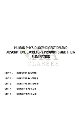
  • 1. UNIT 1 - DIGESTIVE SYSTEM I UNIT 2 - DIGESTIVE SYSTEM II UNIT 3 - DIGESTIVE SYSTEM III UNIT 4 - URINARY SYSTEM I UNIT 5 - URINARY SYSTEM II HUMAN PHYSIOLOGY: DIGESTION AND ABSORPTION, EXCRETORY PRODUCTS AND THEIR ELIMINATION
  • 2. 3 1.1 INTRODUCTION TO THE DIGESTIVE SYSTEM 1.1.1 Main functions  Moves water, nutrients and electrolytes from the external environment to the internal environment.  Breaks down food moving through the tract into its chemical building blocks.  Absorbs these building blocks into the blood.  Eliminates residual indigestible materials. 1.1.2 Basic divisions Alimentary canal  Continuous muscular tube that runs from the mouth to the anus.  Consists of mouth, pharynx, esophagus, stomach, small intestine and large intestine. Accessory organs  Contribute to secretions to the GI tract, but the food doesn’t pass through these organs.  Consists of teeth, tongue, salivary glands, liver, gall bladder and pancreas. 1.1.3 Actions of the gastrointestinal (GI) tract  Ingestion: Occurs when material enters via the mouth.  Mechanical processing: Crushing / shearing make material easier to move through the tract.  Digestion: Chemical breakdown of food into small organic compounds for absorption.  Secretion: Release of water acids, buffers, enzymes and salts by epithelium of GI tract and glandular organs.  Absorption: Movement of organic substrates, electrolytes, vitamins and water across digestive epithelium.  Excretion: Removal of waste products from body fluids. 1.1.4 Movement of digestive materials A. Deglutition Deglutition is the movement of food from the mouth and into the stomach. The entire process takes about four to eight seconds for solid or semisolid food and about one second for every soft food and liquids. Deglutition involves the mouth, pharynx and esophagus. It is facilitated by the secretion of mucus and saliva. There are three stages in deglutition: the voluntary phase, the pharyngeal phase, and the esophageal phase. UNIT 1 - DIGESTIVE SYSTEM I Mouth Esophagus Liver Gall bladder Duodenum Transverse colon Ascending colon Anus Ileum Descending colon Jejunum Pancreas Stomach Parotid gland Salivary glands
  • 3. 4 Tongue Pharynx Windpipe (trachea) Esophagus Larynx closes Food Soft palate Vertebrae Epiglottis Swallowing B. Peristalsis The smooth muscle of the GI tract is arranged in two layers: longitudinal along the length of the tube and circular around the diameter of the tube. Coordinated contraction and relaxation of these two layers create wavelike propulsion of the food called peristalsis. Initial state Longitudinal muscle Circular muscle From mouth To anus Contraction Contraction of circular muscles behind bolus Step 1 Contraction of longitudinal muscles ahead of bolus Contraction Contraction Step 2 Contraction in circular muscle layer forces bolus forward Step 3 1.1.5 Levels of organization  Chemical level – Sodium, potassium, chloride, bicarbonate, and phosphate ions and hydrochloric acid.  Macromolecular level – Enzymes of digestion, bile, and the nutritive molecules such as carbohydrates, proteins, lipids and nucleic acids.  Cellular level – Mucosa cells, secretory cells and immune cells.  Tissue level – Epithelial, connective and smooth muscle tissues.  Organ level – Gastrointestinal tract and accessory organs.  Organ system level – Integration of organs for nutrients and waste movement, digestion and absorption.
  • 4. 5 1.1.6 Nutritive molecules The nutritive organic compounds in our food include carbohydrates, proteins and lipids. These molecules are digested, then absorbed, and reassembled into macromolecules or used as fuel for metabolism in the body. The process of breaking down these macromolecules involves splitting into smaller molecules using water molecule, thus the name hydrolysis. The reverse reaction, called dehydration synthesis, can build macromolecules from the absorbed building blocks. Enzymes can speed up the rate of these reactions. A. Carbohydrates: Our daily food intake usually includes from 200 to 600 grams of carbohydrates. Monosaccharides are absorbed easily and can therefore be a source of quick energy for the body when we consume them in this form. Glucose, galactose, and fructose are the three common monosaccharides in our diet. Disaccharides include sucrose (table sugar), lactose, and maltose. Polysaccharides include polymerized glucose in different forms including glycogen, which is the stored form of glucose in our bodies, and starch. B. Proteins: USDA recommends 5 to 6 ounces of protein in diet per day, although children need less. This dietary protein is usually in the form of polypeptides and must be digested into its amino acid building blocks for absorption. There are numerous enzymes that break large proteins into smaller peptides and then into amino acids. Amino acids are then absorbed from the digestive system into the circulatory system where they are delivered throughout the body. Once amino acids have entered cells throughout the body, they are bonded together to make proteins needed for cell function. As a last resort, proteins and amino acids can also be used as energy; they are metabolically converted to glucose before they are used as energy sources. C. Lipids and Fats: Dietary lipids include fats and oils. While not considered a USDA food group, oils contain some essential nutrients and are recommended as part of a healthy diet, although only in small amounts. Solid fats have more saturated and trans-fatty acids and are considered empty calories when included in a diet because they add calories but not needed nutrients. Most dietary lipids are in the form of triglycerides. Lipids are processed by enzymes secreted from the pancreas (with some enzymes from the stomach and saliva) and are then solubilized for absorption by salts secreted in bile (from the liver). These steps prepare them for absorption in the small intestine. Like proteins, ingested lipids are broken down into smaller parts for absorption and then are either metabolized to make energy or are used to make cellular structures including cell membranes. Lipid absorption is also required for absorption of some fat-soluble vitamins. D. Nucleic acids: When we eat any plant or animal foods, we are able to digest the DNA or RNA nucleic acid that was in their cells. The pancreas produces and releases into the small intestine two enzymes to break down either DNA or RNA into individual nucleotides. Brush border enzymes of the cells lining the small intestine further digest the nucleotide into its molecular components for absorption.
  • 5. 6 1.2 ANATOMY OF THE ALIMENTARY CANAL The alimentary canal is tubular structure which extendsfrom mouth to anus. It develops from ectoderm and endoderm. Ectoderm – Upto hard palate. Endoderm – From soft palate to rectum. Ectoderm – from anal canal to anus. 1.2.1 Mouth Mouth is a horizontal transverse slit like aperture which is surrounded by upper and lower lip; a specific muscle is associated with lip called orbicularis oris muscle. Serous glands are found on the inner part of lip. Serous glands are the modification of mucus glands. Its secretory substance is watery. Mouth opens into Buccopharyngeal cavity. Thiscavity isdivided into two parts. A. Buccal vestibule – The space between the gums and cheeks where the food is stored temporarily for some time. It is a peripheral part. B. Oral cavity – It is inner and central part which is surrounded by upper and lower jaw. Lined by stratified squamous epithelium. Upper jaw is fixed and lower jaw is movable. The roof of oral cavity is called as Palate. Palate is differentiated into two parts: (i) Hard palate – It is the anterior part of the palate. It is made up of maxilla and palatine bone in human. But in rabbit it is made of premaxilla, maxilla, palatine bone. On the ventral surface of hard palate, some projection or transverse ridges are present which are called as palatine rugae. These rugae prevent slip out of the food from buccal cavity during mastication; these rugae are well developed in carnivorous animals. (ii) Soft palate – It is the posterior part of palate. It is made up of involuntary muscle fibrous connective tissue and mucous epithelium. (Stratified squamous epithelium). Upper lip Superior labial frenulum Glossopalatine arch Pharyngopalatine arch Palatine tonsil Salivary duct orifices Sublingual Submandibular Inferior labial frenulum Lower lip Hard palate and transverse palatine folds Soft palate Uvula Fauces Tongue Lingual frenulum Teeth Gingivae
  • 6. 7 The posterior out growth of soft palate which hangs down in the form of finger like process called as uvula or velum palati. On the dorsal side of uvula internal nasal pores are present. One pair of large lymph node is present on the posterolateral surface of soft palate, called as palatine tonsil or tonsils. Soft palate is situated in the pharynx and is divided into two parts. Upper part of pharynx is called nasopharynx which is related to the nasal chamber. The lower part of pharynx is called oropharynx which is related to the oral cavity. One pair of openings of eustachian tube is present in the nasopharynx. This Eustachian tube is related to the middle ear, it maintains air pressure. TARGET POINTS  In rabbit, a small cleft is found in the middle part of upper lip, such type of lip is called as Hare lip. One pair opening of nasopalatine duct is present at the anterior part of hard palate, these connect the buccal cavity to the nasal passage. In rabbit, some olfactory receptors are also found in nasopalatine duct which are called as Jacobson’s organ or vomero nasal organ. It makes them aware of the smell of food while chewing.  Jacobson’s organ is well developed in reptiles like- snake, lizard and sphenodon.  Uvula covers the opening of internal nasal pores during ingestion of food, so food particles cannot move inside nasal chamber. A. Tongue On the floor of oral cavity a muscular, flat, fleshy plate like structure is present which is called tongue. The anterior part of tongue is free while posterior part of tongue is connected to the hyoid bone. The ventral surface of tongue is connected to the floor of buccal cavity through a very flexible membrane / ligamentous fold called as Frenulum linguae. The dorsal surface of tongue is divided into two unequal parts by a V shaped sulcus, called as sulcus terminalis. The two limbs of the ‘V’ meet at a median pit named Foramen caecum. It is divided into two parts: (i) Pharyngeal part – It is the poster 1/3 part of the tongue. Many small lymph nodes are present in this part which are called lingual tonsil. Mouth Hard palate Soft palate Uvula Larynx (with vocal cords) Trachea (windpide) Pharynx Epiglottis Esophagus Median sulcus Vallate papillae Terminal sulcus Palatine tonsil Lingual follicles Root Median glossoepiglottic fold Epiglottis Apex Filiform papillae Fungiform papillae Palatoglossal arch Foramen cecum Lingual tonsil Palatopharyngeal arch Upper surface of human tongue
  • 7. 8 (ii) Oral or papillary part – It is anterior 2/3 part of tongue. Four types of papillae are found in this part in which gustatory or taste receptors are present in the form of taste buds. Fungiform papillae: It is pink coloured, small and spherical in shape. It is found on the entire surface of tongue but mostly present at the anterior part of tongue. It is attached to tongue with the help of small pedicle. It provides pink colour of the tongue. Filiform papillae (conical papillae): They are thread like, white coloured and conical in shape. They are also found on the entire surface of tongue. They are most numerous, but devoid of taste buds. Foliate papillae: They are found on the mid lateral surface of tongue. They are vestigeal in the human. Their structure is leaf like present in rabbit and other mammals. Circumvallate papillae: It is the largest and least existed papillae (8 to 12), they are large spherical shaped papillae which are found near to sulcus terminalis. Taste pore Circumvallate Foliate Taste buds Fungiform TRC a b Bitter Salty Sweet Umami Sour Taste centers in tongue Two types of muscles are present in tongue: (i) Extrinsic muscle: Found on outer and superficial part of tongue, helps in it’s outward and inward movement. (ii) Intrinsic muscle: Situated in the deep part of tongue, helps in the change of shape of tongue.
  • 8. 9 B. Teeth Teeth are ectomesodermal in origin. Major portion of teeth arises from dermis. Part of toothe present outside the gumsonly is derived from ectoderm or epidermis (Enamel part). In human teeth of upper jaw are attached to the maxilla bone. While teeth of lower jaw are attached to mandible bone. But in rabbit upper incisors are attached to premaxilla. While upper pre molars and molars attached to the maxilla bone. While lower teeth attached to dentry bone. Teeth are differentiated in three parts: The crown part of the tooth is made up of very hard substances called the Enamel. It is the hardest material in all animal of animal kingdom. Enamel is ectodermal. It is secreted by ameloblast cells of the ectoderm. It has maximum amount of inorganic salt (96%) in it. Inorganic salt are mainly found in the form of phosphate and carbonate of Ca, Mg, Na and K. 3% of water is found in the enamel. Along with it keratin and ossein protein (1%) are also found in teeth. Ossein is a protein of bones. Remaining part of teeth develop from mesoderm of embryo. Dentine is the main part of tooth. Approximately 69% inorganic salts are present in dentine where as in cemented layer 65% is inorganic salt (62% inorganic salts are present in bones). Dentine surrounds a cavity called pulp cavity. This cavity contains soft connective tissue, blood capillaries and nerve fibers. Pulp cavity is necessary for the nutrition and survival of the teeth. At the base of pulp cavity an aperture is present. Through this aperture, blood capillaries and nerve fibers enter inside the teeth. This aperture is called apical-foramen. A special type of cells form the lining of the pulp-cavity called the odontoblasts cells. These cells are the dentine secreting cells. Cytoplasmic processes of odontoblasts are embedded into dentine in the form of fine tubule. These processes are called canaliculi. These canaliculi secrete dentine. The teeth continue to grow till the odontoblasts cells remain active. In adults, the pulp cavity shrinks and the odontoblasts become inactive so the teeth stop to grow. Cementum PDL Alveolar bone Sharpey’s fibers Pulp cavity Enamel Dentin Gingiva Cementum Periodontal ligament Root canal Alveolar bone Apical foramen Alveolar vessels and nerves Sagittal section of a mandibular (lower) molar Attachment organ Crown It is the outer part of the tooth, exposed outside gums. Neck It is the internal part of the tooth which is embedded inside the gums. Root It is the part of tooth that is inserted inside the socket of jaw bone (Alveoli).
  • 9. 10 The cement layer is made up of the cementocyte cells. Between the root and the bones of the teeth, a periodontal membrane is present. Humans have two sets of teeth (dentitions). The 20 deciduous teeth, or baby teeth, first appear at about 6 months of age. Between ages 6 and 12 years, these teeth are replaced by the 32 permanent teeth. Closest to the midline are the two incisors, chisel shaped teeth we use for cutting into food. The cuspids (canines) are next to the incisors. A cuspid’s pointed edge (cusp) tears and shreds food. Posteriorly, the next teeth are the two premolars (bicuspids) followed by up to three molars. Premolars and molars have broader, flatter surfaces, which we use to crush and grind food. Types of teeth on the basis of appearance in life Jaw bone with tooth sockets Tongue Teeth roots Line of lips Molars Premolars Canine Incisors Specific teeth and structures of the mouth Monophyodont The teeth which appear only once in life. Eg. Premolars and last molar of man. Diphyodont The teeth which appear twice in life eg. Incisors, canines, molars. Polyphyodont The teeth which appear more than twice in life eg. Fish, amphibians. On the basis of position from jaw Thecodont The teeth which are present in bony socket of jaw eg. Man and crocodile. Pleurodont The teeth which are present on the lateral side of jaw bone eg. Reptiles. Acrodont The teeth which are present on the terminal part of jaw bone eg. Fish, amphibian. On the basis of structure and function Heterodont When the teeth are of different type in mammals on the basis of structure and function. Eg. Mammal. Homodont Whether all teeth are of similar type in animal on the basis of structure and function. Eg. Fish, amphibians. On the basis of crown Secodont These are canine teeth of carnivorous animals. In this type of structure canine teeth become long and pointed which is bended towards the backward direction. Hypsodont In this type of teeth the crown part is large root is small (such as incisor and (smiling teeth) canine). These teeth are also called as smiling teeth.
  • 10. 11 Dental formula: TARGET POINTS  Carnessial teeth are modified last premolar of upper jaw and first molar of lower jaw, for shearing and tearing of tendon.  In rabbit and rat, the pulp cavity of the incisor remains wide throughout their life, so these teeth grow continuously throughout their life span; these types of teeth are called open rooted teeth. If one incisor or rabbit and rat is broken then the opposite incisor grows continuously, finally the animal neither can close the mouth nor gnaw the food. So the animal dies due to starvation.  In frog, only upper jaw has teeth.  Hippocampus, tortoise and birds do not have teeth. C. Salivary glands Lying outside the oral mucosa are the three pairs of major salivary glands, which secrete the majority of saliva into ducts that open into the mouth. The parotid glands lie between the skin and the masseter muscle near the ears. They secrete saliva into the mouth through the parotid duct, which is located near the seconds upper molar tooth. The submandibular glands, which are in the floor of the mouth, secrete saliva into the mouth through the submandibular ducts near the lower central incisor. The sublingual glands, which lie below the tongue, use the lesser sublingual ducts to secrete saliva into the oral cavity. Brachyodont In this type of teeth crown part is small root is long (such as premolar and (cheek teeth) molar). Wisdom teeth – these are the last molar teeth of humans which appear in the age of 18 to 25 years.The upper surface of premolar and molar is board. Some small projections are present in the upper surface of premolar and molar. These projections are called lophs or cusps. On the basis of structure of lophs Lophodont In this type of teeth the lophs are large, wide and flat such as rabbit and elephant. Bunodont In this type of teeth. The lophs are small and spherical in shape, such as human. Solenodont In this type of teeth the lophs are large and semilunar shape eg. Ruminant animal (Cow, buffalo). Carnessial In this type of teeth the Lophs are long and pointed eg. Carnivorous animal. Child = 2 1 0 2 5 10 I C PM M = 2 = = 20 2 1 0 2 5 10  Adult = 2 1 2 3 8 16 I C PM M = 2 = = 32 2 1 2 3 8 16  17 years old = 2 1 2 2 7 I C PM M = × 2 = 28 2 1 2 2 7 Rabbit = 2 0 3 3 8 16 I C PM M = 2 = = 28 1 0 2 3 6 12 
  • 11. 12 Parotid glands are the largest salivary glands. Infections of the nasal passages and pharynx can attack any salivary glands. The parotid glands are the usual site of infection with the virus that causes mumps (Paramyxovirus). Mumps manifest with enlargement and inflammation of the parotid glands, causing the characteristic swelling between the ears and the jaw. Symptoms include fever and throat pain, which can be severe when swallowing acidic substances such as orange juice. Wharton’s duct: These are the ducts of submaxillary or submandibular glands; the largest ducts. Sublingual glands are the smallest salivary glands. Ducts arising from these glands are called as the ducts of Rivinus or Bartholin’s ducts. Maximum saliva is secreted by the sublingual glands (smallest salivary duct). Saliva is mainly water, which dissolves chemicals in the food. Only dissolved chemicals can activate the different kinds of taste receptor cells on the tongue, palate and other parts of the mouth and pharynx. Mixed in with the water, saliva also contains mucus, various electrolytes typically found in blood plasma, as well assome digestive molecules, metabolic waste products and immune molecules. Each of these contributes to the various functions of saliva. Bicarbonate and phosphate ions help maintain the pH of saliva as neutral or slightly basic (average 7.4 pH). This not only helps protect the teeth from acidic substances eaten and plaque bacteria that grow best in an acidic environment, but also maintains the optimal pH for the enzyme amylase. Waldeyer’s ring The lymphatic tissues of the pharynx and oral cavity are arranged in a ring like manner, which are collectively called Waldeyer’s ring (Waldeyer’s lymphatic ring). The ring mainly consists of the following:  Nasopharyngeal tonsil (Pharyngeal tonsil): Refer to the nasopharynx. In children nasopharyngeal tonsil may become enlarged and referred asadenoids. The resulting swelling may be a cause of obstruction to normal breathing.  Tubal tonsil: Refer to the nasopharynx.  Palatine tonsils (Faucial tonsils): Refer to the oropharynx. The palatine tonsils are often infected (tonsillitis) leading to sore throat. Such enlarged tonsils may become a focus of infection and their surgical removal (tonsillectomy) becomes necessary.  Lingual tonsil: They are situated on posterior part of tongue. Accessory parotid gland Parotid gland Body of mandible Submandibular gland Submandibular (Wharton’s) duct Sublingual gland Parotid duct Opening of submandibular (Wharton’s) duct Salivary gland locations
  • 12. 13 Adenoid Lingual tonsil Palatine tonsil (“tonsil”) Tubal tonsil Waldeyer's ring Nodules on posterior pharyngeal wall Lateral pharyngeal bond Adenoids Tubal tonsil Palatine tonsil Lingual tonsil TARGET POINTS  Salivary glands are exocrine glands.  Ptyalin is secreted only by the parotid gland. Lysozyme and Thiocyanates mainly kill bacteria. They also check the growth of bacteria in buccopharyngeal cavity.  Salivation is stimulated by cranial nerve VII and IX. 1.2.2 Tissue layers of the GI tract Throughout the gastrointestinal (GI) tract, walls are comprised of the same four fundamental tissue layers. From the lumen of the GI tract, these layers are the mucosa, submucosa, muscularis and serosa. Serosa Circular muscle Longitudinal muscle Submucosa Meissner’s nerve plexus Mesentery Submucosal gland Myenteric nerve plexus Mucosal gland Mucosal muscle Epithelial lining Mucosa Microvillus Mucosa Submucosa Muscularis Serosa Tissue layers of the GI tract Typical cross-section of the gut Serosa: It is outermost layer of gut. It is called tunica adventitia in oesophagus, serosa is made up of visceral peritoneum while tunica adventitia is made up of white fibrous connective tissue.
  • 13. 14 Muscle layer: It is formed by circular inner layer and longitudinal outer layer of smooth muscle. Thickest layer is found in stomach (maximum peristalsis) and thinnest layer in rectum (minimum peristalsis). Submucosa: It is loose connective tissue layer with blood lymph vessels and nerves. Mucosa: It is the inner most layer of gut which contains the secretory and absorptive cells. It is differentiated into the following parts:  Outer part: Called mucosa muscularis, it is made up of longitudinal and circular muscles. It has important role in exposing of surface area for the absorption. They also provide support to the folds of alimentary canal.  Middle part: It is called lamina propria. It contains few modified lymphatic tissue which provides immunity. Eg. Peyer’s patches. It is made up of reticulate and fibrous connective tissue.  Innermost part: Called mucosal layer. In esophagus this layer is made up of non-keratinized stratified squamous epithelium. Except esophagus this layer is single layer thick, which is made up of columnar mucous epithelium. This layer makes the lining of lumen of alimentary canal and makes the folds of alimentary canal. Folds of esophagus are less developed, whereas folds of stomach are finger shaped and develop as gland called gastric gland. Folds of small intestine are conical shaped called villi. Small slit like space is found at the base of villi. These spaces are called crypts of Leiberkuhn. Villi of duodenum are small blunt. Villi of jejunum and ileum are long and pointed. Maximum villi are found jejunum.  Brunner’s gland: They are small spherical multicellular glands. Open into crypts of Lieberkuhn with the help of the tubules. These are found in the submucosa of duodenum, they synthesize and secrete the non-enzymatic alkaline secretion of intestinal juice.  Paneth cells: These cells are found in mucosal layer of crypts of Lieberkuhn of small intestine. They are unicellular glands and secrete antibacterial substances hence it provides immunity. They also synthesize and secretes enzymes of intestinal juices. The secretory substances of Brunner’s glands and Paneth cells are combinedly called intestinal juice or succus entericus.  Peyer’s patches: They are small lymph nodes which are found in the mucosa of small intestine (jejunum and ileum more in number). They are also called intestinal tonsils and provide immunity.  Nerve supply: Two types of nerve plexus are found in muscle of alimentary canal. – Auerbach’s nerve plexus (myentric plexus) this nerve plexus is found between longitudinal muscles and circular muscles. It control muscles contraction. – Meissner’s nerve plexus found between circular muscles and sub mucosa but in stomach it is found between oblique/ muscle and submucosa. It helps in secretion.
  • 14. 15 1.2.3 Organs of the GI tract A. Esophagus Two apertures are found in central part of buccopharyngeal cavity. Ventral of lower aperture is called glottis which is related to the larynx, which is guarded by epiglottis. The dorsal and upper aperture is called gullet which opens into esophagus. Esophagus is simple uniform tube which runs downward and pierces the diaphragm and finally opens into stomach that site of piercing on diaphragm is called hiatus. Longitudinal folds are found on the inner surface of esophagus in which digestive glands are absent, only mucous glands are present in mucosa and submucosa. Voluntary muscles are found on the upper 1/3rd part of esophagus while mid 1/3rd part is formed by voluntary and involuntary muscles where as in lower 1/3rd part of esophagus only involuntary muscles are present. The length of esophagus depends on length of neck so the longest esophagus is present in giraffe. It lacks serosa, but tunica adventitia is present. B. Stomach It is situated on left side of abdominal cavity. It is the widest part of alimentary canal. It is a bag like muscular structure, J-shaped in empty condition. The stomach contains four parts (cardiac, fundus, body, pylorus or antrum). It has two orifices (opening). (i) Cardiac orifice is joined by the lower end of the esophagus. (ii) Pyloric orifice opens into the duodenum. Stomach is covered by layer of peritoneum. Fat tissues and lymph tissue deposits on the peritoneum. Such type of peritoneum are called ommentum. Left curved surface of stomach is called greater ommentum. Right curved surface of stomach is called lesser ommentum. Mucous membrane of the stomach is thick. In empty stomach numerous temporary longitudinal folds are found in mucosa of pyloric antrum called rugae. They disappear when stomach is distended. Esophagus Diaphragm Stomach Lower esophageal sphincter (LES) Upper esophageal sphincter (UES) Throat (pharynx) Trachea
  • 15. 16 Cardia Fundus Body Pyloric antrum Pyloric canal Pylorus Stomach anatomy Esophagus Muscularis - Longitudinal layer - Circular layer - Oblique layer Lesser curvature Duodenum Pyloric sphincter (valve) Greater curvature Rugae of mucosa Serosa The stomach stores the food for 4 – 5 hours. The food mixes thoroughly with the acidic gastric juice of the stomach by the churning movements of its muscular wall and is called the chyme. The mucus and bicarbonates present in the gastric juice play an important role in lubrication and protection of the mucosal epithelium from excoriation by the highly concentrated hydrochloric acid. HCl provides the acidic pH (pH 1.8) optimal for pepsins. Rennin is a proteolytic enzyme found in gastric juice of infants which helps in the digestion of milk proteins. Gastric glands: These are numerous microscopic, simple tubular glands formed by the invagination of epithelium in the stomach. The following types of cells are present in the epithelium of the gastric glands. Chief cells or peptic cells (Zymogen cells) are usually basal in location and secrete gastric digestive enzymes as proenzymes or zymogens called pepsinogen and prorennin. The chief cells also produce small amount of gastric amylase and gastric lipase. Gastric amylase action is inhibited by the highly acid condition. Gastric lipase contributes little to digestion of fat. Prorennin is secreted in young mammals (childhood stage). It is not secreted in adult mammals. Oxyntic cells (Parietal cells) are large and are most numerous on the side walls of the gastric glands. They are called oxyntic cells because they stain strongly with eosin dye. They are called parietal cells as they lie against the basement membrane. They secrete hydrochloric acid and Castle’s intrinsic factor. Mucous cells (Goblet cells) are present through out the surface epithelium and secrete mucus. The epithelium of gastric glands also has the different stem cells and Enteroendocrine cells or argentaffin cells. Enteroendocrine cells are usually present in the basal parts of the gastric glands, which is differentiated in three cells – these cells are D-cells, enterocrinin cells (EC cells) and G cells. D-cells secrete somatostatin, EC-cells secrete serotonin and histamine, whereas G cells secrete gastrin.
  • 16. 17 Somatostatin suppresses the release of hormones from the digestive tract. Serotonin is a vasoconstrictor and stimulates the smooth muscles. Histamine dilates the walls of blood vessels. Gastrin stimulates the gastric glands to release the gastric juice. Surface mucous cell (secretes mucus) Mucous neck cell (secretes mucus) Parietal cell (secretes hydrochloric acid and intrinsic factor) Chief cell (secretes pepsinogen and gastric lipase) G cell (secretes the hormone gastrin) Lamina propria Gastric glands Submucosa Muscularis mucosae Gastric pit Secretory cells Gastric pit Gastric glands Gastric pits Duodenum Hepatic flexure Ascending colon Cecum Ileum Transverse colon Splenic flexure Descending colon Jejunum Sigmoyid colon Anus External sphincter Internal sphincter Anal columns Rectum Intestines of the GI tract C. Small intestine Small intestine is differentiated into three parts: (i) Duodenum (25 cm) (ii) Jejunum (1m) (iii) Ileum (2 m)
  • 17. 18 Duodenum is reteroperitoneal and initial part of small intestine. Duodenum is the shortest, widest and the fixed part of the small intestine. It is C/U shaped. For absorption of digested food a very large surface area is required. Therefore some adaptations are present here. (a) Great length of the intestine. (b) The presence of permanent deep folds in mucosa is called plicae circularis, valvulae conniventae or valves of kerkring, along with villi and microvilli. Microvilli Absorptive cell (absorbs nutrients) Goblet cell (secretes mucus) Enteroendocrine cell (secretes the hormones secretin, cholecystokinin, or GIP) Paneth cell (secretes lysozyme and is capable of phagocytosis) Blood capillary Lamina propria Lacteal Intestinal gland Muscularis mucosae Arteriol Venule Lymphatic vessel Muscularis Submucosa Mucosa C) Enlarged villus showing lacteal, capillaries, intestinal glands, and cell type D. Large intestine Large intestine (larger in diameter) – Large intestine is differentiated into three parts caecum, colon and rectum. In rabbit the dilated last terminal part of ileum iscalled sacculus rotundus. It isconnected with the caecum through an ileocaecal valve. The lower end of the ileum opens on the posteromedial aspect of the ileocaecal junction. The ileocaecal opening is guarded by ileocaecal valve. Caecum is a large blind sac. It is situated in the right iliac fossa. It is 6 cm long and 7.5 cm broad.
  • 18. 19 About 2 cm below the ileocaecal orifice, a worm like structure arises from the caecum called as vermiform appendix. Its length varies from 2 to 20 cm. It is a vestigeal organ. Caecum is well developed in rabbit and other mammals but is vestigial in human. Colon: Caecum continues in colon, which is the middle part of large intestine. The longitudinal muscle coat forms three ribbon like bands called Taeniae coli. Due to the presence of taeniae, pouch like structure develops in Lumen of colon called as Haustra. Rectum: This colon then continues in a uniform tube called rectum (storage chamber for faeces). Rectum open into a small bag like structure called anal canal. Anal canal: Anal canal opens outside by anus. Anus is controlled by anal sphincter. Two types of anal sphincter are found at the opening of anus. Two types of sphincter muscles are found in anal canal. Internal anal sphincter  involuntary. . External anal sphincter  voluntary. . Microvilli Absorptive cell (absorbs water) Goblet cell (secretes mucus) Lymphatic nodule Lamina propria Submucosa Muscularis mucosae Intestinal gland Openings of intestinal glands Sectional view of intestinal glands and cell types E. Liver It develops from endoderm (weight 1.5 kg both exocrine and endocrine). In human it is found in right side of abdominal cavity, below the diaphragm. The liver is the largest gland of body. It is made up of left and right lobe. Left lobe is smaller than right lobe. Right lobe forms 5/6th of the liver and left lobe forms 1/6th of liver. Right and left liver lobe are separate from each other by the falciform ligament (fibrous C T) which is made up of fold of peritoneum. Right and left hepatic duct develops from right and left liver lobe. Both these ducts combine to form a common hepatic duct. Gall bladder is situated below right lobe of liver.
  • 19. 20 Falciform ligament Diaphragm Coronary ligament Right lobe of liver Left lobe of liver Right hepatic duct Cystic duct Gross anatomy of liver Cystic duct of gall bladder is connected to common hepatic duct to form a common bile duct also called ductus choledocus. The functional and structural unit of liver is lobule. Internally liver is made up of numerous polygonal lobules. These lobules are covered by fibrous connective tissue covering called Glisson’s capsule. Each lobule consists of radial rows of hepatic cells which are called as hepatic cord. Each row is one or two cell wide and two cell thick. In between the hepatic cord, a space is present called as hepatic sinusoid. These sinusoids are filled with blood. Sinusoids are lined by the endothelial cells mostly but a few fixed macrophages cells are also present. These are called as Kupffer’s cells (phagocytic cells). Sinusoid Hepatocytes Branch of hepatic artery proper Bile duct A single liver lobule and its cellular components Central vein Kupffer’s cells Bile canaliculi Branch of hepatic portal vein
  • 20. 21 The bile canaliculi run in between the two layers of cells in each cord. Hepatocytes (hepatic cells) pour bile into the canaliculi. Canaliculi open into branch of hepatic duct which is situated at the angular part of lobule in the Glisons capsule. All branches of hepatic duct of right and left lobe are combined to form right and left hepatic duct which come out from the liver and forms a common hepatic duct. Hepatic artery and hepatic portal vein enter into liver and divide to form many branches. These branches are also found at the angular part. Its fine branches open into hepatic sinusoids. Branch of hepatic portal vein, branch of hepatic artery and branch of hepatic duct are collectively called as portal triad. All hepatic sinusoids open into central vein or intralobular vein through fine aperture. All central vein combine to form hepatic vein which comes out from liver and opens into inferior vena cava. Portal triad Sinusoids Portal vein Bile duct Hepatic artery Hepatocyte cords Cholangiocyte Bile duct Bile canaliculi Hepatocyte Stellate cell Space of Disse Portal triad Portal vein Sinusoid Central vein Hepatic artery Kupffer’s cell Endothelial cell CV Liver cells Functions of Liver: Liver is known as biological and chemical factory of the body. Most of the biochemical functions of the body are done by the liver.  Secretion and synthesis of bile: This is the main function of liver. Bile is yellowish green, alkaline fluid. In bile juice, bile salts, sodium bicarbonate, glycocholate, taurocholate, bile pigments, cholesterol, Lecithin etc. are present. Bile salts help in emulsification of fats. Bile prevents the food from decomposition. It kills the harmful bacteria.  Carbohydrate metabolism: The main center of carbohydrate metabolism is liver. Following steps are related with carbohydrate metabolism: (i) Glycogenesis: The conversion and storage of extra amount of glucose into glycogen from the digested food is called glycogenesis. The main stored food in the liver is glycogen. (ii) Glycogenolysis: The conversion of glycogen into glucose agan when glucose level in blood falls down is called glycogenolysis. (iii) Gluconeogenesis: At the time of need, liver converts non carbohydrate compounds (eg. Amino acids, fatty acids) into glucose. This conversion is called gluconeogenesis. This is the neo formative process of glucose.
  • 21. 22 (iv) Glyconeogenesis: Synthesis of glycogen from lactic acid (which comes from muscles) is called glyconeogenesis.  Storage of fats: Liver stores fats in a small amount. Hepatic cells play an important role in fat metabolism. The storage of fat increases in the liver of alcohol addict persons (fatty liver). This stored fat decreases the activity of liver. The damage of liver due to alcohol intake is called alcoholic liver cirrhosis.  Deamination and urea formation: Deamination of amino acids is mainly done by liver (amino acid  NH3 ) liver converts ammonia (more toxic) into urea (less toxic) through ornithine cycle.  Purification of blood: Kupffer cells of liver and splenocytes of spleen are the phagocytic cells, helps in phagocytosis of dead blood cells and bacteria from the blood.  Synthesis of plasma proteins: Many types of proteins are present in blood plasma. All the proteins except gama globulins are synthesized in the liver. Chemically antibodies are gama globulins formed by lymphocytes.  Synthesis of heparin: Heparin is a natural anticoagulant (mucopolysaccharide). Some heparin is also formed by basophils (granulated WBC) and mast cells.  Synthesis of vitamin A: The liver changes -carotene into vitamin A- carotene is a photosynthetic pigment which is obtained from yellow parts of fruits. It is abundantly found in carrot.  Liver stores vitamin A, D, E, K, B12 .  Storage of minerals: Liver stores iron, copper, zinc, cobalt, molybdenum etc. Liver is a good source of iron.  Detoxification: The conversion of toxic substances into non toxic substances is done by liver. The toxic substances are formed by metabolic activities of the body. Eg. Prussic acid is converted into potassium sulfocyanide (it is a non-toxic salt) by the liver.  Haemopoiesis: The formation of blood cells is called haemopoiesis. In embryonic stage R.B.C and WBC are formed by liver.  Yolk synthesis: Most of the yolk is synthesized in liver.  Secretion of enzymes: Some enzymes are secreted by liver, participate in metabolism of proteins, fats and carbohydrates eg. Dehydrogenase, cytochrome oxidase etc.  Prothrombin and fibrinogen proteins are also formed in hepatic cells. These help in blood clotting.  Factors I, II, V, VII, IX and X are formed in liver, which are responsible for blood clotting. F. Pancreas It is developed from endoderm which is soft, lobulated and elongated organ. It is made up of numerous acini. Acini is a group of secretory cells surrounding a cavity. Each acini is lined by pyramidal shaped cells. These acinar cells secrete the enzyme of pancreatic juice. Each acini opens into pancreatic ductule. Many pancreatic ductule combine to form main pancreatic duct (duct of Wirsung).
  • 22. 23 The main pancreatic duct is joined with the bile duct to form the hepatopancreatic ampulla which opens into duodenum. The accessory pancreatic duct (duct of Santorini) opens into duodenum with separate openings located above the opening of major pancreatic duct. Some group of endocrine cells are also found in between groups of acini called islets of Langerhan’s. These islets secrete insulin and glucagon hormone. So this gland is exocrine as well as endocrine (heterocrine). Its 99% part is exocrine while 1%part is endocrine. In humans both bile duct and pancreatic duct combine to form common duct called as hepato-pancreatic duct. The terminal end of common duct is swollen and is called as ampulla of Vater or hepato pancreatic ampulla. Ampulla of Vater opens into middle part of duodenum and is controlled by sphincter of Oddi while bile duct is controlled by sphincter of Boyden. Exocrine acinus Beta cells Alpha cells Pancreatic islet Acinar cells secrete digestive enzymes Pancreatic duct Duodenum of small intesline Digestive enyzmes Common bile duct Bile duct (from gall bladder) Pancreas Splenic artery Pancreatic hormones: - Insulin - Glucagon Pancreatic islets Spleen Gross anatomy of pancreas Pancreatic cells Alpha cell (secretes glucagon) Beta cell (secretes insulin) Delta cell (secretes somatostatin) Exocrine pancreas (acinar cells and duct cells) F cell (secretes pancreatic polypeptide)
  • 23. 24 1. Anxiety and eating spicy food together in an otherwise normal human, may lead to a) Indigestion b) Jaundice c) Diarrhoea d) Vomiting 2. What is the common passage for bile and pancreatic juices? a) Ampulla of Vater b) Ductus Choledochus c) Duct or Wirsung d) Duct of Santorini 3. In the mouth of rabbit, diastema is a gap between the a) Premolar and molar b) Canine and incisor c) Canine and premolar d) Incisor and premolar 4. The longitudinal bands present in colon are known as a) Taeniae b) Sulcie c) Haustra d) None of these 5. Which one of the following pairs of food components in humans reaches the stomach totally undigested? a) Protein and starch b) Starch and fat c) Fat and cellulose d) Starch and cellulose 6. Sphincter of Oddi guards a) Hepato-pancreatic duct b) Common bile duct c) Pancreatic duct d) Cystic duct 7. The diameter of duodenum is about a) 4.0 to 4.5 cm b) 3.0 to 3.2 cm c) 1.1 to 2.1 cm d) None of these 8. The amount of saliva produced per day in humans is a) 200 ml b) 500 ml c) 750 – 1000 ml d) 1.0 – 1.5 L 9. Hepatic portal system connects a) Liver to digestive system b) Digestive system to liver c) Liver to intestine d) Only small intestine to liver 10. In both chordates and non-chordates intestine develops from a) Pharyngeal pouch b) Ectoderm c) Endoderm d) Mesoderm 11. pH of stomach in rabbit is about a) 7 b) 3 c) 8 d) 11 12. Stellate reticulo endothelial cells of liver are a) Mast cells b) Hepatocytes c) Kupffer’s cells d) All of these 13. Peyer’s patches produce a) Enterokinase b) Lymphocyte c) Mucous d) Trypsin 14. Arabbit eats a lot of gram. Then its digestion starts in a) Mouth b) Stomach c) Duodenum d) Ileum 15. In human beings, the three pair of salivary glands and numerous Buccal glands produce about a) 1.0 dm3 of saliva per day b) 1.5 dm3 of saliva per day c) 2.0 dm3 of saliva per day d) 2.5 dm3 of saliva per day 16. Which is not a source of vitamin A? a) Carrot b) Yeast c) Mango d) Apple 17. Submucosa is thickest in a) Oesophagus b) Rectum c) Duodenum d) Stomach Simple Questions
  • 24. 25 18. Dental formula of man is I C Pm M a) 2/2 1/1 2/2 3/3 b) 1/1 2/2 3/3 4/4 c) 1/2 2/2 3/3 4/4 d) 2/3 2/2 3/3 4/4 19. Enamel of teeth is secreted by a) Ameloblast b) Odontoblast c) Osteoblast d) Osteoclast 20. One of the papilla of tongue is not gustatory in function in man? a) Vallate b) Filiform c) Foliate d) Fungiform 21. Crown of teeth is covered by a) Dentine b) Enamel c) Both (a) and (b) d) None of these 22. Brunner’s glands are characteristic of a) Submucosa of duodenum b) Mucosa of jejunum c) Submucosa of stomach d) Mucosa of colon 23. When the teeth are similar cone shaped, it is called a) Heterodont b) Diphyodont c) Acrodont d) Homodont 24. Number of teeth which grow twice are a) 8 b) 14 c) 12 d) 20 25. The amount of gastric juice secreted per day from man’s stomach is about a) 500 ml to 1000 ml b) 2000 ml to 3000 ml c) 100 ml to 500 ml d) 10 ml to 15 ml 26. Pulp cavity of tooth is lined by a) Ameloblasts b) Chondroblasts c) Osteoblasts d) Odontoblasts 27. How many lobes of liver are present in rabbit? a) 2 b) 4 c) 5 d) 6 28. Numbers of pairs of salivary glands present in rabbit a) One b) Three c) Four d) Five 29. Valves of Kerekring’s are found in between a) Right auricle and right ventricle b) Left auricle and left ventricle c) Stomach and duodenum d) Constriction of intestine 30. Match the following columns Codes A B C D A B C D a) 3 1 2 4 b) 3 1 4 2 c) 2 4 3 1 d) 1 3 4 1 31. The diagram shows some food moving along the alimentary canal, by peristalsis X Food bolus Wall of alimentary canal Direction of movement of food In which state are the muscles in the wall of the alimentary canal at point X? Circular Longitudinal muscles muscles a) Contracting Contracting b) Contracting Relaxing c) Relaxing Contracting d) Relaxing Relaxing Column I Column II A Crypts of Lieberkuhn 1 Loop of duodenum B Pancreas 2 Stomach C Adrenal gland 3 Intestine D Gastric gland 4 Kidney
  • 25. 26 32. The digestion of cellulose in rabbit and other herbivorous mammals takes place in a) Vermiform appendix b) Colon c) Caecum d) Ileum 33. The another name for proximate principles of food is a) Macronutrients b) Micronutrients c) Trace elements d) None of the above 34. The mucosal layer in the stomach form irregular folds known as a) Villi b) Lumen c) Rugae d) CryptsofLeiberkuhn 35. The back flow of faecal matter in the large intestine is prevented by the presence of a) Sphincter of Oddi b) Ileo caecal valve c) Gastric oesophageal sphincter d) Pyloric sphincter 36. Among the following four, this one is the shortest part of digestive system a) Large intestine b) Small intestine c) Pharynx d) Oesophagus 37. Match the following columns Codes A B C D A B C D a) 1 2 3 4 b) 4 3 2 1 c) 3 1 4 2 d) 2 4 1 3 Column I Column II A Duodenum 1 A cartilaginous flap B Epiglottis 2 Small blind sac C Glottis 3 `U` shaped structure emerging from the stomach D Caecum 4 Opening of wind pipe Difficult Questions 1. With reference to a normal human being, which one of the following statements is not correct? a) Human saliva is slightly alkaline b) An adult human may secrete 1 to 1.5 L of saliva per day c) Saliva is secreted by six pairs of salivary glands in human beings d) The salivary enzyme (ptyalin) breaks down cooked starch into maltose 2. Which one of the following pairs of the kind of cells and their secretion are correctly matched? a) Oxyntic cells – A secretion with pH between 2.0 and 3.0. b) Alpha cells of islets of Langerhans – Secretionthat decreasesbloodsugarlevel. c) Kupffer’s cells –Adigestive enzyme that hydrolyses nucleic acids. d) Sebaceous glands – A secretion that evaporates for cooling. 3. Match the following columns Codes A B C D A B C D a) 2 5 3 2 b) 4 1 2 3 c) 4 3 1 2 d) 4 5 1 2 4. Besides having C, H and O which of the following also contains N, S and O etc? a) Protein b) Fat c) Carbohydrates d) Vitamin Column I Column II A Jaundice 1 Allergic inflammation of nose B Stenosis 2 Loss of motor functions C Rhinitis 3 Heart value defect D Paralysis 4 Increase in bile pigment in the blood 5 Septal defect of heart
  • 26. 27 5. During rest, metabolic requirements are minimum. This is indicated by a) Pulse b) Breathing c) O2 intake and CO2 output d) All the above 6. Cells of the pancreas is not digested by their own enzymes because a) Enzymes are secreted in inactive form b) Cells are lined by mucus membrane c) Enzymes are released only when needed d) None of the above 7. Hard palate is formed of (in rabbit) a) From premaxilla, vomer and dentary bones b) Premaxilla, maxilla and palatine bones c) Sphenoid, nasal and dentary bones d) From nasal, maxillae and ethanoid bone 8. Characteristic of mammalian liver is a) Kupffer’s cells and leukocytes b) Leukocytes and canaliculae c) Glisson’s capsules and Kupffer’s cells d) Glisson’s capsule and leukocytes 9. What type of teeth are absent in rabbit? a) Molars b) Premolars c) Canines d) Incisors 10. Peristaltic movements found in different parts of alimentary canal, in which one of these there is least peristalsis? a) Stomach b) Duodenum c) Rectum d) Oesophagus 11. If the dental formula of rabbit is 2033/1033. What does it show? a) Total number of teeth in rabbit is 15 b) Number of total incisors in rabbit is 3 c) Diastema is present between incisors and premolars d) In the formula 2033 is for adult and 1033 is for young ones 12. Diastema is a) A part of pelvic girdle in rabbit b) A type of tooth in rabbit c) Space in teeth lines in some mammals d) Structure in eye of rabbit 13. If for some reason the parietal cells of the gut epithelium become partially non functional. What is likely to happen? a) The pH of stomach will fall abruptly b) Steapsin will be more effective c) Proteins will not be adequately hydrolysed by pepsin into proteoses and peptones d) The pancreatic enzymes and specially the trypsin and lipase will not work efficiently 14. Dental formula of adolescent human being before seventeen years: a) 2122 2122 b) 2123 2123 c) 2102 2102 d) 2023 1023 15. The primary function of large intestine in man is a) Storage of waste matter only b) Extraction of water c) Digestion of undigested matter d) Absorption of all digested components 16. The teeth of shark are modified a) Ectoderm b) Keratin substances c) Placoid scales d) Dermis 17. In which animal tongue controls the temperature a) Rabbit b) Dog c) Man d) Cow
  • 27. 28 18. Which of the following teeth in elephant are lophodont? a) Incisor and canine b) Premolar and molar c) Canine and premolar d) Premolar and incisor 19. Weak peristaltic waves pass along stomach wall in every a) 20 s b) 30 s c) 15 s d) 10 s 20. A person is suffering from long standing constipation. It is likely that a) His intestinal bacteria will get killed by poisonous gases produced by accumulated faeces b) He will suffer from piles c) He will feel severe pain in the stomach due to accumulated faeces d) He will suffer from vitamin B deficiency as its absorption is inhibited 21. The center which regulates the amount of food we eat is situated in a) Stomach b) Brain c) Cerebrum d) Cerebellum 22. Chloragen cells help in a) Respiration b) Reproduction c) Circulation d) Nutrition 23. Animals belonging to order Rodentia have a) Long incisors b) Short incisors c) Long canines d) Long molars 24. In humans, digestion is a) Intercellular b) Intracellular c) Extracellular d) Both (a) and (b) 25. Frog and man differ from each other in having a) Parotid glands b) Salivary glands c) Pancreas d) Pituitary gland 26. Liver is ___ in origin a) Ectodermal b) Endodermal c) Mesodermal d) Both (a) and (b) 27. Base or the innermost or backside of tongue gives taste a) Salty b) Sour c) Bitter d) Sweet 28. Argentaffin cells in human beings are found in a) Small intestine b) Stomach c) Large intestine d) Liver 29. Match the following columns Codes A B C D A B C D a) 3 1 4 2 b) 4 5 1 2 c) 2 1 4 5 d) 3 5 4 1 30. Gall bladder is absent in a) Cat b) Horse c) Man d) Dog 31. Liver necrosis and muscular dystrophy are caused by the lack of this trace element a) Arsenic b) Molybdenum c) Zinc d) Selenium Column I Column II A Kupffer’s cells 1 Islets of Langerhans B -cells 2 Liver sinusoids C Brush border cell 3 Thyroid gland D Paneth cells 4 Proximal convoluted tubule 5 Small intestine
  • 28. 29 ANSWER KEYS Simple Questions 1.a 2.a 3.b 4.a 5.c 6.a 7.a 8.d 9.b 10.c 11.b 12.c 13.b 14.b 15.b 16.b 17.b 18.a 19.a 20.b 21.b 22.a 23.d 24.d 25.b 26.d 27.c 28.c 29.c 30.b 31.b 32.c 33.a 34.c 35.c 36.c 37.c Difficult Questions 1.a 2.a 3.c 4.a 5.d 6.a 7.b 8.c 9.c 10.c 11.c 12.c 13.c 14.a 15.b 16.c 17.b 18.b 19.a 20.b 21.d 22.d 23.a 24.c 25.b 26.b 27.c 28.b 29.c 30.b 31.d
  • 29. 30 1. The process of digestion proceeds in this order a) Digestion  Ingestion  Solution  Absorption  Egestion b) Ingestion  Digestion  Absorption  Assimilation  Egestion c) Ingestion  Solution  Absorption  Accumulation  Egestion d) Ingestion  Digestion  Absorption and solution  Egestion 2. Fully digested food reaches the liver by a) Hepatic portal vein b) Hepatic artery c) Hepatic vein d) All the above 3. What are the functions of Goblet cells? a) Production of enzyme b) Production of mucin c) Production of hormone d) Production of HCl 4. In human, teeth are a) Homodont and polyphyodont b) Heterodont and polyphyodont c) Homodont and diphyodont d) Heterodont and diphyodont DPP - 1 5. Dental formula of rabbit is a) 2033 / 1023 b) 2123 / 2023 c) 2023 / 2123 d) 2133 / 2023 6. The canal which connects the anus with rectum is known as a) Anal canal b) Circulatory canal c) Inguinal canal d) Abdominal canal 7. The dentition in rabbit is heterodont and a) Polyphyodont b) Diphyodont c) Acrodont d) Monophyodont 8. Alimentary canal wall contains a) Striated muscles b) Striped muscles c) Smooth muscles d) None of these 9. Kupffer’s cells are a) Phagocytic b) Non-phagocytic c) Myosin d) Fibrin 10. Jaundice is a disease of a) Kidney b) Liver c) Pancreas d) Duodenum
  • 30. 31 2.1 DIGESTION PHYSIOLOGY Digestion is divided in two ways: mechanical digestion and chemical digestion. Mechanical digestion takes place in mouth and small intestine. Teeth, tongue and lips have important role in mechanical digestion through the process of chewing or mastication. Chemical digestion: In this type of digestion, saliva acts with food particles. Saliva contains 99.5% water and 0.5% salts (organic and inorganic type). The main components of saliva are mucin, lysozyme, thiocynate and ptyalin. Mucin: It is a glycoprotein that lubricates the food particles and helps in swallowing. Lysozyme: It kills the harmful bacteria thus, saliva acts as an antiseptic lotion. Thiocynate: It is called bactericidal salt. Ptyalin: Ptyalin Starch Maltose + - Dextrin   Ptyalin is found in human saliva, because human food is mainly made of starch. Ptyalin digests only ripe and cooked starch. It does not digest the raw starch, 30% starch is buccal cavity is digested by ptyalin. Ptyalin is absent in the saliva of rabbit and carnivorous animals as their food is mainly made of cellulose. Bolus is pushed inward through the pharynx into the esophagus by deglutition. It is the coordinated activity of tongue, soft palate, pharynx and esophagus. The tongue blocks the mouth, part of soft palate uvula close off the internal nasal opening nose and larynx rises so that epiglottis closes off the trachea and bolus moves downward into the esophagus by peristalsis. When a peristaltic wave reaches at the end of the esophagus, the cardiac sphincter/ gastroesophageal sphincter opens allowing the passage of bolus to the stomach. Gastroesophageal sphincter normally remains closed and does not allow food contents of the stomach to move back. 2.1.1 Digestion in stomach When bolus enters into stomach, G cells secrete gastrin hormones which stimulate the secretion of gastric juice by gastric glands. Secretion of gastric juice is controlled by nerve, hormones and chemical substances. Some drinking substances also stimulate the secretion such as soup, alcohol, caffeine, histamine. These drinking substances and gastric juice stimulate the desire of appetite, and thus are also called appetiser juice. Composition of gastric juice:  Water = 99.5%  HCl = 0.2 to 0.3%  pH = 1.5 to 2.5 (very acidic)  Mucus water, HCl and gastric enzymes (pepsinogen, prorenin, gastric lipase etc.) UNIT 2 - DIGESTIVE SYSTEM II
  • 31. 32 Functions of HCl The main function of HCl (activator) is to convert inactive enzymes (zymogens) into active enzymes. HCl Pepsinogen Pepsin   HCl Prorenin Renin   It destroys all the bacteria present in the food. HCl stops the action of saliva on food. In stomach, the medium is highly acidic. It dissolves the hard portions of the food and makes it soft. H + K + CI – K+ , CI Conductance – Gastric lumen (pH 1.0) Apical H + K + H+ /K+ ATPase Lateral H CO 2 3 HCO3 – CI – Carbonic anhydrase H CO 2 2 O + 2K + ATP Parietal cell Basolateral membrane 3Na+ Na/K+ ATPase Blood (pH 7.4) Basal Production of HCl HCO3 – CI – ATP Pepsinogen and prorenin are inactive enzymes. Digestion by Renin (Chymosin): Renin is active in the childhood stage of mammals only. It converts milk into curd like substances (clot the milk) and then digests it. In adult stages, it is inactive. Renin, acts on milk protein casein. Casein is a soluble protein. In presence of renin, casein gets converted into insoluble Ca- paracaseinate. This process is termed as curding of milk. After becoming insoluble, milk can remain in the stomach for a longer time. Renin is absent in adult human (clotting/ coagulation/curding of milk is done by HCl, pepsin and chymotrypsin in human). Digestion by pepsin: Inactive pepsinogen on getting proper pH converts into active pepsin.
  • 32. 33 Peptidase: The enzyme which breaks the peptide bond. These peptidase are of two types: Exopeptidase: The peptidase enzyme which breaks the outer and marginal bond of polypeptide called exopeptidase. In this process amino acid and polypeptides are formed. Endopeptidase: The peptidase enzyme which breaks the inner peptide bond of large polypeptide and forms the small polypeptides such as pepton, proteoses and peptides. Pepsin is a strongest endopeptidase. It breaks proteins into smaller molecules. Pepsin Proteins Peptones + Proteoses + Peptides   In stomach, endopeptidases are found so only digestion of proteins can take place properly in the stomach. Digestion by gastric lipase: It converts fats into fatty acids and glycerols. It is secreted in a less amount so less digestion of fats takes place here. Peristalsis continues during the process of digestion so the gastric juice mixes properly with the food. Due to peristalsis the food is converted into a paste. This form of food which is thick, acidic and semidigested in the stomach is called chyme. After short intervals, the pyloric valves keeps on opening and closing so the chyme is fed into the intestine in instalments. 2.1.2 Digestion in small intestine This process of digestion mainly occurs by the help of segmentation. It is a mixing or kind with digestive juice and bring food particle in to contact of mucosa. It starts with contraction of circular muscle this action constrict the intestine in small segment and further each segment constrict from middle and divide each constricted segment again. Finally first contracted fibre relaxes and each small segment unites with adjoining small segment, so large segment form again. A. Digestion in duodenum When food leaves the stomach through its pyloric end and enters the duodenum, it is called chyme (acidic). The HCl of chyme stimulates different enteroendocrine cells which are found in intestinal gland or crypts of lieberkuhn. In these specific modified cells are called K-cells which secrete glucose dependent insulinotropic peptide or GIP, this hormone stimulates the secretion of insulin, including this following hormones also secrete through duodenum. Secretin It is the most important hormone of digestive tract and also first discovered hormone. This hormone stimulates pancreas for synthesis and secretion of non enzymatic part of pancreatic juice. It also stimulates liver for secretion of bile juice and inhibit the gastric juice secretion in stomach and reduce rate of contraction of stomach. Pancreozymin It stimulates the synthesis as well as secretion of pancreatic juices.
  • 33. 34 Bile Juice In the proximal-part of the duodenum bile juice is secreted. The parenchyma cells of the liver produce bile juice and it is stored in the gall bladder. Bile juice does not contain any digestive enzyme. Therefore it is not a true digestive juice (pseudodigestive juice). Composition of bile juice: Bile juice is a greenish (biliverdin), yellow (bilirubin) colored alkaline fluid. Liver bile has pH – 8.0 and H2 O – 98% Organic constituents: Bile acids, bile pigment, cholesterol, lecithin, inorganic constituents, Na+ , K+ etc. Bile pigments: Are the excretory substances of the liver. Bile salts are of two types: Secretin promotes the secretion of the non enzymatic part of the pancreatic juice, while pancreozymin promotes the secretion of enzymatic part of the pancreatic juice. Hepatocrinin Promotes the synthesis and secretion of bile juice in liver. Cholecystokinin Stimulates the liver and the gall bladder (mainly gall bladder) to secrete bile juice. Duocrinin Stimulates the Brunner’s gland for synthesis and secretion of non- enzymatic part of intestinal juice. Enterocrinin Stimulates Paneth cells for synthesis and secretion of enzymatic part of intestinal juice. Villikinin Stimulates the activity of villi. Enterogasterone Inhibits the secretion of HCl in stomach. Gastric inhibitory Inhibits the secretion of gastrin hormone. peptide (GIP) Vasoactive intestinal Inhibits the motility of stomach. peptide and somatostatin Inorganic salts Bile juice contains NaCl, Na2 CO3 , NaHCO3 etc in it. Inorganic salts neutralize the acidity of the food and make the medium basic. It is necessary for the medium to become basic because the pancreatic juices enzymes can act only in basic medium. Organic salts Organic salts like Na-glycocholate and Na-taurocholate are found in bile juice. The main function of these salts is the emulsification of fats. Because pancreatic Lipase can act only in emulsified fats.
  • 34. 35 Bile salts also help in the absorption of fats and fat soluble vitamins (A, D, E, K). Bile salts combine with fats, cholesterol, phospholipid (lecithin) and these vitamins to form compounds called micelles, which are absorbed rapidly. In the form of micelles, cholesterol and phospholipid (lecithin) remain soluble. Functions of bile juice  Neutralization of HCl: Its sodium neutralizes HCl of chyme (semifluid food found in the stomach).  Emulsification: Sodium glycocholate and sodium taurocholate are bile salts which break the large fat droplets into the smaller ones.  Absorption of fat and fat soluble vitamins: Its salts help in the absorption of fat (fatty acids and glycerol) and fat soluble vitamin (A, D, E and K)  Activation of lipase: Bile contains no enzyme but activates the enzyme lipase. Bile pigments, cholesterol and lecithin are the excretory substances found in bile juice.  Gall stones: Sometimes the passage inside the bile duct gets blocked or becomes narrow, so the cholesterol gets deposited or precipitated in the gall bladder. This is termed as the gall stone (cholelithiasis).  Obstructive jaundice: If the passage of bile is blocked then the amount of bilirubin increases in the blood. So the yellowish coloration of body like skin, cornea and nails appear yellow. Urine also becomes yellow.  Excretion: Bile pigments (bilirubin and biliverdin) are excretory products.  Prevention of decomposition: Bile is alkaline hence it prevents the decomposition of food preventing the growth of bacteria on it.  Stimulation of peristalsis: Bile increases peristalsis of the intestine. Pancreatic juice Pancreozymin stimulates the acini and glandular cells so pancreatic juice is secreted. The pancreatic juice is secreted by the exocrine cells of the pancreas. Pancreatic juice is highly odoriferous, colourless basic fluid which contains enzymes and salts. Composition of pancreatic juice: Total amount in man = 1 to 1.5 liter/day. Water = 98% pH = 7.5 to 8.3 Salts and enzyme = 2% Pancreatic juice contains only inorganic salts. The action of enzymes present in the pancreatic juice is as follows: Pancreatic, -amylase: Amylase and amylopsin dissociates starch into maltose. Majority of starch breaks up into the duodenum. Trypsinogen and chymotrypsinogen: The step by step action of these enzymes is as follows:
  • 35. 36 Trypsinogen Enterokinase or Enteropeptidase Trypsin Ca++ Chymotrypsinogen Chymotrypsin Trypsin Trypsin and chymotrypsin are Endopeptidase type of enzymes. They dissociate proteins into peptones and proteoses. Majority of proteins are broken into the stomach and the remaining are broken into the duodenum. Proteins Chymotrypsin Large peptide Trypsin and Procarboxy peptidase: These are also called zymogens. Trypsin converts it into active carboxy- peptidase. Large peptide Peptidase Amino-acid Carboxy Large peptides Amino-acid Amino peptidase Fat digesting enzyme: In pancreatic juices various fat digesting enzymes are found which are collectively called steapsin.  Pancreatic lipase: It converts triglyceride into monoglyceride, fatty acid, glycerol.  Cholesterol esterase: It digests cholesterol esters. These esters are made up of cholesterol and fatty acids like- Lanolin (cholesterol and palmitic acid).  Phospholipase: These digest phospholipids. Nucleases = DNase and RNase – Digestion of DNA and RNA. B. Digestion in jejunum and ileum These hormones stimulate the crypts of Lieberkuhn to secrete succus-entericus or intestinal juice. This succus entericus mainly contains water (99%) and digestive enzymes (<1%). Intestinal juice act on food. The intestinal mucosal epithelium has goblet cells which secrete mucus. The secretions of the brush border cells of the mucosa alongwith the secretions of the goblet cells constitute the intestinal juice or succus entericus. This juice contains a variety of enzymes like disaccharidases (eg. maltase), dipeptidases, lipases, nucleosidases, etc. The mucus alongwith the bicarbonates from the pancreas protects the intestinal mucosa from acid as well as provide an alkaline medium (pH 7.8) for enzymatic activities. Sub mucosal glands (Brunner’s glands) also help in this. Succus-entericus mainly contains the following enzymes: Peptidase or Erepsin – This is a type of Exopeptidase. It converts oligopeptides into amino acids. Disaccharidases Sucrase It is also known as invertase. It converts sucrose into glucose and fructose. Maltase It converts maltose sugar into glucose molecules. Lactase This enzyme is found only in mammals. It converts milk sugar lactose into glucose and galactose. Intestinal lipase This fat digesting enzyme converts fats into fatty acids and glycerol.
  • 36. 37 Nucleotidase and Nucleosidase: These act in the following way: Nucleotides Nucleosides + Phosphate Nucleotidase Nucleosides Pentose + Nitrogen base Nucleosidase Maximum digestion of carbohydrates is done in duodenum, but its digestion is completed in jejunum. 2.1.3 Digestion in large intestine No significant digestive activity occurs in the large intestine. The functions of large intestine are: (i) Absorption of some water, minerals and certain drugs; (ii) Secretion of mucus which helps in adhering the waste (undigested) particles together and lubricating it for an easy passage. The undigested, unabsorbed substances called faeces enters into the caecum of the large intestine through ileocaecal valve. It is temporarily stored in the rectum till defecation. There are many species of bacteria that populate the large intestine. They digest food product for which we do not have enzymes (such as plant materials) and we absorb some of the nutrient products. They can also produce some vitamins like vitamin K and B vitamins. A by-products of bacterial fermentation (digestion) is gas (flatus). Although the bacteria provide additional nutrients from the food we ingest, they can also infect us. Because of the large and diverse bacterial population that resides in the large intestine, the large intestine also contains plentiful lymphatic tissue to protect us from potentially harmful effects of resident bacteria. In herbivores, the symbiotic bacteria and protozoans present in the caecum help in digestion of cellulose into glucose by decomposition. This decomposition process is very slow, very less amount of cellulose is digested at a time in caecum. GLP-1 CCK Secretin G cells of stomach Pancreas Pancreas Gall bladder Pancreas Insulin (blood) Enzymes Bile salts HCO3 – Chyme Duodenum CHO Fats and peptides Acid Control of digestion
  • 37. 2.2 ABSORPTION OF DIGESTED FOOD The mechanical and chemical digestive processes that begin in the mouth and continue through the small intestine have one endpoint; to convert food into substances that can be absorbed from the lumen by epithelial cells in the lining of the GI tract and then enter blood or lymphatic vessels. Absorbable substances are the monosaccharides, glucose, galactose, and fructose from carbohydrates; single amino acids, dipeptides, and tripeptides from proteins; and monoglycerides, glycerol, and fatty acids from lipids such as triglycerides. Most nutrients are absorbed into digestive epithelial cells by active transport mechanism. Nutrients not absorbed through active transport include lipids, lipid-soluble vitamins, and most water soluble vitamins. With the help of bile salts, the breakdown products of lipids are transported into intestinal cells in structures called micelles. These absorbed fats then aggregate into chylomicrons for transport. These substances absorbed into the epithelial cells of the digestive tract are then absorbed into either the circulatory or lymphatic capillaries for transport where needed by body cells for metabolism. Absorption from the GI lumen is enhanced by increasing the surface area. Most absorption occurs in the small intestine, the longest organ of the GI tract. Several anatomical structures at the organ, tissue, and cell level also improve nutrient, water and electrolyte absorption. 2.2.1 Anatomical structures for absorption A. Plicae circulares On the organ scale, the inside lining of the small intestine lumen is not smooth. Plicae circulares, are permanent ridges or folds forming successive rings along the length of the small intestine. These folds increase the surface area of the lining exposed to contents in the lumen for a given length of small intestine, which allows increased absorption. B. Villi The villi increase the surface area of the epithelial tissue exposed to contents moving through the lumen. This allows more superficial epithelial cells to absorb within a square millimetre of the lining than if this same millimetre surface were flat. Within each villi, deep to the epithelial basement membrane, the connective tissue supports the villi structure as it extends into the lumen. Capillary networks that pick up and transport absorbed substances through the body extend from the deeper connective tissue into each villus (singular of villi). These include blood capillaries and a central specialized lymph capillary, called a lacteal, for lipid absorption. C. Microvilli At the cellular level, the apical surface of each epithelial cell has cell membrane extensions into the lumen called microvilli. These cells are collectively known as brush border cells because their combined microvilli along the surface of each villus looks like the surface of a brush under the microscope. Because of the microvilli, each epithelial cell has a longer cell membrane exposed to the lumen contents than it would if its apical surface were smooth, improving cellular absorption. Embedded in the cell’s folded plasma membrane are different cell membrane proteins: transporters and enzymes. The transporters absorb by active transport, but the brush border enzymes are catalysts 38
  • 38. 39 that complete the digestion of only partially digested proteins and carbohydrates at the cell surface, so they can pass through the adjacent transporters. Capillaries Brush border Mucous cells Lacteal Circular or spiral folds Microvillus Villi Lymph vessel Vein Artery Capillary Lacteal Microvilli Anatomy of small intestine for absorption 2.2.2 Absorption of biomolecules A. Carbohydrates The principal carbohydrate of our food is usually starch (from rice or wheat) which is broken down by the pancreatic amylase. Disaccharides are broken down to their monosaccharides by enzymes of the succus-entericus. Monosaccharides are absorbed via the capillary blood within the villus to finally reach into portal vein. Absorption of glucose molecules occurs along with Na+ by active symport (co-transportation) and fructose is absorbed by facilitated diffusion, because concentration of glucose is higher in cells whereas concentration of fructose is low in cells. B. Amino acids All these proteins are exposed to pepsin, trypsin, chymotrypsin, carboxypeptidases etc. and as a result they are converted into tri and dipeptides or free amino acids. Amino acids are of two types L-amino acids and D-amino acids. The L-amino acids are naturally occurring and are absorbed by active process against the concentration gradient while D-amino acids are absorbed passively by diffusion. Di and tripeptides enter the enterocytes where they are hydrolysed to amino acids by dipeptidases and then absorbed via portal blood. a.a Na+ H+ Na + H + Di/tri-peptide a.a a.a glc Na+ glc Na+ K+ ATP Lumen Blood Absorption of nutrients and water
  • 39. 40 D. Fats One molecule of triglyceride is hydrolysed into one molecule of monoglycerides and two molecules of fatty acids by pancreatic lipase. After hydrolysis, the bile salt, monoglycerides and the fatty acid together produce a complex called a mixed micelle. These are water soluble and enter in the enterocytes. Monoglycerides and fatty acids are resynthesized within enterocyte to form a molecule of triglyceride (TG ). TG combines with a small amount of protein and resultant complex is called chylomicron (150 white). Chylomicron enters the lacteal. Fat soluble vitamins are absorbed along with dietary fat whereas water soluble vitamins are absorbed by passive diffusion vitamin B12 is absorbed with intrinsic factor by forming a complex. In ileum, vitamin B12 and bile salts are absorbed. In colon, only water is absorbed. All lymph capillaries coming out of the alimentary canal unite to form lymph vessels. All lymph vessels coming from the alimentary canal open into the left thoracic lymph duct. This duct now opens into the left subclavian vein. Through the blood, fats reaches the heart and from here it is distributed throughout the body. Fat drops + bile salts from liver + lipase + colipase Monoglycerides + ffa Cholesterol Micelles Lumen Triglycerides + Cholesterol + protein Golgi Chylomicron Lymphatic drainage Blood Absorption of fat ISF Fat globule Emulsification Bile salt Nonpolar region (hydrophobic and lipophilic) Polar (charged) regions (hydrophobic and lipophilic) Fat droplets coated with bile salts are suspended in water Emulsification of a fat globule by bile salts Besides fats, other substances of the digested food like sugars, amino-acids, vitamins, minerals, salts after being absorbed, enter the blood capillaries. All blood capillaries coming out of the alimentary
  • 40. 41 canal, join together to form the hepatic portal vein. This vein takes the digested food materials into the liver. From the liver, the hepatic vein and the superior postcaval vein takes them to the heart. Heart distributes them throughout the body. Liver performs some necessary and important actions on the digested food. Colon absorbs water from the undigested food. Due to Haustra, the water – absorbing surface of colon increases and it efficiently increases absorption of water. Undigested food goes into rectum where it gets converted into faeces contains – water and solid matter. Solid matter contains dead bacteria 30%, fat 10 – 12%, proteins 2 – 4% and others. These faeces ejected outside through anus. Brown colour of the excreta is due to 2 pigments- stercobilin and urobilin. Both of them are formed due to the degradation of bilirubin. Foul smell of the excreta is due to indole, scatole, tryptophan, CH4 , NH3 and H2 S. These are found in the colon due to the decomposition of amino acids by bacteria. Absorbed Saliva (1-1.5 liters) Ingestion of liquids (2.5 liters) Gastric juice (2 liters) Bile (500-700 ml) Pancreatic juice (1-1.5 liters) Intestinal juice (1.5-2 liters) Total ingested and secreted = 8.5 liters Excreted in feaces (0.1 liters) Small intestine (7.5 liters) Large intestine (0.9 liters) Total absorted = 8.4 liters Fluid balance in GI tract
  • 41. 42 TARGET POINTS  The excreta of rabbit are given out of the body in the form of small pellets. The process of removal of undigested food from the body is termed as the defecation. This process of defecation is involuntary in rabbit, though it is voluntary in most animals.  In the morning the excreta of rabbit is in the form of semi solid pellets. It has more amount of undigested cellulose in it. Cellulose is a colloid substance, colloids have the capacity to bind water on their surface, so complete absorption of water is not possible in intestine; to completely digest the cellulose rabbit again ingests the semi-solid excreta so again digestion of cellulose takes place in the caecum.  In the evening the excreta of rabbit is in the form of solid, dry pellets. These have less amount of undigested cellulose in them. This nature of rabbit to eat is own excreta is termed as coprophagy or caecotrophy or also pseudorumination. Double circulation of food through the alimentary canal is termed as caecotrophy. Food of rabbit mainly consists of cellulose so this activity is necessary for rabbit.  Pellets of rabbit do not have a foul smell because it has a minimum amount of proteins in its diet. Carnivores have excess protein rich diet so their excreta is highly foul smelling. 2.3 ASSIMILATION OF ABSORBED FOOD The absorbed substances finally reach the tissues which utilise them for their activities. This process is called assimilation. Amino acids are not stored but taken up by the cells in connection with the synthesis of proteins. Excess amino acids can be converted into glucose and then to fat and are thus stored. This is an Irreversible reaction. The liver is chief site for deamination, i.e. process by which the amino group is removed from the amino acid resulting in the production of ammonia. The ammonia is soon converted into urea, which is filtered from the blood in the kidney. The excess of the monosaccharides; the glucose, fructose and galactose are usually stored in the liver and muscle cells in the form of glycogen (glycogenesis). Whenever, there is a deficiency of glucose in the blood the glycogen is converted into glucose (glycogenolysis). Muscle glycogen is utilized during muscle contraction. Glucose is utilized in the production of energy for various body activities. A considerable amount of glucose is converted into fat and stored as such. The fat is stored in the fat deposit of the body. Such as subcutaneous layers, mesenteries etc. The fat stored is a readily available source of fuel for the cells. Fat has important insulating properties in connection with the conservation of heat and maintenance of body temperature. Fat also plays a protective role as filling or around packing material and between organs. In the liver phospholipids are formed which are returned to the blood to be used by all the cells. In the liver cells the fats are converted into amino acids and carbohydrates. Vitamins, salts and water are also useful for various metabolic processes.
  • 42. 43 2.4 DEFECATION The digestive wastes, solidified into coherent faeces in the rectum initiate a neural reflex causing an urge or desire for its removal. The egestion of faeces to the outside through the anal opening (defecation) is a voluntary process and is carried out by a mass peristaltic movement. Peristalsis gradually pushes the indigestible materials of the small intestine into the large intestine or colon. Normally 1500 ml of chyme passes into the large intestine per day. The colon absorbs most of the water. It also absorbs electrolytes, including sodium and chloride from the chyme. The epithelial cells of the colon also excrete certain salts such as iron and calcium from the blood. Escherichia coli (bacterium) lives in the colon which feeds on undigested matter. This bacterium, in turn produces vitamin – B12 , B1 , B2 and K that are absorbed by the wall of the colon. Consequently, the chyme converts into semisolid faeces. As the pellets of faeces enter the rectum, distension of rectal wall induces the feeling of defecation due to a defecation reflex. This reflex initiates peristalsis in the last part of the colon (sigmoid colon) and the rectum, forcing the faeces towards anus. As the faeces reach anus the anal sphincters relax to allow its discharge (defecation). The external anal sphincter is under voluntary control whereas the internal anal sphincter is involuntary relaxation of the internal anal. In infants the defecation occurs by reflex action without the voluntary control of the external anal sphincter. 2.5 GI TRACT DISORDER A. Celiac disease: Also known as gluten intolerance. Gluten is made up of amino acids is found in wheat. When the enzyme transglutaminase reacts with gluten in the digestive system, it can cause an autoimmune response that destroys the lining of the small intestine. B.Lactose intolerance: In this genetic disorder, the person does not produce enough lactase to digest lactose in their diet, so lactose is not absorbed in the small intestine. When it moves on to the large intestine, lactose upsets the water homeostasis and bacteria can metabolize it. C.Jaundice: The liver is affected; skin and eyes turn yellow due to the deposit of bile pigments. D.Vomiting: It is the ejection of stomach contents through the mouth. This reflex action is controlled by the vomit centre in the medulla. A feeling of nausea precedes vomiting. E. Diarrhea: The abnormal frequency of bowel movement and increased liquidity of the faecal discharge is known as diarrhea. It reduces the absorption of food. F. Constipation: In constipation, the faeces are retained within the rectum as the bowel movements occur irregularly. G. Indigestion: In this condition, the food is not properly digested leading to a feeling of fullness. The causes of indigestion are inadequate enzyme secretion, anxiety, food poisoning, overeating, and spicy food. H.Ulcers: Breakdown of the mucosal barrier.
  • 43. 44 I. Peritonitis: Homeostatic imbalances of the peritoneum. J. GERD: If this control of gastric juice secretion is not working properly and too much hydrochloric acid is produced, conditions such as gastroesophageal reflux disease (GERD) can occur. The acidic pH irritates the lining of the esophagus near the cardiac or lower esophageal sphincter and can lead to esophageal cancer. Antacids provide only temporary relief. Some drugs known as histamine – 2 antagonists (H2 blockers) or proton-pump inhibitors (PPIs) may be needed to control the amount of stomach acid produced. However, the two drugs act on different parts of the parietal cell metabolism.
  • 44. 45 1. Emulsification is the function of a) Bile b) Lipases c) Esterases d) Proteases 2. Herbivorous animals can digest cellulose because a) Their molar and premolar teeth can crush and grind the food b) Bacteria present in their caecum help in digestion of cellulose c) Gastric juice has a digestive enzyme for cellulose digestion d) Alimentary canal is very long 3. Casein present in milk, which is a) Bacterium b) Sugar c) Protein d) Fat 4. The lactase enzyme acts on pH a) 7.0 to 7.5 b) 3.4 to 4.5 c) 4.0 to 4.5 d) 5.0 to 5.5 5. Which is different? a) Gastrin b) Secretin c) Ptyalin d) Glucagon 6. The full form of GIP is a) Gastric inhibitory peptide b) Growth inhibitory protein c) Germ inhibitory protein d) None of the above 7. The another name for salivary amylase enzyme is a) Ptyalin b) Pepsin c) Ribozyme d) Peptidase 8. The pH of pancreatic juice is a) 7.5 to 8.5 pH b) 6.0 to 6.5 c) 4.0 to 4.5 d) 3.0 to 3.5 9. The contraction of gall bladder is due to a) Gastrin b) Secretin c) Cholecystokinin d) Enterogasterone Simple Questions 10. Absorption of digested food chiefly occurs in a) Stomach b) Colon c) Small intestine d) Large intestine 11. pH of succus entericus is a) 6.6 b) 5.6 c) 2.0 d) 7.8 12. If pancreas is removed, the compound which remains undigested is a) Carbohydrate b) Fat c) Protein d) All of these 13. Excess amino acids are deaminated and converted into urea in a) Kidneys b) Liver c) Spleen d) Pancreas 14. If we take food rich in lime juice, then action of ptyalin on starch a) Is enhanced b) Is reduced c) Is unaffected d) Stops 15. In whose milk percentage of lactose is highest a) Human mother b) Cow c) She buffalo d) She goat 16. Which of the following does not produce any digestive enzyme? a) Mouth b) Liver c) Pancreas d) Gastric mucosa 17. If pH of the stomach is 1.6, then which enzyme will digest protein? a) Trypsin b) Pepsin c) Amylase d) Erypsin 18. Rennin acts on a) Milk changing casein into calcium paracaseinate at 7.2 to 8.2 pH b) Proteins in stomach c) Fat in intestine d) Milk changing casein into calcium paracascinate at 1 to 3 pH.
  • 45. 46 19. During prolonged starvation, body derives nutrition from storage of a) Liver and adipose tissue b) Spleen c) Liver and lungs d) Subcutaneous fat and pancreas 20. The enzyme that catalyse the changing of emulsified oils to fatty acids and glycerol is a) Pepsin b) Lipase c) Amylase d) Sucrose 21. Glycogen is stored in a) Liver b) Muscles c) Both (a) and (b) d) Blood 22. Formation of glucose from lipid is a) Gluconeogenesis b) Glycogenesis c) Glycogenolysis d) Lipogenesis 23. Excessive intake of alcohol causes a) Jaundice b) Dermatitis c) Liver cirrhosis d) Lung fibrosis 24. Chymotrypsin is a) Proteolytic enzyme b) Fat digestive enzyme c) Vitamin d) Hormone 25. Which reserve food is consumed by man during starvation? a) Fat b) Protein c) Glycogen d) Vitamin 26. Enterokinase stimulates which of the following? a) Pepsinogen b) Trypsin c) Pepsin d) Trypsinogen 27. Enzyme maltase in human gut acts on food at a pH of a) More than seven to change starch into maltose b) Less than 7 to change starch into maltose c) More than 7 to change maltose into glucose d) Less than 7 to change maltose into glucose 28. Alkaline chyme is a) More diluted than chyme b) Concentrated than chyme c) Similar to chyme d) Similar to saliva in pH 29. Amino acids are absorbed in a) Blood capillaries of villi b) Wall of rectum c) Lacteals and blood capillaries of villi d) Lacteals of villi 1. A woman takes dairy products, she complains of diarrhea and stomach ache. She does not complain when she consumes food other than the dairy product. She is suffering with deficiency of which of these enzymes? a) Rennin b) Lactase c) Lipase d) Trypsin Difficult Questions 2. Green colour of bile is derived from a) Chlorophyll of various vegetable we consume b) Fatty acid metabolism c) Tryptophan metabolism d) Breakdown products of red pigment from decomposing RBCs
  • 46. 47 3. Removal of stomach produces a) Dumping syndrome b) Turner’s syndrome c) Emphysema d) Midget 4. The diagram shows the effect of pH on the activity of two enzymes, a protease and an amylase, in the alimentary canal Protease Amylase Enzyme activity Strongly acid Neutral Strongly alkaline In which regions of the alimentary canal would these enzymes be most active? 5. Harmful Prussic acid changed into potassium sulfo cyanide in a) Bone marrow b) Liver c) Spleen d) Lymph glands 6. Cellulose digestion in herbivorous mammals occur in a) Villi of intestine b) Pyloric caeca c) Vermiform appendix d) Appendix and caecum 7. Milk casein Y paracasein  + M  Calcium paracaseinate (curdling of milk) In the above question, letter ‘Y’ and ‘M’ denote a) Rennin and Ca2+ respectively b) Ca2+ and Rennin respectively c) Rennin, HCl and Ca2+ respectively d) Rennin and Ca2+ respectively 8. If the chyme of a person who had orally consumed only starch as food is analysed before it enters the duodenum, it will show the presence of a) Dextrin and maltose b) Maltose and glucose c) Starch, dextrin and maltose d) Starch, dextrin and glucose 9. The hormone involved in the discharge of pancreatic juice in mammal is called a) Gastrin b) Secretin c) Secretin and CCK d) Enterogasterone 10. Absence of which of these in bile will make fat digestion difficult a) Cholesterol b) Bile salts c) Pigment d) Acids 11. Just as hydrochloric acid is for pepsinogen, so is the a) Haemoglobin for oxygen b) Enterokinase for Trypsinogen c) Bile juice for fat d) Glucagon for glycogen 12. The process of conversion of excess of carbohydrates into fat is known as a) Lipogenesis b) Glycogenesis c) Glycogenolysis d) None of these Protease Amylase a) Duodenum Colon b) Duodenum Stomach c) Stomach Colon d) Stomach Duodenum
  • 47. 48 13. The ions involved in active transport are a) Ca2+ b) Fe2+ c) K+ d) All of these 14. Castle’s intrinsic factor is connected with internal absorption of a) Pyridoxine b) Riboflavin c) Thiamine d) Cobalamine 15. Which of these will leave the stomach first in man? a) Beer b) Proteins c) Fats d) Carbohydrates 16. One of the digestive juices that lacks enzyme, but aids digestion is a) Bile b) Succus-entericus c) Chyle d) Chyme 17. Which food substance is absorbed, without digestion? a) Carbohydrates b) Proteins c) Vitamins d) Fats 18. Animals which eat their own faeces are called. a) Omnivorous b) Herbivorous c) Coprophagus d) Carnivorous 19. Pepsin acts in a) Basic medium b) Acidc medium c) Neutral medium d) All types of media 20. Lactase hydrolyses lactose into a) Glucose b) Glucose and galactose c) Fructose d) Glucose and fructose 21. Main cation of extracellular fluid is a) Iron b) Potassium c) Calcium d) Sodium 22. Alkaline nature of bile is due to a) NaCI b) Na2 CO3 b) KOH c) NaOH 23. Gall stones cause a) Anaemia b) Obstructive jaundice c) Kidney failure d) Dysentery 24. Fat digestion is facilitated by a) Bile Juice b) Pancreatic juice c) Gastric juice d) All of these 25. Ptyalin prefers....... media a) Strongly acidic b) Slightly acidic c) Slightly neutral d) Strongly alkaline 26. HCI of the gastric juice a) Activates ptyalin and inactivates pepsin b) Inactivates ptyalin and activates pepsin c) Activates both ptyalin and pepsin d) Inactivates both ptyalin and pepsin 27. Starch is converted to maltose by a) Invertase b) Diastase c) Maltase d) Hydrogenase 28. Glucose is transported to cell by a) Na+ symport b) K+ Symport c) Na+ Antiport d) K+ Antiport 29. Intrinsic factor is required for a) Production of gastric juice b) Absorption of vitamin-B12 c) Peristalsis d) Feeling of hunger 30. Amount of fat increases in the body due to excess intake of a) Vitamins b) Minerals c) Carbohydrates d) None fo these 31. Mumps is infection of salivary glands a) Sub mandibular b) Sub maxillary c) Sub lingual d) Parotid
  • 48. 49 ANSWER KEYS Simple Questions 1.a 2.b 3.c 4.a 5.c 6.a 7.a 8.a 9.c 10.c 11.d 12.d 13.b 14.d 15.a 16.b 17.b 18.d 19.a 20.b 21.c 22.a 23.c 24.a 25.c 26.d 27.c 28.a 29.a Difficult Questions 1.b 2.d 3.a 4.d 5.b 6.c 7.d 8.b 9.c 10.b 11.b 12.a 13.d 14.d 15.a 16.a 17.c 18.c 19.d 20.b 21.d 22.b 23.b 24.a 25.a 26.b 27.b 28.a 29.b 30.c 31.d
  • 49. 50 1. The diagram shows part of the human alimentary canal. When food is present, which two structures usually contain bile? A B C D a) A and C b) A and D c) B and C d) B and D 2. Bacteria entering with contaminated food are killed in stomach by a) Pepsin b) Renin c) Sodium bicarbonate d) HCl 3. If all bacteria of intestine die, then what will be the effect on the body a) Man will feel tired all the day b) It causes blindness c) There will be no synthesis of vitamin B complex d) Excretion will be affected DPP - 2 4. Curdling of milk in small intestine occursdue to a) Trypsin b) Erypsin c) Rennin d) Chymotrypsin 5. Microvilli in intestine serve to a) Release digestive enzyme b) Increase membrane area of absorption c) Protects cells from invading microorganisms d) Establish inter-cellular contact 6. Which one is incorrectly matched? a) Rennin – Liver b) Ptyalin – Mouth c) Pepsin – Stomach d) Trypsin – Intestine 7. Which part of the body does secrete the hormone secretin? a) Ileum b) Stomach c) Duodenum d) Esophagus 8. The major site of protein digestion is a) Gullet b) Stomach c) Duodenum d) Oesophagus 9. Trypsinogen is activated by a) Renin b) Enterokinase c) HCl d) Pepsin 10. Digestion is brought about by a) Enzymes b) Hormones c) Water d) Mucus
  • 50. 51 3.1 REVIEW Organs of the digestive system UNIT 3 - DIGESTIVE SYSTEM III Organ Major functions Other functions Mouth Ingests food; mechanical chewing of Salivary mucus helps dissolve food; food; salivary amylase begins release of flavors stimulates taste buds chemical breakdown of starch; allowing us to appreciate its taste; saliva swallows food and propels it into moistens food, and tongue helps create a pharynx. bolus that can be swallowed; saliva cleans and lubricates the teeth and oral cavity. Pharynx Propels bolus from oral cavity to Mucus lubricates food passageways. esophagus. Esophagus Peristaltic waves propel food bolus Mucus lubricates food passageways. to stomach. Stomach Peristaltic waves combine food with Hydrochloric acid neutralizes ingested gastric juice and move it into the pathogens and stimulates protein digesting duodenum; pepsin begins protein enzymes; mucus lubricates and protects the digestion; absorbs some fat soluble stomach; intrinsic factor allows vitamin substances (eg. Alcohol, aspirin). B12 to be absorbed in intestines. Small Mixes contents with digestive juices Alkaline mucus helps neutralize acidic intestine for digestion and absorption; brush chyme from the stomach. border enzymes digest food; absorbs breakdown products of carbohydrates, protein, fat, and nucleic acid digestion, along with vitamins, water and electrolytes. Large Enteric bacteria digest some food Residues are concentrated and temporarily intestine residue and vitamins; absorbs most stored prior to defecation; mucus smoothens residual water; electrolytes and passage of faeces through colon. vitamins produced by enteric bacteria; propels faeces toward rectum; defecation reflex eliminates feces.
  • 51. 52 Review of mouth Review of esophagus: Accessory Liver: produces bile; Gall bladder: Gall bladder releases bile, which emulsifies organ stores and concentrates bile; fat and stimulates the digestion of fat and Pancreas: produces enzymes that the absorption of fatty acids, monoglycerides, digests food. cholesterol, phospholipids and fat soluble vitamins; bicarbonate rich pancreatic juice helps neutralize acidic chyme (from the stomach) and provide optimal environment for enzymatic activity. Action Outcome Upper esophageal sphincter relaxation Allows bolus to move from the laryngopharynx to the esophagus. Peristalsis Propels bolus down the esophagus Lower esophageal sphincter relaxation Allows bolus from the esophagus to enter the stomach. Mucus secretion Lubricates the esophagus, allowing easy passage of bolus. Structure Action Outcome Lips and Confine food Foods are chewed evenly during mastication. cheeks between teeth Salivary Secrete saliva Moistens and lubricates the lining of mouth and pharynx; glands moistens, softens and dissolves food so we can taste; cleans mouth and teeth; salivary amylase breaks down starch. Tongue- Tongue moves Maneuvers food for chewing; shapes food into bolus. Maneuvers extrinsic sideways, in and food for deglutition (swallowing) muscles out and elevates posteriorly Tongue- Tongue changes Maneuvers food for deglutition (swallowing). Intrinsic shape muscles Tongue- Sense taste and Nerve impulses from taste buds are conducted to salivatory taste buds presence of food nuclei in brain stem and then to salivary glands, stimulating in mouth saliva secretion. Lingual Secrete lingual Breaks down triglycerides into fatty acids and diglycerides. glands lipase Teeth Cut, tear and Breaks down solid food into smaller particles for deglutition crush food (swallowing).
  • 52. 53 Overview of stomach Overview of small intestine Structure Action Outcome Muscularis Mixing waves; Formschyme by liquefying food and mixing it with gastric juice; externa peristalsis propels chyme through pyloric sphincter. Pyloric Controls passage Delivers chyme to the intestine at a rate optimal for digestion sphincter of chyme from the and absorption; prevents chyme from flowing back to stomach stomach to the from duodenum. duodenum. Chief cells Secrete pepsinogen; Pepsin (activated form) breaks proteins down into peptides; secrete gastric lipase breaks down triglycerides into fatty acids and monoglycerides. G cells Secrete gastrin Activates secretion of HCl by parietal cells and pepsinogen by chief cells; contracts lower esophageal sphincter, enhances stomach motility, and relaxes pyloric sphincter. Parietal Secrete HCl; secrete Kills food microbes; denatures proteins, and transforms cells intrinsic factor pepsinogen into pepsin; enables vitamin B12 absorption for red blood cell formation. Surface Secrete mucus; Protects stomach wall from digestive processes; allows limited mucus cells absorption. amount of water, ions, short chain fatty acids and certain drugs and mucus to enter blood stream. neck cells Cell type Location in Function the mucosa Columnar epithelium Lining Digestion and absorption of nutrients in chyme with microvilli Goblet Intestinal crypts Secretion of mucus Paneth Intestinal crypts Secretion of the bactericidal enzyme lysozyme and other defensive proteins; phagocytosis Enteroendocrine S cells Intestinal glands Secretion of the hormone secretin I (or CCK) cells Intestinal glands Secretion of the hormone cholecystokinin K cells Intestinal glands Secretion of the hormone gastric inhibitory peptide (GIP)
  • 53. 54 Digestive function of the large intestine Review of accessory structures Cells of the pancreas Structure Action Outcome Lumen Bacterial degradation of Breaks down undigested proteins, carbohydrates and chyme components amino acids into substances that can be absorbed and detoxified by the liver or eliminated in faeces; synthesizes vitamin K and some B vitamins. Mucosa Mucus secretion Lubricates colon, protects mucosa absorbs water, absorption solidifies faeces, and help maintain water balance in body; absorbed solutes include ions and certain vitamins. Muscularis Haustral churning; Muscle contractions move contents between haustrums; externa peristalsis defecation contractions of circular and longitudinal muscles propel reflex contents along length of colon; propels contents into sigmoid colon and rectum; contractions in sigmoid colon and rectum eliminate faeces. Organ Function in the digestive process Outcome Liver Produces bile, which includes bile salts. Bile salts emulsify lipids for digestion and absorption. Pancreas Produces pancreatic juice and sends it to Pancreatic juice contains variety of the duodenum via the pancreatic duct digestive enzymes. Gall bladder Stores and concentrates bile Sends bile to the duodenum via the common bile duct. Salivary Produce saliva Begin to break down carbohydrates glands and to cleanse the mouth. Cell type Percent of islet cells Endocrine material Downstream effect Alpha cells 15 – 20% Glucagon Increases blood glucose of glycogen Beta cells 65 – 80% Insulin and amylin Lowers blood glucose Delta cells 3 – 10% Somatostatin Restricts absorption PP cells 0 – 5% Pancreatic polypeptide Regulates pancreatic function.