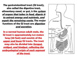
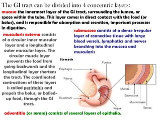
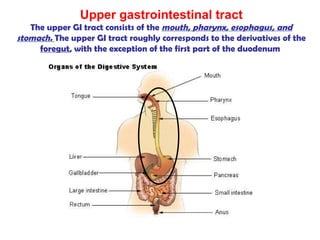
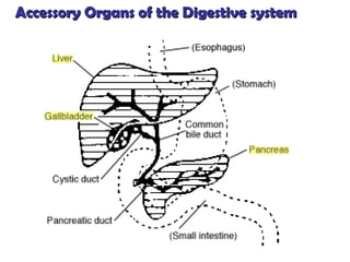
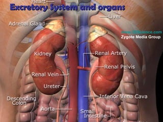
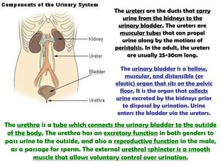
SlideShare a Scribd company logo
Digestive System Digestive System
The  gastrointestinal tract  ( GI tract ), also called the  digestive tract ,  alimentary canal , or  gut , is the  system of organs that takes in food, digests it to extract energy and nutrients, and expels the remaining waste.  The major functions of the GI tract are  digestion and excretion . In a normal human adult male, the GI tract is approximately 6.5 meters long (20 feet) and consists of the  upper and lower  GI tracts. The tract may also be divided into  foregut, midgut, and hindgut, reflecting the embryological origin of each segment of the tract.
The GI tract can be divided into 4 concentric layers: mucosa  the innermost layer of the GI tract, surrounding the lumen, or space within the tube. This layer comes in direct contact with the food (or bolus), and is responsible for absorption and secretion, important processes in digestion. submucosa   consists of a dense irregular layer of connective tissue with large blood vessels, lymphatics and nerves branching into the mucosa and muscularis muscularis externa   consists of a circular inner muscular layer and a longitudinal outer muscular layer. The circular muscle layer prevents the food from going backwards and the longitudinal layer shortens the tract. The coordinated contractions of these layers is called  peristalsis  and propels the bolus, or balled-up food, through the GI tract.  adventitia   (or serosa)   consists of several layers of epithelia.
Upper gastrointestinal tract The upper GI tract consists of the  mouth, pharynx, esophagus, and stomach.  The upper GI tract roughly corresponds to the derivatives of the  foregut , with the exception of the first part of the duodenum
The primary function of teeth is  to tear, scrape, and chew food  (=mechanical digestion).  The mouth contains the  buccal mucosa , which contains the openings of the  salivary glands; the tongue; and the teeth .
Summary of salivary gland features Parotid gland (#1) – secretes a watery fluid rich in amylase. Sublingual gland (#3) – secretions are thick and stringy which binds food particles and lubricates during swallowing.   Submandibular gland (#2) – secretes watery fluid rich in amylase but  more viscous. Salivary Glands – moistens food particles, helps bind them together and begins the chemical digestion of food. Saliva contains enzymes that initiate the breakdown of carbohydrates.
Behind the mouth lies the pharynx (refer to our discussion of the respiratory system), which  leads to  a hollow muscular tube, the esophagus. The esophagus extends through the chest and  pierces the diaphragm  to reach the stomach.  Food is passed through the esophagus by using the process of  peristalsis . Specifically,  it connects the pharynx with the stomach, where the  second stage of digestion is initiated  (the first stage of digestion is in the mouth, with teeth and tongue masticating food and mixing it with saliva).
The esophagus is lined with a  mucous membrane , and is more deeply lined with muscle that acts with peristaltic action to move swallowed food down to the stomach.
The  stomach  is a bean-shaped hollow muscular organ involved in the  second phase of digestion , following mastication. The stomach is usually a  highly acidic environment due to gastric acid production which produces a pH range usually between 2 and 4  depending on food intake and other factors (a pH lower than 2 generally indicates a health problem). Such an environment is able to break down large molecules (such as from food) to smaller ones so that  they can eventually be absorbed from the small intestine.  The stomach can produce and secrete about 2 to 3 liters of gastric acid per day.
Two smooth muscle bands, or sphincters, keep the contents of the stomach contained. They are the  Cardiac or  esophageal sphincter  dividing the tract above, and the  Pyloric sphincter  dividing the stomach from the small intestine. In humans, the stomach has a volume of about  50 mL  when empty. After a meal, it generally expands to hold about  1 litre  of food, but it can actually expand to hold as much as 4 litres. The human stomach has more nerve endings than the human brain .  Chyme  is the liquid substance found in the stomach before passing through the pyloric valve and entering the duodenum.
The movement and the flow of chemicals into the stomach are controlled by both the  autonomic nervous system  and by various digestive system hormones: Pepsin  is a necessity in  protein digestion . Other functions include  absorbing water, some ions, and some lipid soluble compounds such as alcohol, aspirin, and caffeine. Gastrin  causes an increase in the  secretion of  HCl  and pepsinogen. The stomach is divided into four sections, each of which has different cells and functions. The sections are: Cardia where the contents of the esophagus empty into the stomach. Fundus formed by the upper curvature of the organ. Body or corpus the main, central region. Pylorus  the lower section of the organ that facilitates emptying the contents into the small intestine.
Most cases of stomach ulcers, gastritis, and stomach cancer are caused by  Helicobacter pylori  infection.
[object Object],[object Object],[object Object],[object Object],[object Object],[object Object],[object Object],[object Object],[object Object],[object Object],[object Object],[object Object]
The intestinal tract can be broadly divided into two different parts,  the small and large intestine.  Grayish-purple in color, the small intestine is the  first and longest , measuring  twenty feet  on average in an adult man.
The  lumen  is the cavity where digested material passes through and from where  nutrients are absorbed . Along the whole length of the small intestine are  goblet cells . These  secrete mucus  which lubricates the passage of food along and protects it from digestive enzymes.  Villi  are extensions of the mucosa and  increase the overall surface  area of the intestine.  Micro villi  are present on the epithelium of a villus and further increase the surface area over which absorption can take place.
The small intestine is divided into three structural parts: A. duodenum  25 cm (9.84 in) in length  B. jejunum  2.5 m (8.2 ft)  C. ileum  3.5 m (11.5 ft)  Although the small intestine is much longer than the large intestine (typically 4-5 times longer), it is referred to as such due to its comparatively  smaller diameter . On average, the diameter of the small intestine of an adult human measures approximately  2.5-3cm , and the large intestine measures about 7.6 cm in diameter. Absorption of the  majority of nutrients takes place in the jejunum , with the following notable exceptions: Iron is absorbed in the duodenum   Vitamin B12 is absorbed in the terminal ileum .  Water and lipids are absorbed by passive diffusion throughout
The digestion of  proteins into peptides and amino acids  principally occurs in the stomach but some also occurs in the small intestine. The small intestine is where the most chemical digestion takes place: lipids (fats) are degraded into fatty acids and glycerol.  Lipid digestion is the sole responsibility of the small intestine.  Pancreatic lipase  breaks down triglycerides into free fatty acids and monoglycerides.   carbohydrates are degraded into simple sugars  (e.g., glucose). In the small intestine pancreatic  amylase  breaks down carbohydrates into oligosaccharides. Chemical digestion in the duodenum
The duodenum is largely responsible for the  breakdown of food in the small intestine .   The pH in the jejunum is usually between 7 and 8 (neutral or slightly alkaline). The  jejunum and the ileum are suspended by mesentery which gives the bowel great mobility within the abdomen. It also contains muscles to help move the food along.
The function of the ileum is to  absorb the products of digestion.  The wall itself is made up of folds, each of which has many tiny finger-like projections known as  villi , on its surface. In turn, the epithelial cells which line these villi possess even larger numbers of  microvilli . Therefore the ileum has an extremely large surface area both for the absorption (attachment) of enzyme molecules and for the absorption of products of digestion.
[object Object],[object Object],[object Object],[object Object],[object Object],[object Object],[object Object],[object Object],[object Object],[object Object],[object Object],[object Object],[object Object],[object Object],The  large intestine , or the organ which is now more commonly referred to as the  colon , is the  last part of the digestive system : the final stage of the alimentary canal. Its function is to  absorb the remaining water  from indigestible food matter, store these unusable food matter (wastes) and then  eliminates the wastes  from the body.
The  large intestine  hosts several kinds of  bacteria (i.e. ecoli)  that deal with molecules the human body is not able to breakdown itself. This is an example of symbiosis. These bacteria also account for the production of gases inside our intestine (this gas is released as  flatulence  when removed through the anus). The large intestine houses over 700 species of bacteria that perform a variety of functions including synthesis of  vitamin B complex vitamins and vitamin K.
The large intestine  takes 12 to 24 hours  to finish up the remaining processes of the digestive system. Food  is not  broken down any further in this stage of digestion. The large intestine  absorbs vitamins that are created by the bacteria.  The most obvious difference between the small intestine and the large intestine is that the large intestine  is wider and lacks villi.  Medical literature shows that the appendix is rich in infection-fighting lymphoid cells, and therefore,  has an important role in immunity.   Appendicitis  is the result of a blockage that traps infectious material in the lumen. The appendix can be removed with no damage or consequence to the patient
The  cecum  is a pouch connected to the  ascending colon of the large intestine and the ileum.  It is considered to be the  beginning  of the large intestine.
Ascending colon The ascending colon, on the right side of the abdomen, is about 12.5 cm long.  Transverse colon The transverse colon hangs off the stomach, attached to it by a wide band of tissue called the greater omentum. More cancers form as the large intestine goes along and the  contents become more solid (water is removed) in order to form feces.  Descending colon The descending colon is the part of the colon from the splenic flexure to the beginning of the sigmoid colon.  Sigmoid colon The  sigmoid colon  is the part of the large intestine after the descending colon and before the rectum. The name  sigmoid  means S-shaped. The walls of the sigmoid colon are muscular, and  contract to increase the pressure inside the colon, causing the stool to move into the rectum . Sigmoidoscopy  is a common diagnostic technique used to examine the sigmoid colon.
The  rectum  is the final straight portion of the large intestine, terminating in the anus. The human rectum is about 12 cm long.  The rectum intestinum acts as a  temporary storage facility for feces.  As the rectal walls expand due to the materials filling it from within, stretch receptors from the nervous system located in the rectal walls stimulate the desire to defecate. If the  urge is not acted upon, the material in the rectum is often returned to the colon where more water is absorbed.  If defecation is delayed for a prolonged period, constipation and hardened feces results. The rectum shortens as material is forced into the anal canal and peristaltic waves propel the feces out of the rectum. The  internal and external sphincter allow the feces to be passed by muscles pulling the anus up over the exiting feces. Anus- terminal end of the digestive system
Accessory Organs of the Digestive system
The  liver  is an organ that plays a major role in  metabolism  and has a number of functions in the body,  including glycogen storage, protein synthesis, and drug detoxification.  This organ also is the  largest gland  in the human body. It  produces bile , an alkaline compound which aids in digestion.  The liver is the  second largest organ  (the largest organ being the skin). The liver lies on the right of the stomach and makes a kind of bed for the gallbladder (which stores bile).
The bile produced in the liver is collected in bile canaliculi, which merge to form  bile ducts. The  gallbladder   stores about 50 ml of bile (or "gall") until the body needs it for digestion.  The bile, produced in the liver,  emulsifies fats and neutralizes acids in partly digested food. After being stored in the gallbladder, the bile becomes  more concentrated than when it left the liver, increasing its potency and intensifying its effect on fats.
The  pancreas  is an organ in the  digestive and endocrine system . It is both  exocrine  (secreting pancreatic juice containing digestive enzymes) and  endocrine  (producing several important hormones, including insulin and glucagon).
Excretory System and organs www.3DScience.com  Zygote Media Group
In anatomy, the  kidneys  are bean-shaped  excretory organs . Part of the urinary system, the kidneys  filter wastes (such as urea) from the blood and excrete them, along with water, as urine.  The medical field that studies the kidneys and diseases of the kidney is called  nephrology . The basic functional unit of the kidney is the  nephron , of which there are more than a million within the cortex and medulla of each normal adult human kidney.  Nephrons regulate water and soluble matter (especially electrolytes) in the body by first filtering the blood under pressure, and then reabsorbing some necessary fluid and molecules back into the blood  while secreting other, unneeded molecules.
Nephron – Glomerulus/Bowman’s Capsule A  glomerulus  is a capillary tuft surrounded by  Bowman's capsule  in nephrons of the kidney. The resistance of the arterioles results in high pressure in the glomerulus aiding the process of  ultrafiltration  where fluids and soluble materials in the blood are forced out of the capillaries and into Bowman's capsule. A glomerulus and its surrounding Bowman's capsule constitute a  renal corpuscle , the basic filtration unit of the kidney.
The kidney is one of the major organs involved in  whole-body homeostasis.  Among its homeostatic functions are  acid-base  balance, regulation of electrolyte concentrations,  control of blood volume, and regulation of blood pressure.  The kidneys accomplish these homeostatic functions independently and through coordination with other organs, particularly those of the endocrine system. The kidney communicates with these organs through hormones secreted into the bloodstream. Congenital absence of one or both kidneys, known as unilateral or bilateral  renal agenesis  can occur.
The  ureters  are the ducts that  carry urine from the kidneys to the urinary bladder.  The ureters are  muscular tubes  that can propel urine along by the motions of  peristalsis . In the adult, the ureters are usually 25-30cm long. The  urinary bladder  is a  hollow, muscular, and distensible (or elastic) organ that sits on the pelvic floor.  It is the organ that  collects urine  excreted by the kidneys prior to disposal by urination. Urine enters the bladder via the ureters. The  urethra  is a  tube which connects the urinary bladder to the outside of the body.  The urethra has an  excretory function  in both genders to pass urine to the outside, and also a  reproductive function  in the male, as a passage for sperm. The external  urethral sphincter is a smooth muscle that allows voluntary control over urination.

More Related Content

What's hot

The digestive system
The digestive systemThe digestive system
The digestive system
rajender arutla
 
ANATOMY OF PANCREAS
ANATOMY OF PANCREASANATOMY OF PANCREAS
ANATOMY OF PANCREAS
Aamir Hela
 
Stomach anatomy and physiology
Stomach anatomy and physiologyStomach anatomy and physiology
Stomach anatomy and physiology
Ashish Tripathi
 
Gastrointestinal physiology
Gastrointestinal physiologyGastrointestinal physiology
Gastrointestinal physiology
Kern Rocke
 
large intestine
large intestinelarge intestine
large intestine
Muni Venkatesh
 
Acid secretion
Acid secretionAcid secretion
Acid secretion
Sharath !!!!!!!!
 
Gall bladder Anatomo physiology
Gall bladder Anatomo physiology Gall bladder Anatomo physiology
Gall bladder Anatomo physiology
AbdullahIhsaas
 
Stomach anatomy
Stomach anatomy Stomach anatomy
Stomach anatomy
Mawara Ali
 
abdominal cavity
 abdominal cavity abdominal cavity
abdominal cavity
Jay Patel
 
Anatomy and physiology of the gastrointestinal tract (git)
Anatomy and physiology of the gastrointestinal tract (git)Anatomy and physiology of the gastrointestinal tract (git)
Anatomy and physiology of the gastrointestinal tract (git)
Dr. Armaan Singh
 
Liver
LiverLiver
Kidney
KidneyKidney
Liver
LiverLiver
The pancreas
The pancreasThe pancreas
The pancreas
Brisso Mathew Arackal
 
Small intestine
Small intestine Small intestine
Small intestine
Brisso Mathew Arackal
 
Anatomy & Physiology of large intestine
Anatomy & Physiology of large intestineAnatomy & Physiology of large intestine
Anatomy & Physiology of large intestine
iqraIrshad14
 
Structure of Human Stomach by Prabeen
Structure of Human Stomach by PrabeenStructure of Human Stomach by Prabeen
Structure of Human Stomach by Prabeen
Prabeen Mahato
 
anatomy of male urethra
anatomy of male urethraanatomy of male urethra
anatomy of male urethra
madhu sunkara
 
Gall Bladder & Pancreas.
Gall Bladder & Pancreas.Gall Bladder & Pancreas.
Gall Bladder & Pancreas.
Saadiyah Naeemi
 

What's hot (20)

The digestive system
The digestive systemThe digestive system
The digestive system
 
ANATOMY OF PANCREAS
ANATOMY OF PANCREASANATOMY OF PANCREAS
ANATOMY OF PANCREAS
 
Stomach anatomy and physiology
Stomach anatomy and physiologyStomach anatomy and physiology
Stomach anatomy and physiology
 
Gastrointestinal physiology
Gastrointestinal physiologyGastrointestinal physiology
Gastrointestinal physiology
 
large intestine
large intestinelarge intestine
large intestine
 
Acid secretion
Acid secretionAcid secretion
Acid secretion
 
Gall bladder Anatomo physiology
Gall bladder Anatomo physiology Gall bladder Anatomo physiology
Gall bladder Anatomo physiology
 
Stomach anatomy
Stomach anatomy Stomach anatomy
Stomach anatomy
 
abdominal cavity
 abdominal cavity abdominal cavity
abdominal cavity
 
Anatomy and physiology of the gastrointestinal tract (git)
Anatomy and physiology of the gastrointestinal tract (git)Anatomy and physiology of the gastrointestinal tract (git)
Anatomy and physiology of the gastrointestinal tract (git)
 
Liver
LiverLiver
Liver
 
Digestive system
Digestive systemDigestive system
Digestive system
 
Kidney
KidneyKidney
Kidney
 
Liver
LiverLiver
Liver
 
The pancreas
The pancreasThe pancreas
The pancreas
 
Small intestine
Small intestine Small intestine
Small intestine
 
Anatomy & Physiology of large intestine
Anatomy & Physiology of large intestineAnatomy & Physiology of large intestine
Anatomy & Physiology of large intestine
 
Structure of Human Stomach by Prabeen
Structure of Human Stomach by PrabeenStructure of Human Stomach by Prabeen
Structure of Human Stomach by Prabeen
 
anatomy of male urethra
anatomy of male urethraanatomy of male urethra
anatomy of male urethra
 
Gall Bladder & Pancreas.
Gall Bladder & Pancreas.Gall Bladder & Pancreas.
Gall Bladder & Pancreas.
 

Viewers also liked

Chapter 15 the uirnary system
Chapter 15 the uirnary systemChapter 15 the uirnary system
Chapter 15 the uirnary systemjpolos
 
Heart and lung development
Heart and lung developmentHeart and lung development
Heart and lung development
Harmeet Kaur Brar
 
Embryology of heart and fetal circulation
Embryology of heart and fetal circulationEmbryology of heart and fetal circulation
Embryology of heart and fetal circulation
Archana Koshy
 
Development of Kidney
Development of KidneyDevelopment of Kidney
Development of Kidney
Garima Aggarwal
 
Saliva and salivary glands
Saliva and salivary glandsSaliva and salivary glands
Saliva and salivary glands
Dr Arpita Dutta
 
Introduction of gastrointestinal tract
Introduction of gastrointestinal tractIntroduction of gastrointestinal tract
Introduction of gastrointestinal tractRajesh Goit
 
Oral Histology "tooth development"
Oral Histology "tooth development"Oral Histology "tooth development"
Oral Histology "tooth development"
Ŵĕssam Osama
 
Upper GI bleeding
Upper GI bleedingUpper GI bleeding
Upper GI bleeding
Abed El-Rahman Sawalmeh
 
Approach to gastrointestinal bleeding
Approach to gastrointestinal bleedingApproach to gastrointestinal bleeding
Approach to gastrointestinal bleedingSamir Haffar
 
Upper Gastrointestinal bleeding
Upper Gastrointestinal bleedingUpper Gastrointestinal bleeding
Upper Gastrointestinal bleeding
Ammar L. Aldwaf
 
Gi hormone
Gi hormoneGi hormone
Gi hormone
Atai Rabby
 
Anatomy and Physiology: Gastrointestinal Tract
Anatomy and Physiology: Gastrointestinal TractAnatomy and Physiology: Gastrointestinal Tract
Anatomy and Physiology: Gastrointestinal TractKatherine 'Chingboo' Laud
 
Development of tooth
Development of toothDevelopment of tooth
Development of toothPiyush Verma
 

Viewers also liked (20)

Gastrointestinal tract
Gastrointestinal tractGastrointestinal tract
Gastrointestinal tract
 
Chapter 15 the uirnary system
Chapter 15 the uirnary systemChapter 15 the uirnary system
Chapter 15 the uirnary system
 
11.3 kidney
11.3 kidney11.3 kidney
11.3 kidney
 
6 development of the heart akd
6 development of the  heart akd6 development of the  heart akd
6 development of the heart akd
 
Heart and lung development
Heart and lung developmentHeart and lung development
Heart and lung development
 
Embryology of heart and fetal circulation
Embryology of heart and fetal circulationEmbryology of heart and fetal circulation
Embryology of heart and fetal circulation
 
Embryology of brain
Embryology of brainEmbryology of brain
Embryology of brain
 
Brain Development
Brain DevelopmentBrain Development
Brain Development
 
Development of Kidney
Development of KidneyDevelopment of Kidney
Development of Kidney
 
Gastrointestinal physiology
Gastrointestinal physiologyGastrointestinal physiology
Gastrointestinal physiology
 
Saliva and salivary glands
Saliva and salivary glandsSaliva and salivary glands
Saliva and salivary glands
 
Introduction of gastrointestinal tract
Introduction of gastrointestinal tractIntroduction of gastrointestinal tract
Introduction of gastrointestinal tract
 
Oral Histology "tooth development"
Oral Histology "tooth development"Oral Histology "tooth development"
Oral Histology "tooth development"
 
Upper GI bleeding
Upper GI bleedingUpper GI bleeding
Upper GI bleeding
 
Approach to gastrointestinal bleeding
Approach to gastrointestinal bleedingApproach to gastrointestinal bleeding
Approach to gastrointestinal bleeding
 
Heart embryology
Heart embryologyHeart embryology
Heart embryology
 
Upper Gastrointestinal bleeding
Upper Gastrointestinal bleedingUpper Gastrointestinal bleeding
Upper Gastrointestinal bleeding
 
Gi hormone
Gi hormoneGi hormone
Gi hormone
 
Anatomy and Physiology: Gastrointestinal Tract
Anatomy and Physiology: Gastrointestinal TractAnatomy and Physiology: Gastrointestinal Tract
Anatomy and Physiology: Gastrointestinal Tract
 
Development of tooth
Development of toothDevelopment of tooth
Development of tooth
 

Similar to Digestive system

Digestive system notes
Digestive system notesDigestive system notes
Digestive system notes
Tia Hohler
 
HAP 4 SEMESTER 2 BPHARMACY AS PER PCI SYLLABUS
HAP 4 SEMESTER 2 BPHARMACY AS PER PCI SYLLABUSHAP 4 SEMESTER 2 BPHARMACY AS PER PCI SYLLABUS
HAP 4 SEMESTER 2 BPHARMACY AS PER PCI SYLLABUS
christinajohn24
 
Digestive system
Digestive systemDigestive system
Digestive system
Krishna Varshney
 
Digestion and Absorption - Class 11 ppt
Digestion and Absorption - Class 11 pptDigestion and Absorption - Class 11 ppt
Digestion and Absorption - Class 11 ppt
Karthik Bharadwaj
 
Human Digestive Anatomy
Human Digestive Anatomy Human Digestive Anatomy
Human Digestive Anatomy
Aditi Tanwar
 
Digestion
DigestionDigestion
Digestion
GraciaIssac
 
Study Human Physiology for MH-CET 2014
Study Human Physiology for MH-CET 2014 Study Human Physiology for MH-CET 2014
Study Human Physiology for MH-CET 2014
Ednexa
 
Nutrition in mammals
Nutrition in mammalsNutrition in mammals
Nutrition in mammals
Saad Adil
 
Digestive system
Digestive systemDigestive system
Digestive system
malkeetsingh104
 
Digestive system 1 ehs unit 6
Digestive system 1 ehs unit 6Digestive system 1 ehs unit 6
Digestive system 1 ehs unit 6
RHSHealthScience
 
anatomy: Human digestive system and its parts
anatomy: Human digestive system and its partsanatomy: Human digestive system and its parts
anatomy: Human digestive system and its parts
drparul6375
 
Human digestive system
Human digestive systemHuman digestive system
Human digestive systemSimren Cena
 
Anatomy and physiology ; gastrointestinal.pdf
Anatomy and physiology ; gastrointestinal.pdfAnatomy and physiology ; gastrointestinal.pdf
Anatomy and physiology ; gastrointestinal.pdf
taquqi
 
182-Anatomy-Digestive-System-of-human-body.ppt
182-Anatomy-Digestive-System-of-human-body.ppt182-Anatomy-Digestive-System-of-human-body.ppt
182-Anatomy-Digestive-System-of-human-body.ppt
RatnaWulandari52
 
Human digestive system
Human digestive systemHuman digestive system
Human digestive system
Anamika Gupta
 
(HUMAN PHYSIOLOGY) DIGESTIVE SYSTEM.docx
(HUMAN PHYSIOLOGY) DIGESTIVE SYSTEM.docx(HUMAN PHYSIOLOGY) DIGESTIVE SYSTEM.docx
(HUMAN PHYSIOLOGY) DIGESTIVE SYSTEM.docx
samakshiverma1
 
Digestive system.pptx
Digestive system.pptxDigestive system.pptx
Digestive system.pptx
ahishamohire
 
Digestive system 2
Digestive system 2Digestive system 2
Digestive system 2
skyporter
 

Similar to Digestive system (20)

Digestive system notes
Digestive system notesDigestive system notes
Digestive system notes
 
HAP 4 SEMESTER 2 BPHARMACY AS PER PCI SYLLABUS
HAP 4 SEMESTER 2 BPHARMACY AS PER PCI SYLLABUSHAP 4 SEMESTER 2 BPHARMACY AS PER PCI SYLLABUS
HAP 4 SEMESTER 2 BPHARMACY AS PER PCI SYLLABUS
 
Digestive system
Digestive systemDigestive system
Digestive system
 
Digestion and Absorption - Class 11 ppt
Digestion and Absorption - Class 11 pptDigestion and Absorption - Class 11 ppt
Digestion and Absorption - Class 11 ppt
 
Human Digestive Anatomy
Human Digestive Anatomy Human Digestive Anatomy
Human Digestive Anatomy
 
Digestion
DigestionDigestion
Digestion
 
Snsd world
Snsd worldSnsd world
Snsd world
 
Study Human Physiology for MH-CET 2014
Study Human Physiology for MH-CET 2014 Study Human Physiology for MH-CET 2014
Study Human Physiology for MH-CET 2014
 
Nutrition in mammals
Nutrition in mammalsNutrition in mammals
Nutrition in mammals
 
Digestive system
Digestive systemDigestive system
Digestive system
 
Digestive system
Digestive systemDigestive system
Digestive system
 
Digestive system 1 ehs unit 6
Digestive system 1 ehs unit 6Digestive system 1 ehs unit 6
Digestive system 1 ehs unit 6
 
anatomy: Human digestive system and its parts
anatomy: Human digestive system and its partsanatomy: Human digestive system and its parts
anatomy: Human digestive system and its parts
 
Human digestive system
Human digestive systemHuman digestive system
Human digestive system
 
Anatomy and physiology ; gastrointestinal.pdf
Anatomy and physiology ; gastrointestinal.pdfAnatomy and physiology ; gastrointestinal.pdf
Anatomy and physiology ; gastrointestinal.pdf
 
182-Anatomy-Digestive-System-of-human-body.ppt
182-Anatomy-Digestive-System-of-human-body.ppt182-Anatomy-Digestive-System-of-human-body.ppt
182-Anatomy-Digestive-System-of-human-body.ppt
 
Human digestive system
Human digestive systemHuman digestive system
Human digestive system
 
(HUMAN PHYSIOLOGY) DIGESTIVE SYSTEM.docx
(HUMAN PHYSIOLOGY) DIGESTIVE SYSTEM.docx(HUMAN PHYSIOLOGY) DIGESTIVE SYSTEM.docx
(HUMAN PHYSIOLOGY) DIGESTIVE SYSTEM.docx
 
Digestive system.pptx
Digestive system.pptxDigestive system.pptx
Digestive system.pptx
 
Digestive system 2
Digestive system 2Digestive system 2
Digestive system 2
 

More from Andrew McCaskill (20)

Human reproduction and development
Human reproduction and developmentHuman reproduction and development
Human reproduction and development
 
Respiratory syst
Respiratory systRespiratory syst
Respiratory syst
 
Cardiovascular system
Cardiovascular systemCardiovascular system
Cardiovascular system
 
37 plantnutrition text
37 plantnutrition text37 plantnutrition text
37 plantnutrition text
 
53 communities text
53 communities text53 communities text
53 communities text
 
48 nervous text
48 nervous text48 nervous text
48 nervous text
 
41 animalnutrition text
41  animalnutrition text41  animalnutrition text
41 animalnutrition text
 
51 behavior text
51 behavior text51 behavior text
51 behavior text
 
46 reproduction text
46 reproduction text46 reproduction text
46 reproduction text
 
44 excretion text
44 excretion text44 excretion text
44 excretion text
 
40 animalform&function text
40 animalform&function text40 animalform&function text
40 animalform&function text
 
32 animaldiversity text
32  animaldiversity text32  animaldiversity text
32 animaldiversity text
 
35 plantstructure text
35  plantstructure text35  plantstructure text
35 plantstructure text
 
28 protists
28  protists28  protists
28 protists
 
29 plants ii text
29  plants ii text29  plants ii text
29 plants ii text
 
31 fungi text
31  fungi text31  fungi text
31 fungi text
 
24 originofspecies text
24  originofspecies text24  originofspecies text
24 originofspecies text
 
27 prokaryotes text
27  prokaryotes text27  prokaryotes text
27 prokaryotes text
 
23 evolutionofpopulations text
23  evolutionofpopulations text23  evolutionofpopulations text
23 evolutionofpopulations text
 
22 darwin text
22  darwin text22  darwin text
22 darwin text
 

Digestive system

  • 3. The gastrointestinal tract ( GI tract ), also called the digestive tract , alimentary canal , or gut , is the system of organs that takes in food, digests it to extract energy and nutrients, and expels the remaining waste. The major functions of the GI tract are digestion and excretion . In a normal human adult male, the GI tract is approximately 6.5 meters long (20 feet) and consists of the upper and lower GI tracts. The tract may also be divided into foregut, midgut, and hindgut, reflecting the embryological origin of each segment of the tract.
  • 4. The GI tract can be divided into 4 concentric layers: mucosa the innermost layer of the GI tract, surrounding the lumen, or space within the tube. This layer comes in direct contact with the food (or bolus), and is responsible for absorption and secretion, important processes in digestion. submucosa consists of a dense irregular layer of connective tissue with large blood vessels, lymphatics and nerves branching into the mucosa and muscularis muscularis externa consists of a circular inner muscular layer and a longitudinal outer muscular layer. The circular muscle layer prevents the food from going backwards and the longitudinal layer shortens the tract. The coordinated contractions of these layers is called peristalsis and propels the bolus, or balled-up food, through the GI tract. adventitia (or serosa) consists of several layers of epithelia.
  • 5. Upper gastrointestinal tract The upper GI tract consists of the mouth, pharynx, esophagus, and stomach. The upper GI tract roughly corresponds to the derivatives of the foregut , with the exception of the first part of the duodenum
  • 6. The primary function of teeth is to tear, scrape, and chew food (=mechanical digestion). The mouth contains the buccal mucosa , which contains the openings of the salivary glands; the tongue; and the teeth .
  • 7. Summary of salivary gland features Parotid gland (#1) – secretes a watery fluid rich in amylase. Sublingual gland (#3) – secretions are thick and stringy which binds food particles and lubricates during swallowing. Submandibular gland (#2) – secretes watery fluid rich in amylase but more viscous. Salivary Glands – moistens food particles, helps bind them together and begins the chemical digestion of food. Saliva contains enzymes that initiate the breakdown of carbohydrates.
  • 8. Behind the mouth lies the pharynx (refer to our discussion of the respiratory system), which leads to a hollow muscular tube, the esophagus. The esophagus extends through the chest and pierces the diaphragm to reach the stomach. Food is passed through the esophagus by using the process of peristalsis . Specifically, it connects the pharynx with the stomach, where the second stage of digestion is initiated (the first stage of digestion is in the mouth, with teeth and tongue masticating food and mixing it with saliva).
  • 9. The esophagus is lined with a mucous membrane , and is more deeply lined with muscle that acts with peristaltic action to move swallowed food down to the stomach.
  • 10. The stomach is a bean-shaped hollow muscular organ involved in the second phase of digestion , following mastication. The stomach is usually a highly acidic environment due to gastric acid production which produces a pH range usually between 2 and 4 depending on food intake and other factors (a pH lower than 2 generally indicates a health problem). Such an environment is able to break down large molecules (such as from food) to smaller ones so that they can eventually be absorbed from the small intestine. The stomach can produce and secrete about 2 to 3 liters of gastric acid per day.
  • 11. Two smooth muscle bands, or sphincters, keep the contents of the stomach contained. They are the Cardiac or esophageal sphincter dividing the tract above, and the Pyloric sphincter dividing the stomach from the small intestine. In humans, the stomach has a volume of about 50 mL when empty. After a meal, it generally expands to hold about 1 litre of food, but it can actually expand to hold as much as 4 litres. The human stomach has more nerve endings than the human brain . Chyme is the liquid substance found in the stomach before passing through the pyloric valve and entering the duodenum.
  • 12. The movement and the flow of chemicals into the stomach are controlled by both the autonomic nervous system and by various digestive system hormones: Pepsin is a necessity in protein digestion . Other functions include absorbing water, some ions, and some lipid soluble compounds such as alcohol, aspirin, and caffeine. Gastrin causes an increase in the secretion of HCl and pepsinogen. The stomach is divided into four sections, each of which has different cells and functions. The sections are: Cardia where the contents of the esophagus empty into the stomach. Fundus formed by the upper curvature of the organ. Body or corpus the main, central region. Pylorus the lower section of the organ that facilitates emptying the contents into the small intestine.
  • 13. Most cases of stomach ulcers, gastritis, and stomach cancer are caused by Helicobacter pylori infection.
  • 14.
  • 15. The intestinal tract can be broadly divided into two different parts, the small and large intestine. Grayish-purple in color, the small intestine is the first and longest , measuring twenty feet on average in an adult man.
  • 16. The lumen is the cavity where digested material passes through and from where nutrients are absorbed . Along the whole length of the small intestine are goblet cells . These secrete mucus which lubricates the passage of food along and protects it from digestive enzymes. Villi are extensions of the mucosa and increase the overall surface area of the intestine. Micro villi are present on the epithelium of a villus and further increase the surface area over which absorption can take place.
  • 17. The small intestine is divided into three structural parts: A. duodenum 25 cm (9.84 in) in length B. jejunum 2.5 m (8.2 ft) C. ileum 3.5 m (11.5 ft) Although the small intestine is much longer than the large intestine (typically 4-5 times longer), it is referred to as such due to its comparatively smaller diameter . On average, the diameter of the small intestine of an adult human measures approximately 2.5-3cm , and the large intestine measures about 7.6 cm in diameter. Absorption of the majority of nutrients takes place in the jejunum , with the following notable exceptions: Iron is absorbed in the duodenum Vitamin B12 is absorbed in the terminal ileum . Water and lipids are absorbed by passive diffusion throughout
  • 18. The digestion of proteins into peptides and amino acids principally occurs in the stomach but some also occurs in the small intestine. The small intestine is where the most chemical digestion takes place: lipids (fats) are degraded into fatty acids and glycerol. Lipid digestion is the sole responsibility of the small intestine. Pancreatic lipase breaks down triglycerides into free fatty acids and monoglycerides. carbohydrates are degraded into simple sugars (e.g., glucose). In the small intestine pancreatic amylase breaks down carbohydrates into oligosaccharides. Chemical digestion in the duodenum
  • 19. The duodenum is largely responsible for the breakdown of food in the small intestine . The pH in the jejunum is usually between 7 and 8 (neutral or slightly alkaline). The jejunum and the ileum are suspended by mesentery which gives the bowel great mobility within the abdomen. It also contains muscles to help move the food along.
  • 20. The function of the ileum is to absorb the products of digestion. The wall itself is made up of folds, each of which has many tiny finger-like projections known as villi , on its surface. In turn, the epithelial cells which line these villi possess even larger numbers of microvilli . Therefore the ileum has an extremely large surface area both for the absorption (attachment) of enzyme molecules and for the absorption of products of digestion.
  • 21.
  • 22. The large intestine hosts several kinds of bacteria (i.e. ecoli) that deal with molecules the human body is not able to breakdown itself. This is an example of symbiosis. These bacteria also account for the production of gases inside our intestine (this gas is released as flatulence when removed through the anus). The large intestine houses over 700 species of bacteria that perform a variety of functions including synthesis of vitamin B complex vitamins and vitamin K.
  • 23. The large intestine takes 12 to 24 hours to finish up the remaining processes of the digestive system. Food is not broken down any further in this stage of digestion. The large intestine absorbs vitamins that are created by the bacteria. The most obvious difference between the small intestine and the large intestine is that the large intestine is wider and lacks villi. Medical literature shows that the appendix is rich in infection-fighting lymphoid cells, and therefore, has an important role in immunity. Appendicitis is the result of a blockage that traps infectious material in the lumen. The appendix can be removed with no damage or consequence to the patient
  • 24. The cecum is a pouch connected to the ascending colon of the large intestine and the ileum. It is considered to be the beginning of the large intestine.
  • 25. Ascending colon The ascending colon, on the right side of the abdomen, is about 12.5 cm long. Transverse colon The transverse colon hangs off the stomach, attached to it by a wide band of tissue called the greater omentum. More cancers form as the large intestine goes along and the contents become more solid (water is removed) in order to form feces. Descending colon The descending colon is the part of the colon from the splenic flexure to the beginning of the sigmoid colon. Sigmoid colon The sigmoid colon is the part of the large intestine after the descending colon and before the rectum. The name sigmoid means S-shaped. The walls of the sigmoid colon are muscular, and contract to increase the pressure inside the colon, causing the stool to move into the rectum . Sigmoidoscopy is a common diagnostic technique used to examine the sigmoid colon.
  • 26. The rectum is the final straight portion of the large intestine, terminating in the anus. The human rectum is about 12 cm long. The rectum intestinum acts as a temporary storage facility for feces. As the rectal walls expand due to the materials filling it from within, stretch receptors from the nervous system located in the rectal walls stimulate the desire to defecate. If the urge is not acted upon, the material in the rectum is often returned to the colon where more water is absorbed. If defecation is delayed for a prolonged period, constipation and hardened feces results. The rectum shortens as material is forced into the anal canal and peristaltic waves propel the feces out of the rectum. The internal and external sphincter allow the feces to be passed by muscles pulling the anus up over the exiting feces. Anus- terminal end of the digestive system
  • 27. Accessory Organs of the Digestive system
  • 28. The liver is an organ that plays a major role in metabolism and has a number of functions in the body, including glycogen storage, protein synthesis, and drug detoxification. This organ also is the largest gland in the human body. It produces bile , an alkaline compound which aids in digestion. The liver is the second largest organ (the largest organ being the skin). The liver lies on the right of the stomach and makes a kind of bed for the gallbladder (which stores bile).
  • 29. The bile produced in the liver is collected in bile canaliculi, which merge to form bile ducts. The gallbladder stores about 50 ml of bile (or "gall") until the body needs it for digestion. The bile, produced in the liver, emulsifies fats and neutralizes acids in partly digested food. After being stored in the gallbladder, the bile becomes more concentrated than when it left the liver, increasing its potency and intensifying its effect on fats.
  • 30. The pancreas is an organ in the digestive and endocrine system . It is both exocrine (secreting pancreatic juice containing digestive enzymes) and endocrine (producing several important hormones, including insulin and glucagon).
  • 31. Excretory System and organs www.3DScience.com Zygote Media Group
  • 32. In anatomy, the kidneys are bean-shaped excretory organs . Part of the urinary system, the kidneys filter wastes (such as urea) from the blood and excrete them, along with water, as urine. The medical field that studies the kidneys and diseases of the kidney is called nephrology . The basic functional unit of the kidney is the nephron , of which there are more than a million within the cortex and medulla of each normal adult human kidney. Nephrons regulate water and soluble matter (especially electrolytes) in the body by first filtering the blood under pressure, and then reabsorbing some necessary fluid and molecules back into the blood while secreting other, unneeded molecules.
  • 33. Nephron – Glomerulus/Bowman’s Capsule A glomerulus is a capillary tuft surrounded by Bowman's capsule in nephrons of the kidney. The resistance of the arterioles results in high pressure in the glomerulus aiding the process of ultrafiltration where fluids and soluble materials in the blood are forced out of the capillaries and into Bowman's capsule. A glomerulus and its surrounding Bowman's capsule constitute a renal corpuscle , the basic filtration unit of the kidney.
  • 34. The kidney is one of the major organs involved in whole-body homeostasis. Among its homeostatic functions are acid-base balance, regulation of electrolyte concentrations, control of blood volume, and regulation of blood pressure. The kidneys accomplish these homeostatic functions independently and through coordination with other organs, particularly those of the endocrine system. The kidney communicates with these organs through hormones secreted into the bloodstream. Congenital absence of one or both kidneys, known as unilateral or bilateral renal agenesis can occur.
  • 35. The ureters are the ducts that carry urine from the kidneys to the urinary bladder. The ureters are muscular tubes that can propel urine along by the motions of peristalsis . In the adult, the ureters are usually 25-30cm long. The urinary bladder is a hollow, muscular, and distensible (or elastic) organ that sits on the pelvic floor. It is the organ that collects urine excreted by the kidneys prior to disposal by urination. Urine enters the bladder via the ureters. The urethra is a tube which connects the urinary bladder to the outside of the body. The urethra has an excretory function in both genders to pass urine to the outside, and also a reproductive function in the male, as a passage for sperm. The external urethral sphincter is a smooth muscle that allows voluntary control over urination.