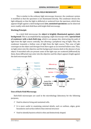
GENERAL MICROBIOLOGY UNIT 2
Arthi Mohan Page 1
DARK-FIELD MICROSCOPY
This is similar to the ordinary light microscope; however, the condenser system
is modified so that the specimen is not illuminated directly. The condenser directs the
light obliquely so that the light is deflected or scattered from the specimen, which then
appears bright against a dark background. Live, unstained specimens can be observed
more readily with dark-field than with bright-field microscopy.
Working Principle
In a dark field microscope the object is brightly illuminated against a dark
background. This is accomplished by equipping a light microscope with a special kind
of condenser with a dark field stop, which is an opaque disc obstructing the path of
light from the light source centrally, but allowing a peripheral ring of light. Thus, the
condenser transmits a hollow cone of light from the light source. This cone of light
converges on the object and diverges from there again as an inverted hollow cone. Thus,
no light enters into the objective and the background remains dark in the absence of any
object. If microbial cells are present some of the light rays are scattered (diffracted) by
them, these diffracted rays enter into the objective and the object appears bright against
the dark microscopic field.
.
Uses of Dark-Field Microscopy
Dark-field microscopes are used in the microbiology laboratory for the following
purposes,
 Used to observe living and unstained cells.
 It is more useful in examining external details, such as outlines, edges, grain
boundaries and surface defects than internal structure
 Used to identify bacteria which is thin and distinctively shaped.
or Annular stop or
Dark field stop
GENERAL MICROBIOLOGY UNIT 2
Arthi Mohan Page 2
 Used to identify bacteria which are thin and distinctively shaped such as
spirochetes (cannot be seen by light microscopy because of their thin
dimensions) such as Treponema pallidum (syphilis), Borrelia burgdorferi (lyme
borreliosis) and Leptospira interrogans (leptospirosis) in clinical samples.
 To observe microbial motility; tufts of bacterial flagella can often be seen in
unstained cells by dark-field or phase-contrast microscopy
 To observe internal structure in larger eukaryotic microorganisms such as algae,
yeasts, etc.
 Used to observe the blood cells.
 A stereo dark-field microscope used to observe the shrimp or other
invertebrates.
Advantages of Dark-Field Microscopy
 Resolution by dark-field microscopy is somewhat better than bright-field
microscopy
 Improves image contrast without the use of stain, and thus do not kill cells.
 It is useful for those specimens that are transparent and absorb little or no light.
 Direct detection of non-culturable bacteria present in patient samples.
 No sample preparation is required
 Requires no special set up, even a light microscope can be converted to dark
field.
 Marine organisms such as algae, plankton, diatoms, insects, fibers, hairs, yeast,
and protozoa can observe under a dark-field microscope.
 Dark-field microscope can use to study for minerals and crystals, thin polymers,
and some ceramics.
Limitations of Dark-Field Microscopy
o Necessity to examine wet, moist specimens containing living organisms very
quickly, because visualization of the moving bacteria is essential to detection.
o An intense amount of light is required for a dark field microscope to work.
o The sample must be very strongly illuminated, which can cause damage to the
sample.
o Besides the sample, dust particles also scatter the light and appear bright.
GENERAL MICROBIOLOGY UNIT 2
Arthi Mohan Page 3
o Sample material needs to be spread thinly, dense preparations can grossly affect
the contrast and accuracy of the image.
References
Madigan Michael T, Bender, Kelly S, Buckley, Daniel H, Sattley, W. Matthew, & Stahl,
David A. (2018). Brock Biology of Microorganisms (15th Edition). Pearson.
Willey, Joanne M, Sherwood, Linda M, & Woolverton, Christopher J. (2016). Prescott’s
Microbiology (10 edition). McGraw-Hill Education.

3. Dark-field microscopy.pdf

  • 1.
    GENERAL MICROBIOLOGY UNIT2 Arthi Mohan Page 1 DARK-FIELD MICROSCOPY This is similar to the ordinary light microscope; however, the condenser system is modified so that the specimen is not illuminated directly. The condenser directs the light obliquely so that the light is deflected or scattered from the specimen, which then appears bright against a dark background. Live, unstained specimens can be observed more readily with dark-field than with bright-field microscopy. Working Principle In a dark field microscope the object is brightly illuminated against a dark background. This is accomplished by equipping a light microscope with a special kind of condenser with a dark field stop, which is an opaque disc obstructing the path of light from the light source centrally, but allowing a peripheral ring of light. Thus, the condenser transmits a hollow cone of light from the light source. This cone of light converges on the object and diverges from there again as an inverted hollow cone. Thus, no light enters into the objective and the background remains dark in the absence of any object. If microbial cells are present some of the light rays are scattered (diffracted) by them, these diffracted rays enter into the objective and the object appears bright against the dark microscopic field. . Uses of Dark-Field Microscopy Dark-field microscopes are used in the microbiology laboratory for the following purposes,  Used to observe living and unstained cells.  It is more useful in examining external details, such as outlines, edges, grain boundaries and surface defects than internal structure  Used to identify bacteria which is thin and distinctively shaped. or Annular stop or Dark field stop
  • 2.
    GENERAL MICROBIOLOGY UNIT2 Arthi Mohan Page 2  Used to identify bacteria which are thin and distinctively shaped such as spirochetes (cannot be seen by light microscopy because of their thin dimensions) such as Treponema pallidum (syphilis), Borrelia burgdorferi (lyme borreliosis) and Leptospira interrogans (leptospirosis) in clinical samples.  To observe microbial motility; tufts of bacterial flagella can often be seen in unstained cells by dark-field or phase-contrast microscopy  To observe internal structure in larger eukaryotic microorganisms such as algae, yeasts, etc.  Used to observe the blood cells.  A stereo dark-field microscope used to observe the shrimp or other invertebrates. Advantages of Dark-Field Microscopy  Resolution by dark-field microscopy is somewhat better than bright-field microscopy  Improves image contrast without the use of stain, and thus do not kill cells.  It is useful for those specimens that are transparent and absorb little or no light.  Direct detection of non-culturable bacteria present in patient samples.  No sample preparation is required  Requires no special set up, even a light microscope can be converted to dark field.  Marine organisms such as algae, plankton, diatoms, insects, fibers, hairs, yeast, and protozoa can observe under a dark-field microscope.  Dark-field microscope can use to study for minerals and crystals, thin polymers, and some ceramics. Limitations of Dark-Field Microscopy o Necessity to examine wet, moist specimens containing living organisms very quickly, because visualization of the moving bacteria is essential to detection. o An intense amount of light is required for a dark field microscope to work. o The sample must be very strongly illuminated, which can cause damage to the sample. o Besides the sample, dust particles also scatter the light and appear bright.
  • 3.
    GENERAL MICROBIOLOGY UNIT2 Arthi Mohan Page 3 o Sample material needs to be spread thinly, dense preparations can grossly affect the contrast and accuracy of the image. References Madigan Michael T, Bender, Kelly S, Buckley, Daniel H, Sattley, W. Matthew, & Stahl, David A. (2018). Brock Biology of Microorganisms (15th Edition). Pearson. Willey, Joanne M, Sherwood, Linda M, & Woolverton, Christopher J. (2016). Prescott’s Microbiology (10 edition). McGraw-Hill Education.