
More Related Content
What's hot
What's hot (20)
Similar to Dark ground microscopy
Similar to Dark ground microscopy (20)
More from 9925752690
More from 9925752690 (12)
Recently uploaded
Recently uploaded (20)
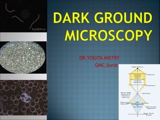
Dark ground microscopy
- 2. Historical aspect Principle How to convert other microscope into dark field microscope Uses Advantages Disadvantages
- 3. Dark – field microscopy is an optical microscopy illumination technique used to enhance the contrast in unstained samples . In this the specimens is illuminated by inclined rays and the field of vision is rendered dark by preventing the axial rays with use of a circular stopper in the light path . The result is a bright image of the object against a dark background.
- 4. In this the specimens is illuminated by inclined rays and the field of vision is rendered dark by preventing the axial rays with use of a circular stopper in the light path . The result is a bright image of the object against a dark background. This produces the classic appearance of a dark, almost black, background with bright objects on it.
- 5. In 1830, J.J. Lister (the father of Joseph Lister) invented the darkfield microscope, in which the standard brightfield (Abbe) condenser is replaced with a single or double-reflecting dark field condenser. In 1906 in Vienna, Karl Landsteiner and Viktor Mucha were the first to use darkfield microscopy to visualize T pallidum from syphilis lesions.
- 7. Light reaches the object mounted on the stage through the condenser . The condenser lens focuses the light towards the sample. The patch stop provided in the condenser lens blocks light around the central region allowing light to pass only the periphery of the lens Of the light that enters the specimen , most is directly transmitted , while some is scattered from the sample .
- 8. The scattered light alone enters the objective lens and produce the image . The directly transmitted light is not collected and is omitted . Thus the field of vision is rendered dark and the image created is by the scattered light and therefore , cell component that reflect light are clear in the image .
- 9. Light enters the microscope for illumination of the sample. A specially sized disc, the patch stop (see figure) blocks some light from the light source, leaving an outer ring of illumination. A wide phase annulus can also be reasonably substituted at low magnification. The condenser lens focuses the light towards the sample. The light enters the sample. Most is directly transmitted, while some is scattered from the sample. The scattered light enters the objective lens, while the directly transmitted light simply misses the lens and is not collected due to a direct illumination block (see figure). Only the scattered light goes on to produce the image, while the directly transmitted light is omitted.
- 11. BRIGHT FIELD MICROSCOPY DARK FIELD MICROSCOPY 1.Light from a plane-wave source is focused through an object by a condenser. 2. Some light is blocked (absorbed) by opaque parts of the object, or reflected away at boundaries between components/materials of different refractive indices. 3. The remainder passes through the objective lens, to the observer. 1.An opaque disc is put between the source and the condenser, blocking out the middle of the beam. 2. The condenser focuses the beam onto the sample. 3. No light enters the objective directly from the source. Light from the beam is scattered by the sample – some scattered into the objective.
- 12. 4.This produces a bright background, with object details appearing darker in the image than their surroundings. The brightness of the brightest parts of the image is determined by the source brightness and block size. 5. This results in poorer contrast compared to darkfield, as the dark areas are generally grey rather than black. 4.Only light scattered by the object enters the objective. This produces a dark background, with sample details appearing brighter than surroundings. The brightness of the brightest parts of the image is determined by the amount of light scattered by the object. 5. This results in superior contrast to bright-field, as dark areas may be completely black, while increasing the brightness of the light source brightens the bright areas
- 13. When viewing a specimen with the 10x objective while slowly raising and lowering the condenser, a point will be reached where a bright spot will appear in the field of view as illustrated in Figure 2(a). As the condenser is slightly raised or lowered, a dark spot similar to the one shown in Figure 2(b) will appear, if the condenser is properly centered. In cases where the condenser is not properly aligned and centered, a typical field of view might look like that shown in Figure 2(c), and if the microscope is not properly adjusted for , the viewfield might appear as it does in Figure 2(d).
- 14. Rheinberg illumination is a special variant of dark – field illumination. It is named after its inventor , Julis Rheinberg . In this variant , transparent coloured filters are inserted just before the condenser so that light rays at high aperature are differently coloured than those at low aperature .
- 16. There is no special microscope model as a dark-field microscope. The light passing through the central part of the condenser lens system is to be screened off allowing only the light in the periphery of the lens to pass . This ensures illumination of the object by inclined rays only. This can be achieved by placing a circular disc called patch stop in between the iris diaphragm and the lens in the condenser. This along with low-power objectives will suffice for ordinary dark-field observation.
- 17. A specimen is placed on the microscope stage as usual, and the illumination should be made as uniform as possible. An aperture diaphragm in the condenser (contrast lever), it should be opened up wide. After focusing at low power, the slide with occulting disks is placed in the light path between source and condenser, bringing it as close to the bottom of the condenser as it will go.
- 18. In reflected darkfield microscopy, an opaque occluding disk is placed in the path of the light traveling through the vertical illuminator so that only the peripheral rays of light reach the deflecting mirror. These rays are reflected by the mirror and pass through a hollow collar surrounding the objective to illuminate the specimen at highly oblique angles.
- 19. Mechanism involved : Each of these reflecting devices (housed in mirror blocks or cubes) is tilted at a 45 degree angle facing the light traveling along the vertical illuminator and, simultaneously, at a 45 degree angle to the optical axis of the microscope. Both of the respective mirrors direct the light downward at 90 degrees toward the specimen and also permit the upward- traveling reflected light to pass through to the viewing tubes and eyepieces for observation. The best-designed vertical illuminators include condensing lenses to gather and control the light, an aperture iris diaphragm and a pre-focused, centerable field iris diaphragm to permit the desirable Köhler illumination.
- 20. Affixed to the back end of the vertical illuminator is a lamp house containing the light bulb, usually a high-performance tungsten-halogen lamp. For very faint darkfield samples, the lamp house can be replaced with one containing a mercury burner. The burner lamp may be powered by the electronics built into the microscope stand, or (in simpler models) by means of an external transformer.
- 21. Within the vertical illuminator, light emitted by a 50 or 100-watt low voltage-high intensity tungsten- halogen lamp passes through a collector lens and then through the aperture and field diaphragms before striking the opaque stop in the opening port of the darkfield mirror block located above the objective at the front of the illuminator. The opaque stop blocks the central portion of the light beam allowing only a hollow cylinder of light to pass into the mirror block, as illustrated in Figure . The field and aperture diaphragms are opened to their maximum positions to avoid blocking peripheral rays of light from the source.
- 22. Dark-field microscopy produce a clear image in a dark back background. It is ideal for viewing objects that are unstained, transparent and absorb little or no light. It is very simple but effective technique for observing live and unstained biological samples . It clearly shows even transparent objects . It is very simple to set up dark – field microscope with only basic equipments. No sample preparation is required . Hence allows viewing of the live cells.
- 25. Not only the specimen, but dust and other particles scatter the light and are easily observed. Glass slides need to be thoroughly cleaned of extraneous dust and dirt. It may be necessary to filter sample media (agar, water, saline) to exclude confusing contaminants. The sample has to be adequately illuminated. sometimes strong illumination may become necessary and care must be exercised , so that the specimen is not damaged by strong illumination
- 26. Sample materials need to be spread thinly; too much material on the slide creates many overlapping layers and edges making it difficult to interpret structures. To create the image, this technique relies on scattered light from specimens. Color is lacking or minimal; this can be disappointing to the viewer. The actual size of specimens is also impacted; the width of objects becomes exaggerated. Numerous problems can arise when adapting and using a dark field microscope. The amount and intensity of light, the position, size and placement of the condenser and stop need to be correct to avoid any aberrations.
- 27. The internal structure of organisms cannot be studied as the light passes around, rather than through the organism. Risk of HIV transmission from infectious specimens. Sometimes strong illumination is required for visualization– may damage specimen. Images can be difficult to interpret to those unfamiliar with dark field microscopy. A specimen that is not thin enough or its density differs across the slide, may appear to have artifacts throughout the image. The preparation and quality of the slides can grossly affect the contrast and accuracy of a dark field image.
- 28. Determination of motility in cultures. Useful in diagnosis of Treponema, borrelia and leptospira infection. Demonstration of live fresh blood and its components- ability to for function and the cellular resistance of the leucocytes--- value for immune disorders and tumours. Therapeutic tests can also be carried out by adding the medicine directly to the blood sample and observing the reaction. The examination is extremely motivating for the patient since he can witness the diagnostic findings directly alongside the physician. You can be useful in study marine organisms such as algae and plankton, diatoms, insects, fibers, hairs, yeast and protozoa as well as some minerals and crystals, thin polymers and some ceramics. It is more useful in examining external details, such as outlines, edges, grain boundaries and surface defects than internal structure.
- 29. The major companies are: Nikon Olympus Ziess Leica Meiji
- 31. How to turn any light microscope into a dark field scope.mp4
- 32. Microscopy and microtechnique. R.Marimuthu. MJP publishers. Koneman: Textbook of Diagnostic Microbiology Dark field microscopy. www.ruf.rice.edu/~bioslabs/methods/microscopy/d