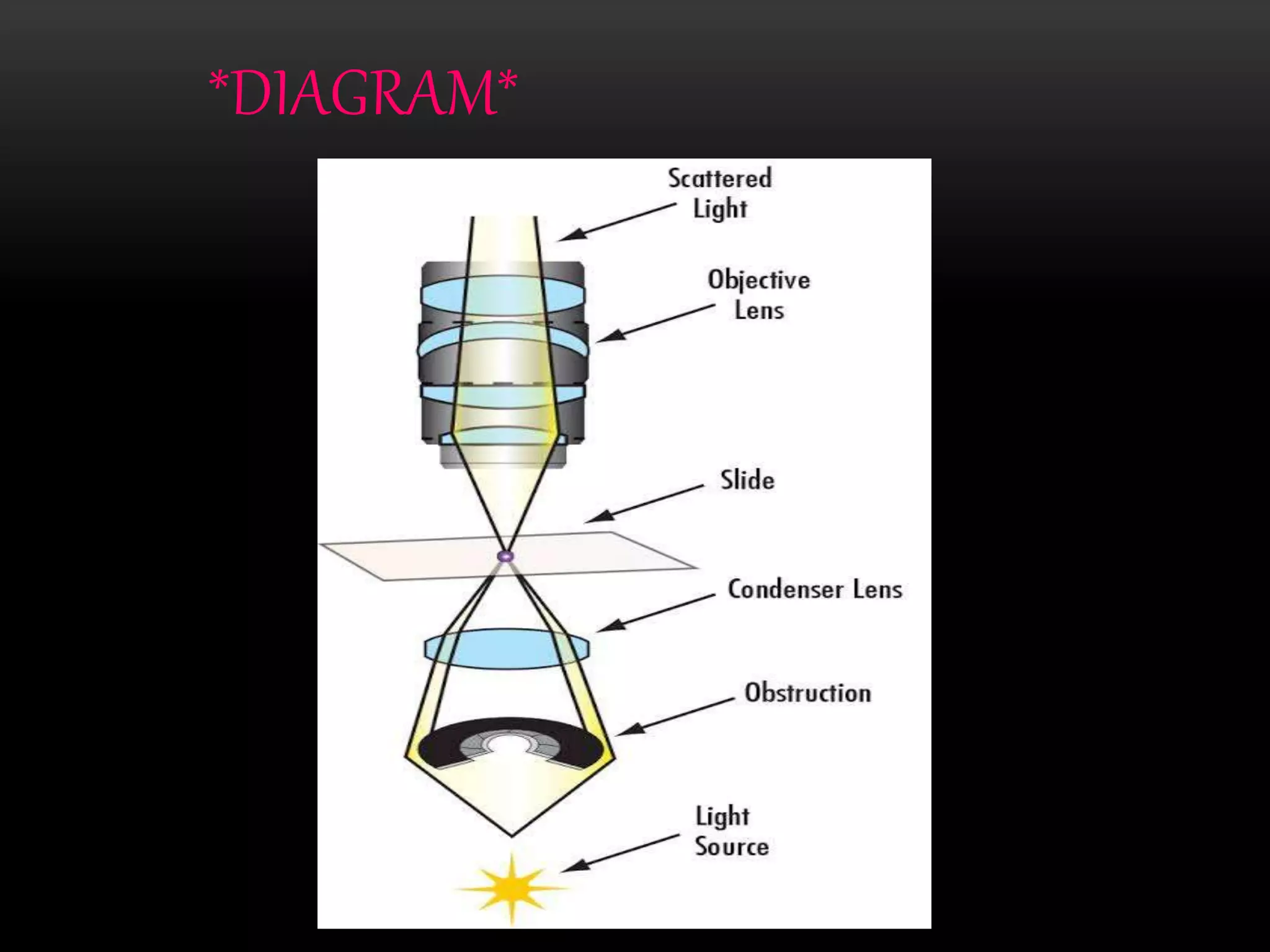
VISHAL RAJ K
HCA20M10
DARK FIELD MICROSCOPE
*CONTENT*
INTRODUCTION
DIAGRAM
WORKING PRINCIPLE
COMPONENTS
APPLICATION
ADVANTAGES & DISADVANTAGES
RELATED IMAGES
*INTRODUCTION*
Dark field microscope is a technique that
can be used for the observation of living,
unstained cells and microorganisms.
In this microscope, the specimen is brightly
illuminated while the background is dark.
The dark field microscope can reveal
considerable internal structure in
microorganisms.
*DIAGRAM*
*WORKING PRICIPLE*
A dark field microscope is arranged so that
the light source enters the microscope to
illuminate the specimen.
A special disc, called patch stop blocks
some light from the light source, leaving
an outer ring of illumination.
This is ideal for making objects with
refractive value similar to the back ground.
When light hits an object, rays are
scattered in all directions. The design
of dark field microscope is such that it
removes the dispersed light, so that
only scattered beams hit the sample.
The introduction of a condenser below
the stage ensures that these light rays
will hit the specimen at different
angles, rather than as a direct light
source towards the object.
Only scattered light started to enter the
objective lens and creates an image of
the specimen.
The result is a “cone of light” where
rays are diffracted, reflected and
refracted off the object ultimately,
allowing the individual to view a
specimen in dark field.
*COMPONENTS*
Dark field microscope consists of three key
components :~
Light source : enters the microscope and hits
the dark field path stop, which is a disc used
to block light from entering the condenser and
leaves a circular ring of illumination.
Condenser lens : collects outer ring of
illumination and focuses it on the sample.
Objective lens : light hits the sample, and
is transmitted or scattered from it.
Scattered light enters the objective lens,
whereas transmitted light is not collected
by the lens. The direct illumination block
assists in this step.
*APPLICATION*
The dark field microscope is used to
identify bacteria like thin and
distinctively shaped.
We can observe the living and
unstained cells using a dark field
microscope.
Considerable internal structure in
microorganisms can be revealed by the
dark field microscope.
A biological dark field microscope
used to identify algae.
A stereo dark field microscope used to
observe the shrimp or other
invertebrates.
It helps to characterize the embedded
nanomaterials in cells by combined
with hyperspectral imaging.
*ADVANTAGES*
No need to stain the specimen.
It is useful for those specimens that are
transparent and absorb little or no light.
Marine organisms such as algae,
plankton, insects, yeast and protozoa
can observe under a dark field
microscope.
It can be used for the research of live
bacteria and mounted cells and tissues.
*DISADVANTAGES*
Before use make sure slide, stage, nose
and light source are free from dust.
It cannot measure the specimen
accurately.
An intense amount of light is required
for a dark field microscope to work.
*RELATED IMAGES*
Blood cells Bacterial colony
Biomaterials
Nanoparticals in cancer cells
Cancer cells
THANK YOU

Dark field microscope

  • 1.
  • 2.
  • 3.
    *INTRODUCTION* Dark field microscopeis a technique that can be used for the observation of living, unstained cells and microorganisms. In this microscope, the specimen is brightly illuminated while the background is dark. The dark field microscope can reveal considerable internal structure in microorganisms.
  • 4.
  • 5.
    *WORKING PRICIPLE* A darkfield microscope is arranged so that the light source enters the microscope to illuminate the specimen. A special disc, called patch stop blocks some light from the light source, leaving an outer ring of illumination. This is ideal for making objects with refractive value similar to the back ground.
  • 6.
    When light hitsan object, rays are scattered in all directions. The design of dark field microscope is such that it removes the dispersed light, so that only scattered beams hit the sample. The introduction of a condenser below the stage ensures that these light rays will hit the specimen at different angles, rather than as a direct light source towards the object.
  • 7.
    Only scattered lightstarted to enter the objective lens and creates an image of the specimen. The result is a “cone of light” where rays are diffracted, reflected and refracted off the object ultimately, allowing the individual to view a specimen in dark field.
  • 8.
    *COMPONENTS* Dark field microscopeconsists of three key components :~ Light source : enters the microscope and hits the dark field path stop, which is a disc used to block light from entering the condenser and leaves a circular ring of illumination. Condenser lens : collects outer ring of illumination and focuses it on the sample.
  • 9.
    Objective lens :light hits the sample, and is transmitted or scattered from it. Scattered light enters the objective lens, whereas transmitted light is not collected by the lens. The direct illumination block assists in this step.
  • 10.
    *APPLICATION* The dark fieldmicroscope is used to identify bacteria like thin and distinctively shaped. We can observe the living and unstained cells using a dark field microscope. Considerable internal structure in microorganisms can be revealed by the dark field microscope.
  • 11.
    A biological darkfield microscope used to identify algae. A stereo dark field microscope used to observe the shrimp or other invertebrates. It helps to characterize the embedded nanomaterials in cells by combined with hyperspectral imaging.
  • 12.
    *ADVANTAGES* No need tostain the specimen. It is useful for those specimens that are transparent and absorb little or no light. Marine organisms such as algae, plankton, insects, yeast and protozoa can observe under a dark field microscope. It can be used for the research of live bacteria and mounted cells and tissues.
  • 13.
    *DISADVANTAGES* Before use makesure slide, stage, nose and light source are free from dust. It cannot measure the specimen accurately. An intense amount of light is required for a dark field microscope to work.
  • 14.
  • 15.
  • 16.
  • 17.