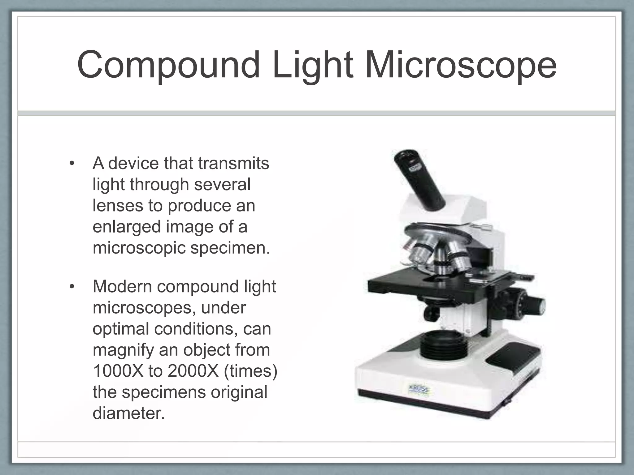
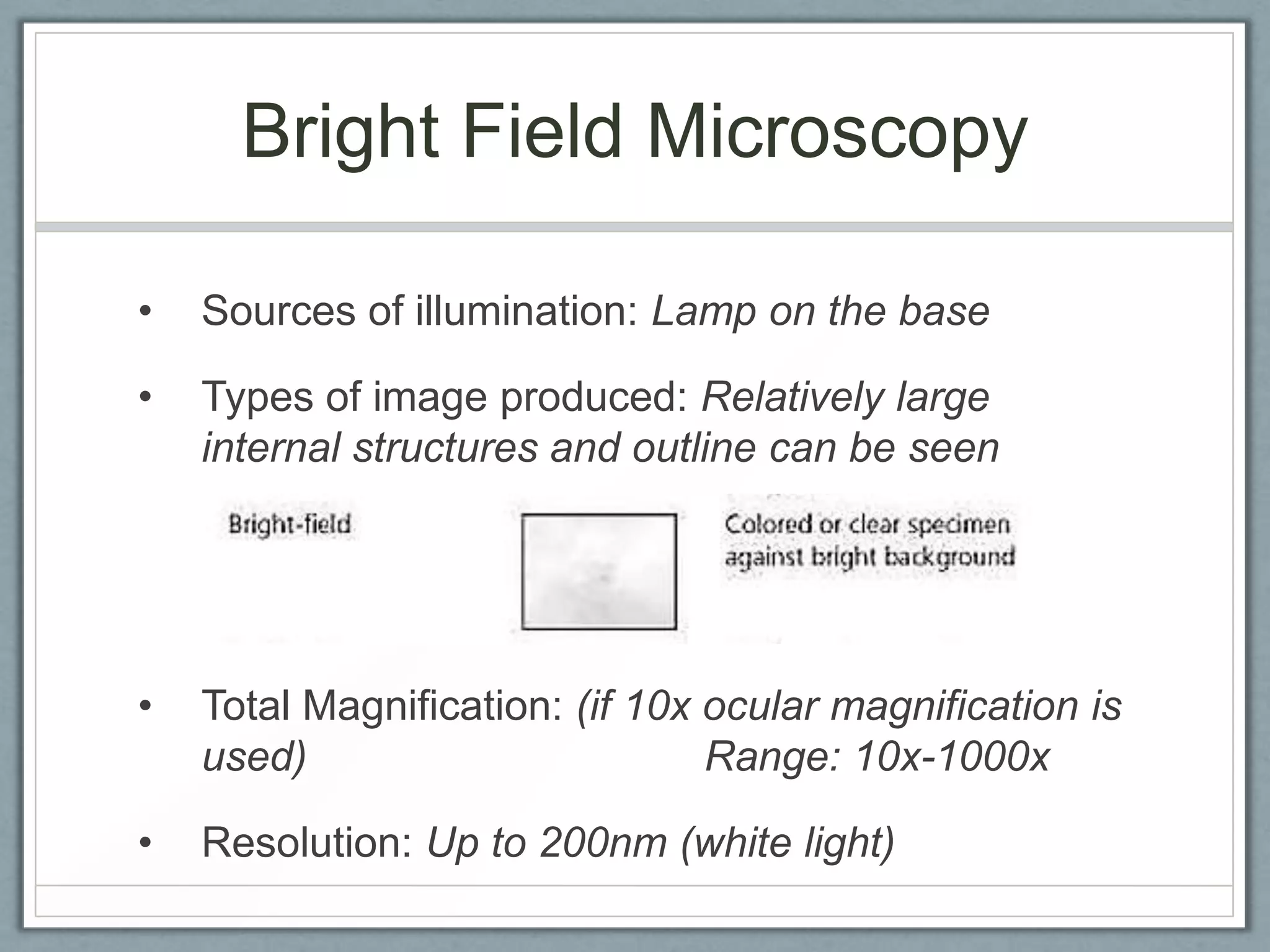
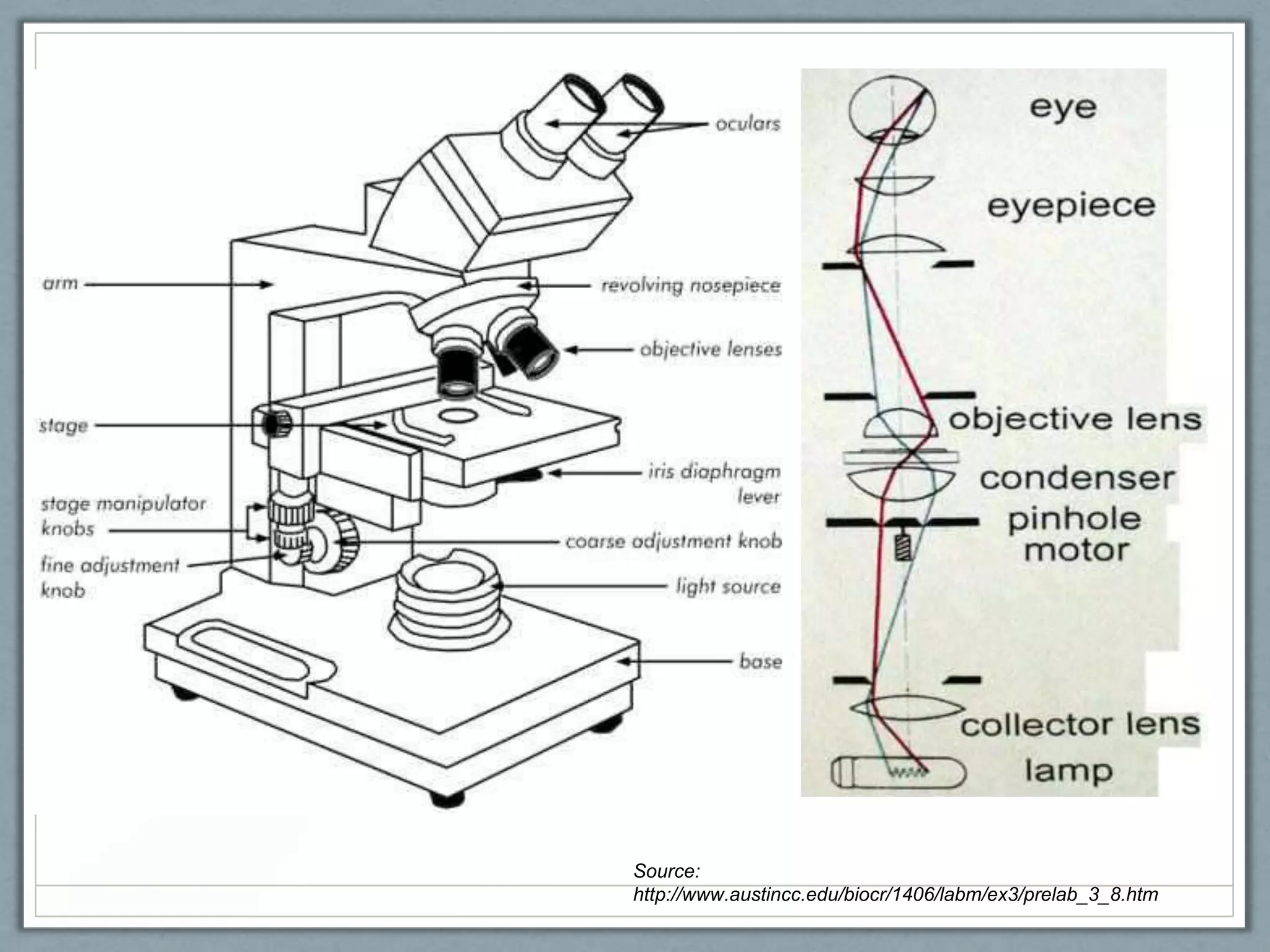
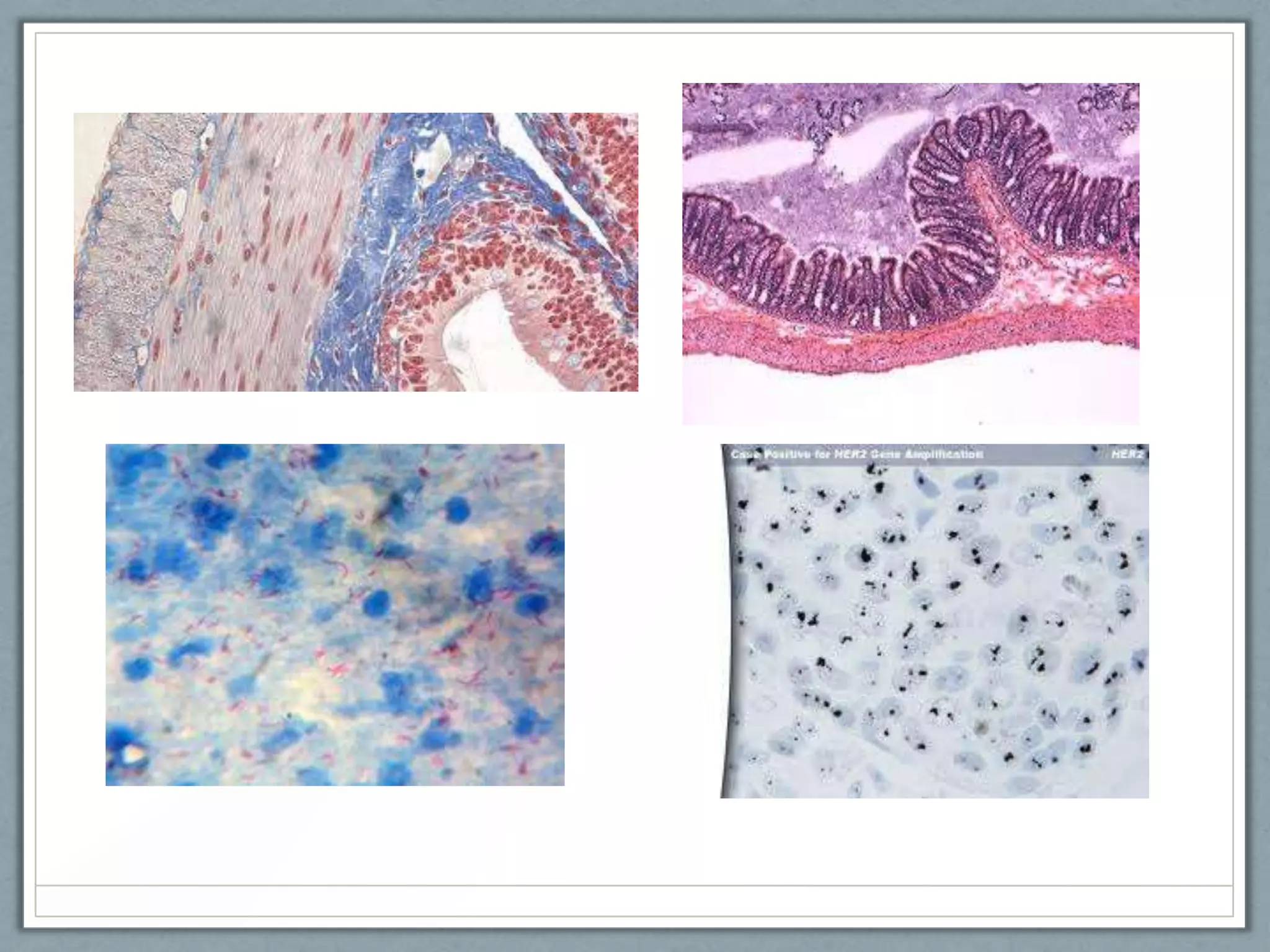
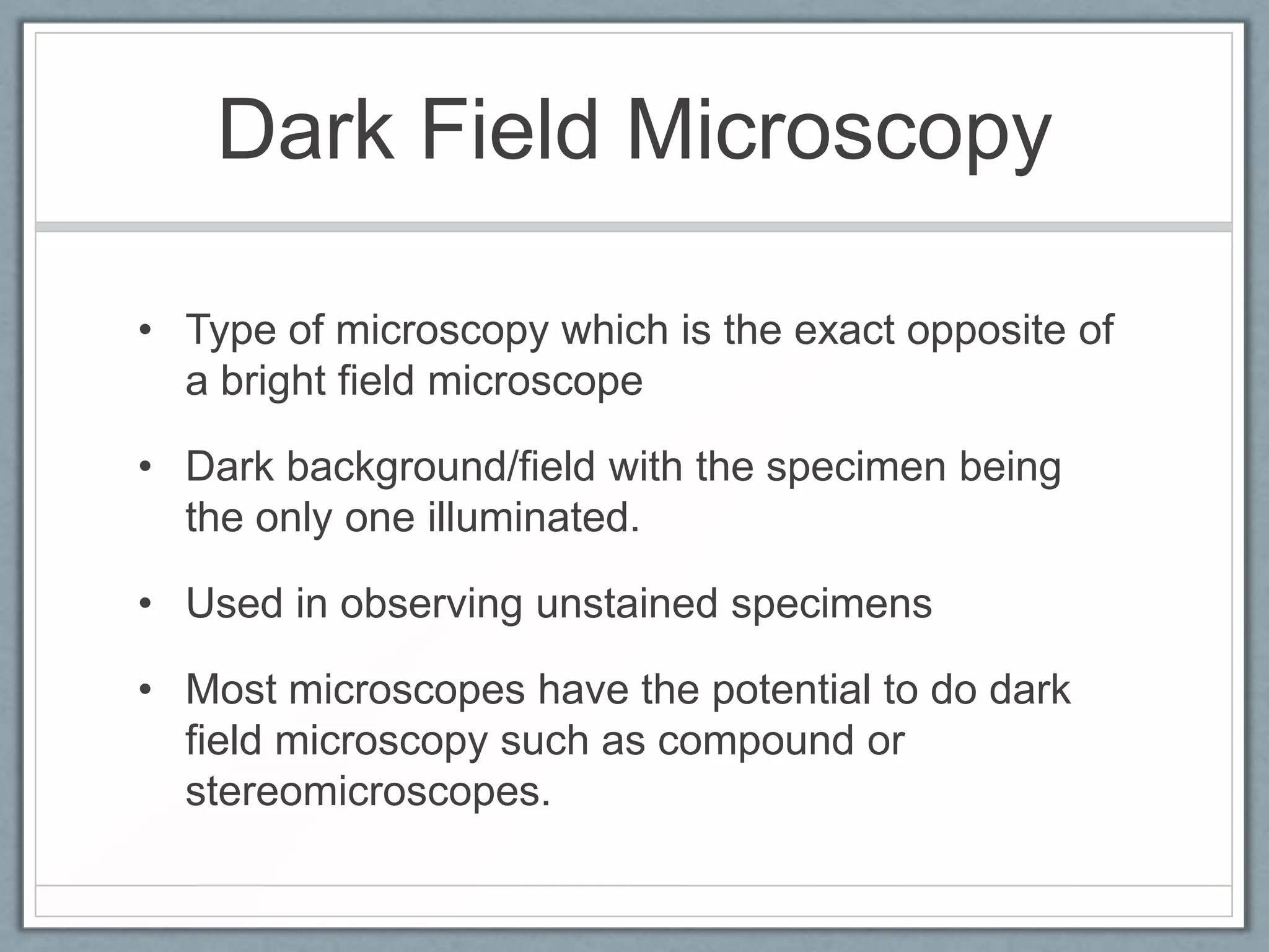
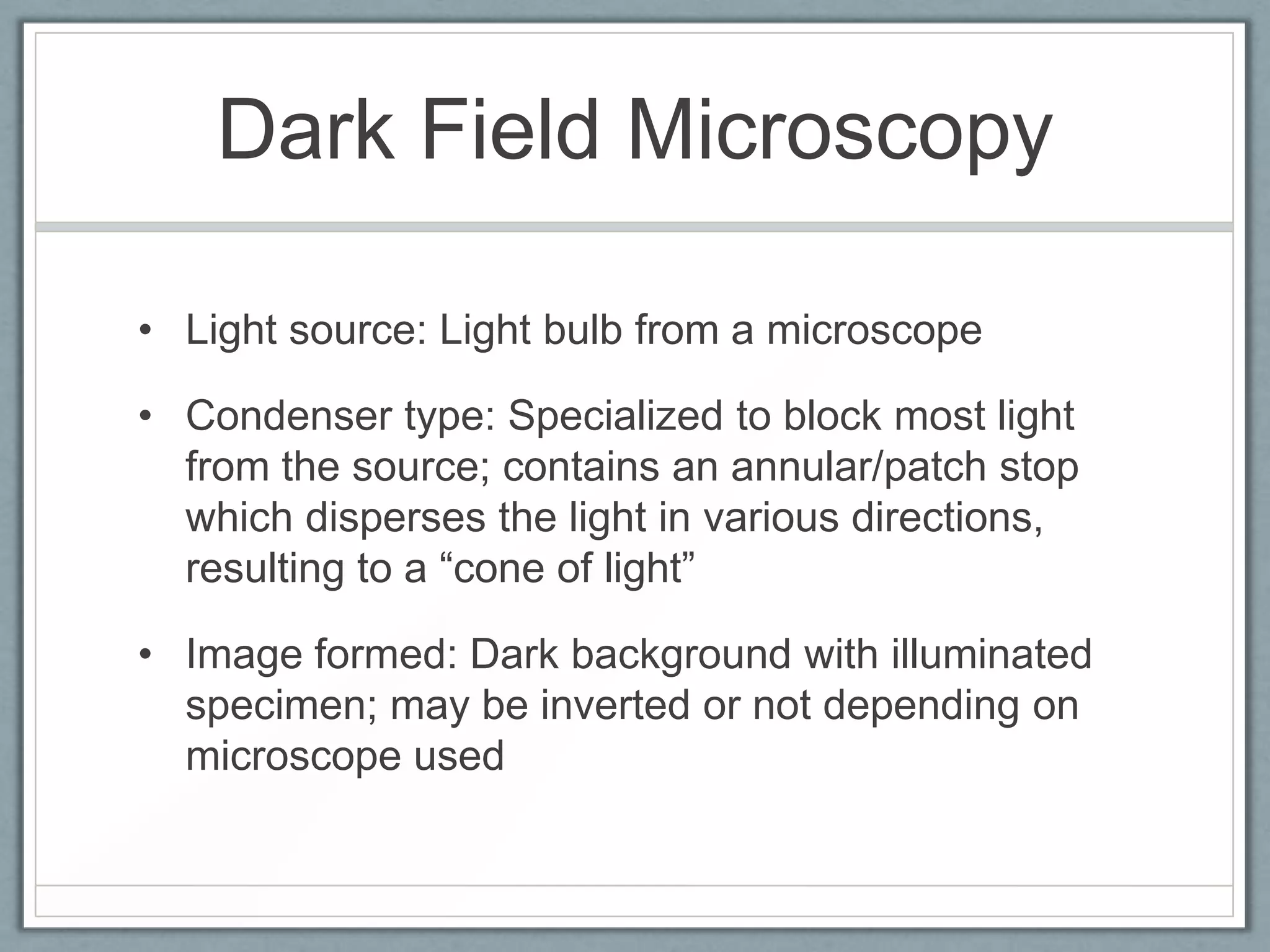
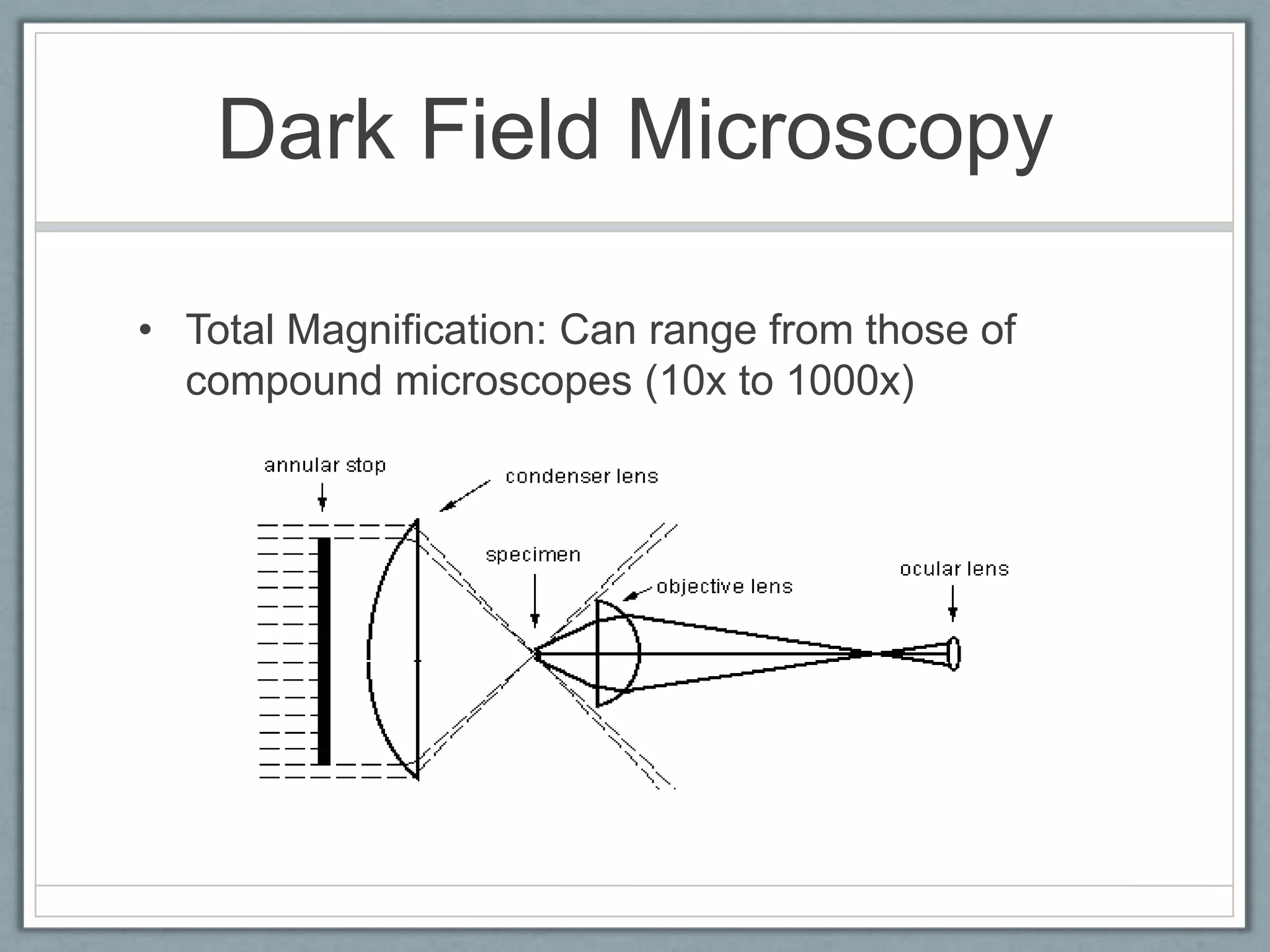
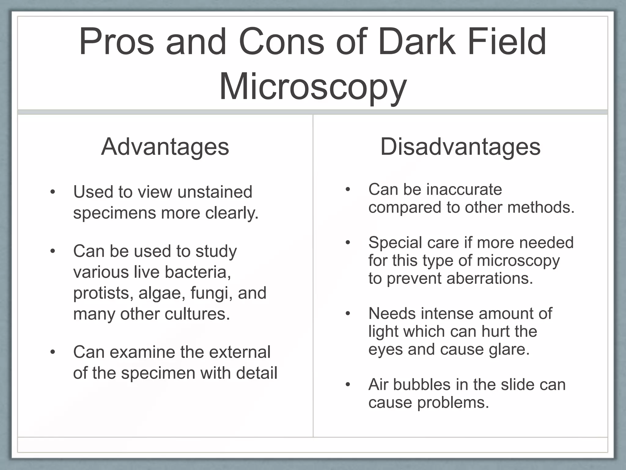
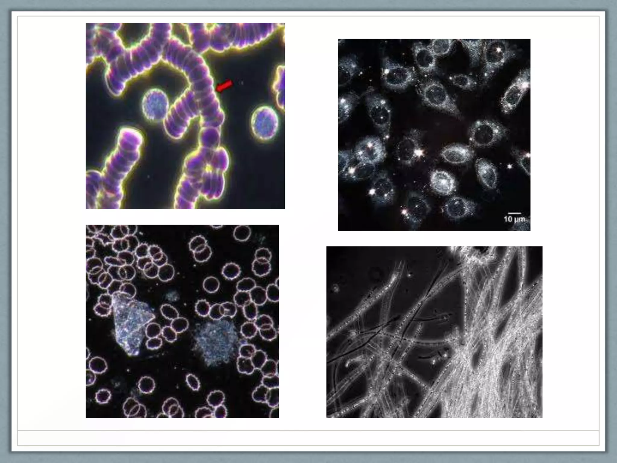
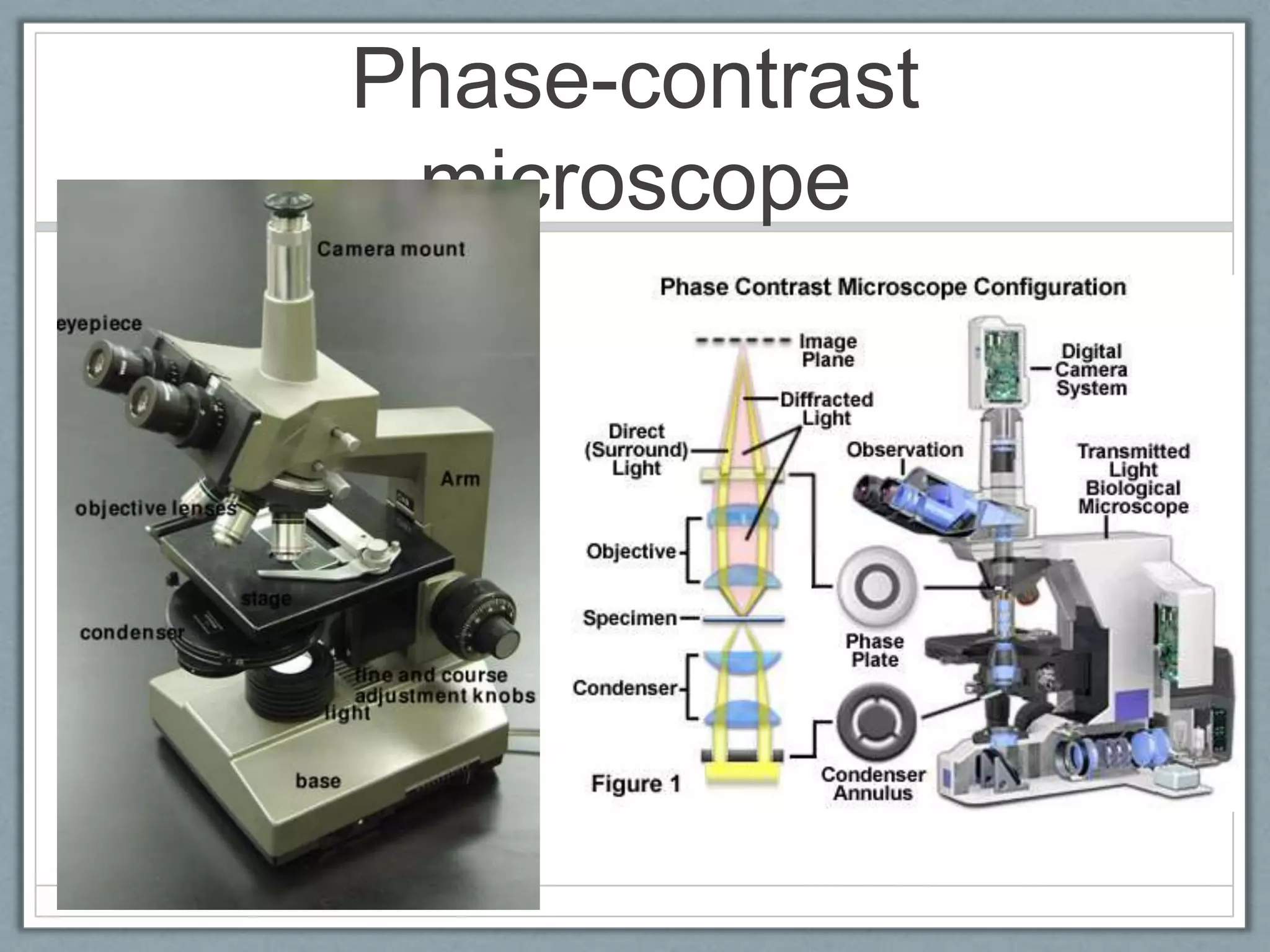
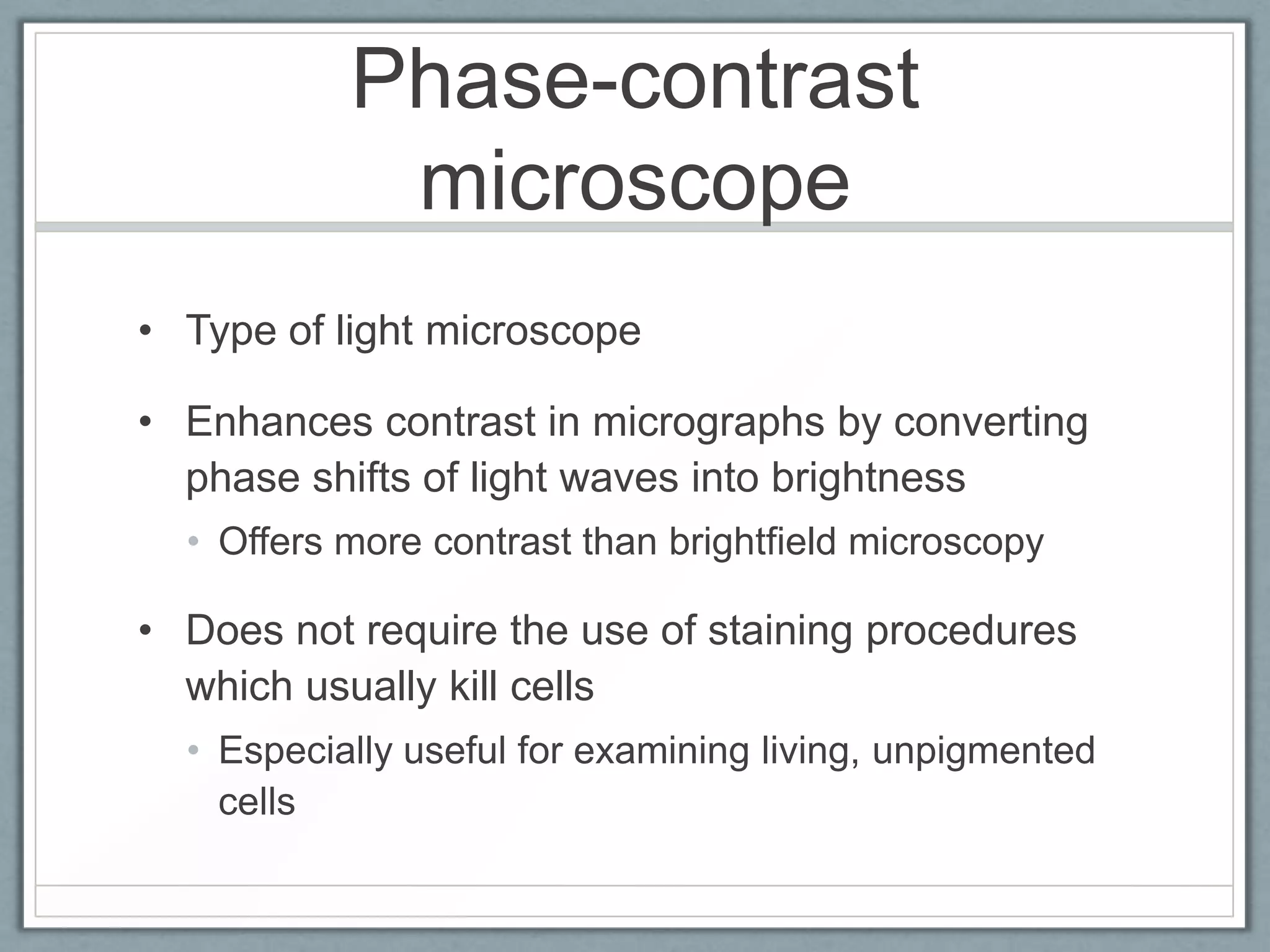
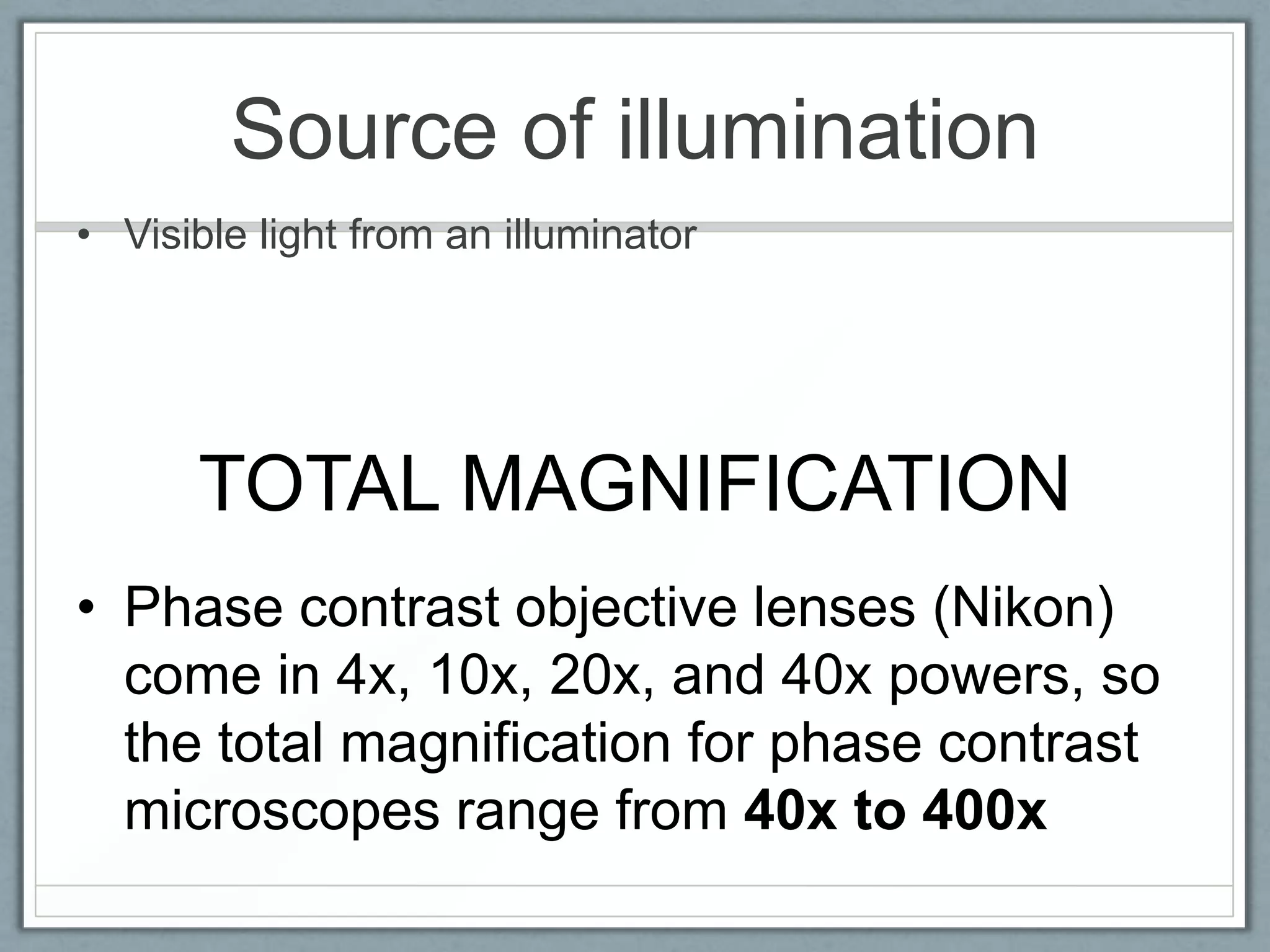
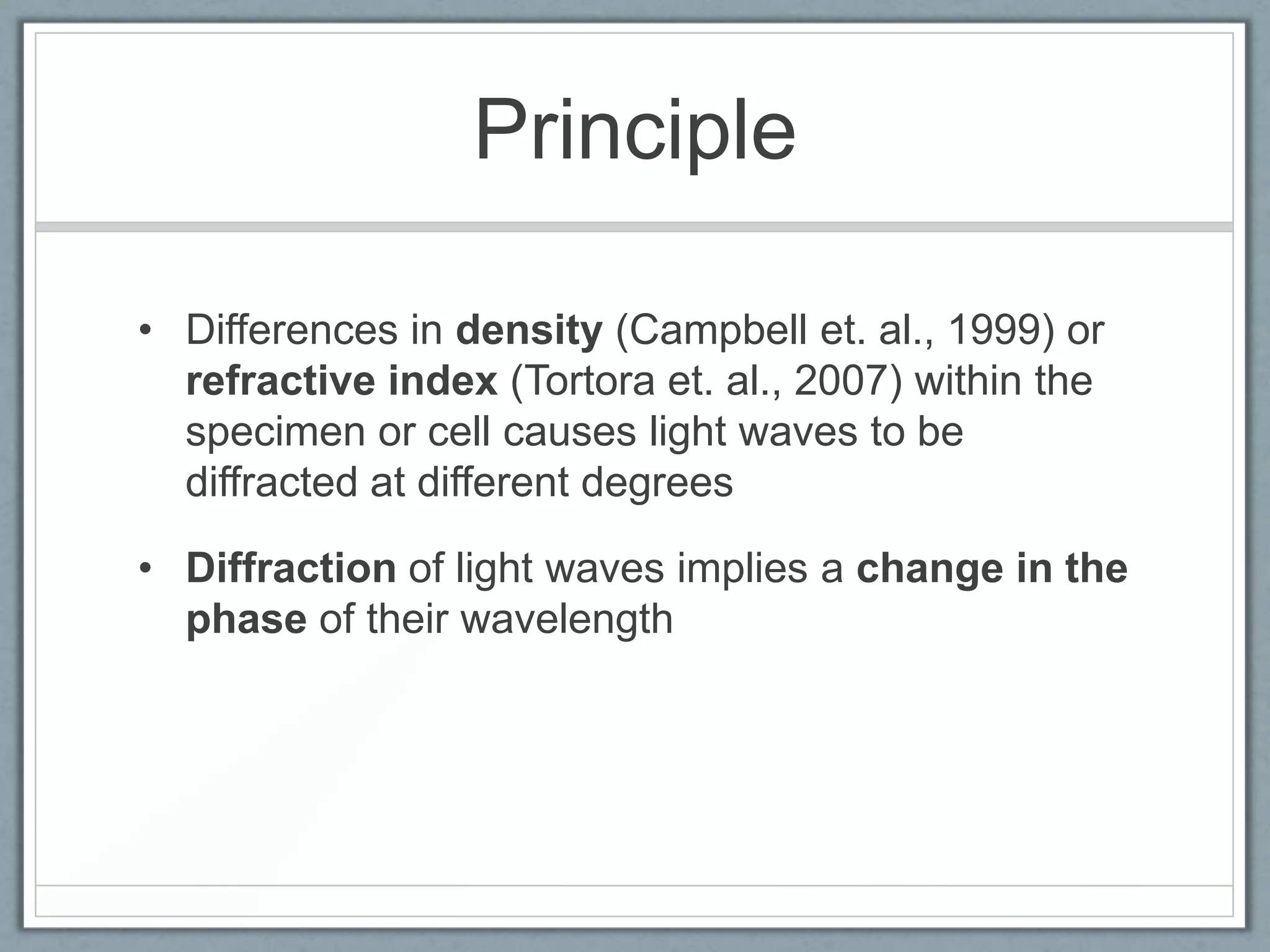
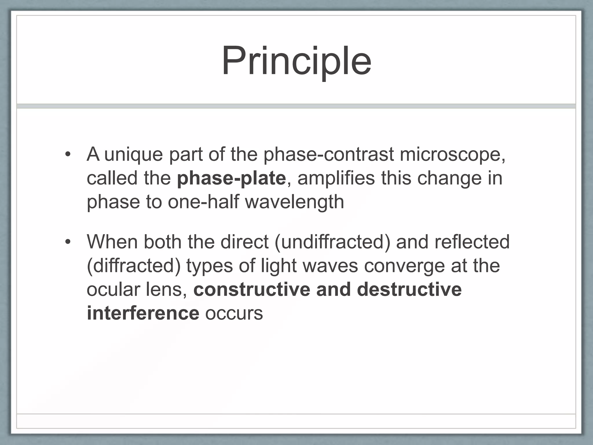
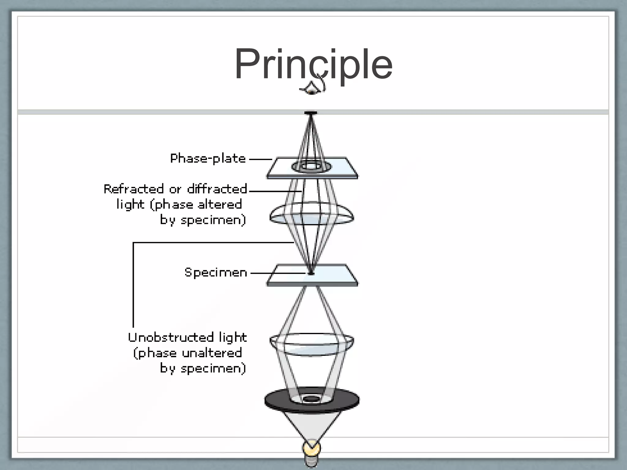
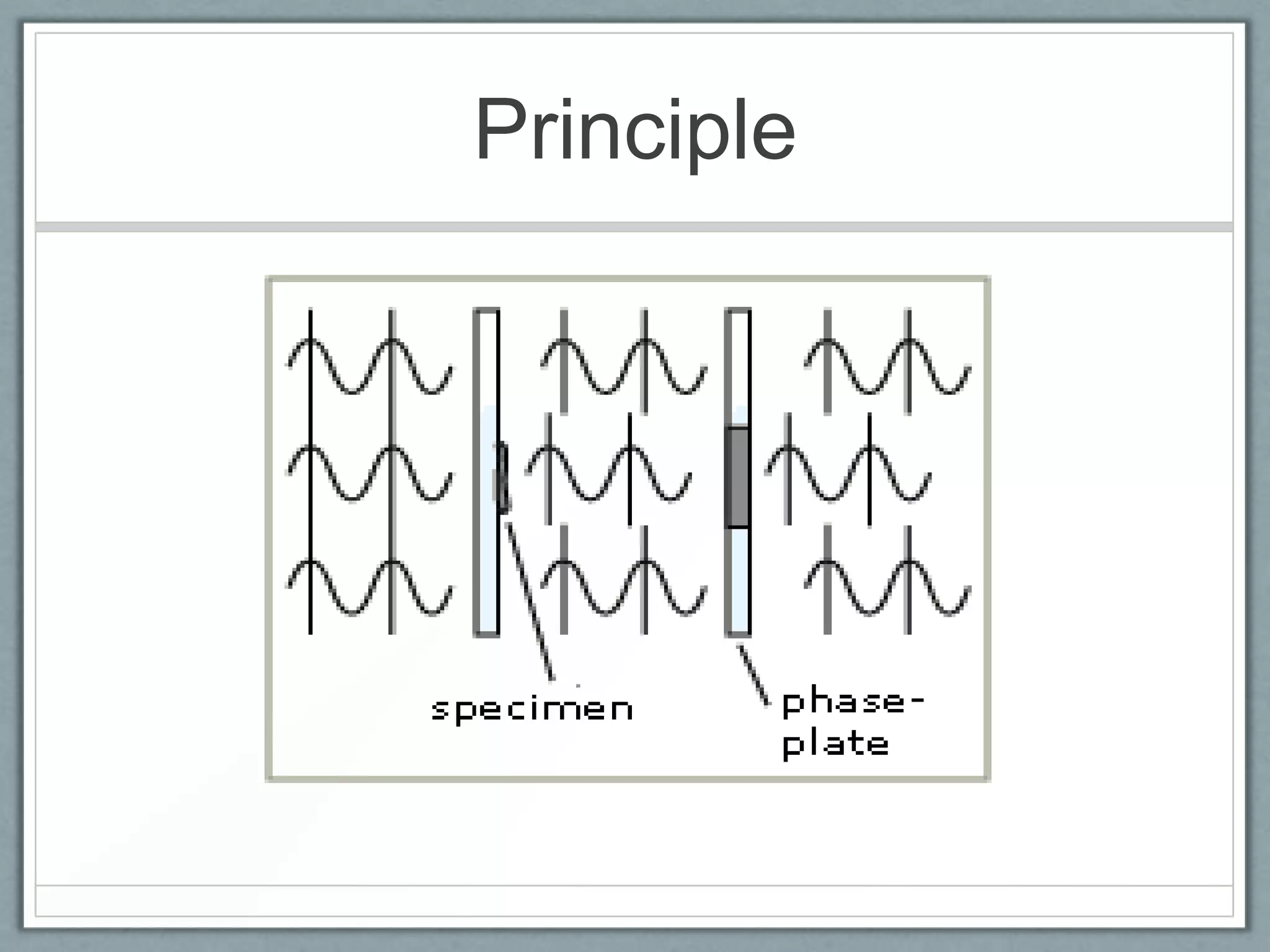
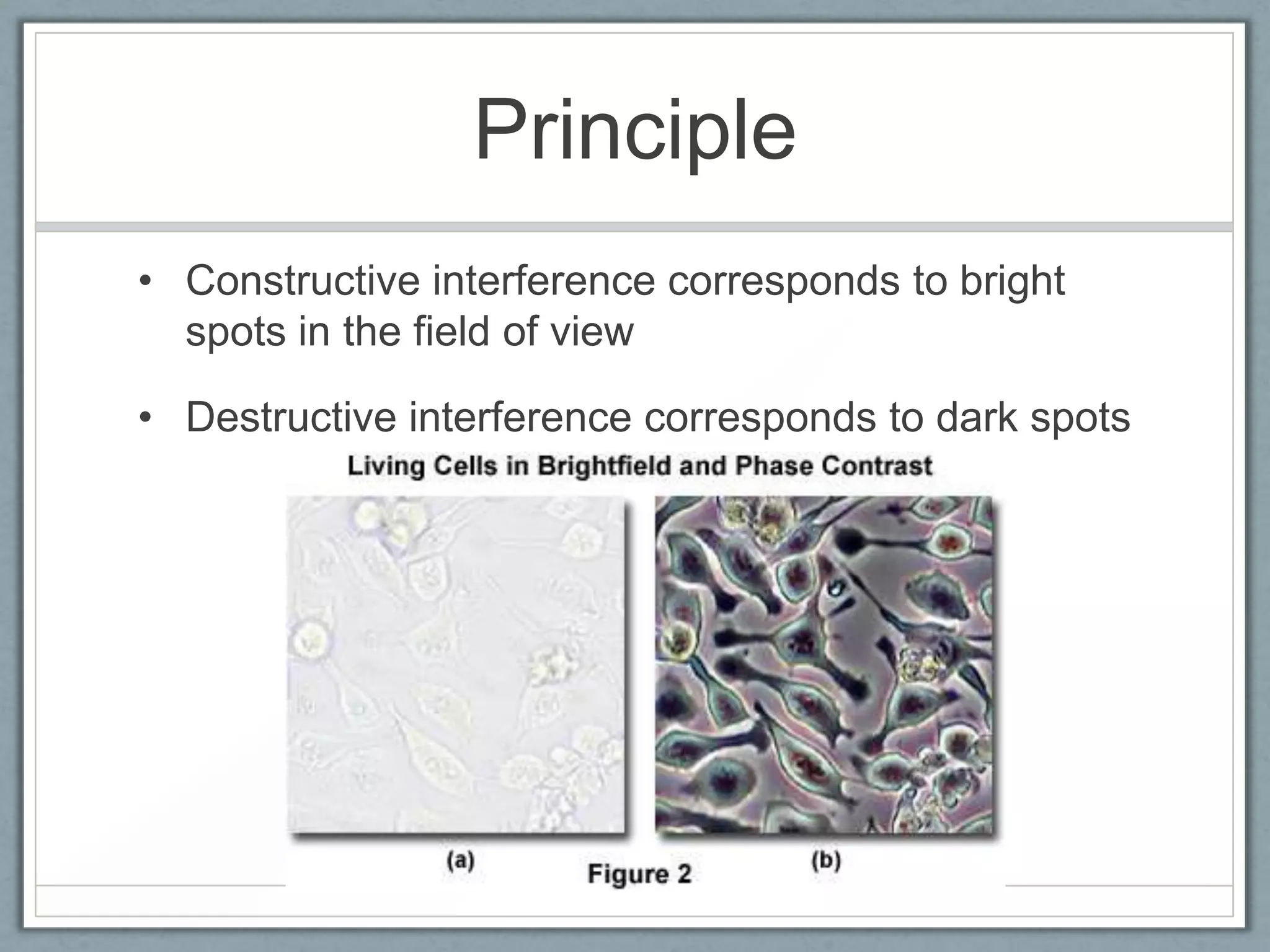
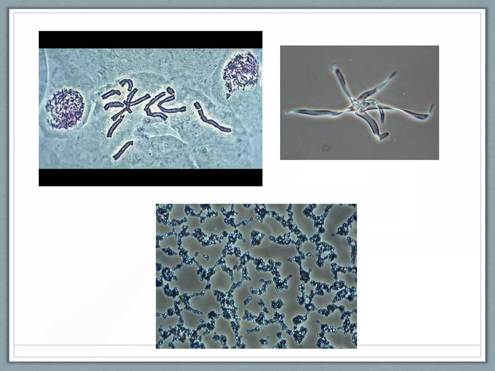
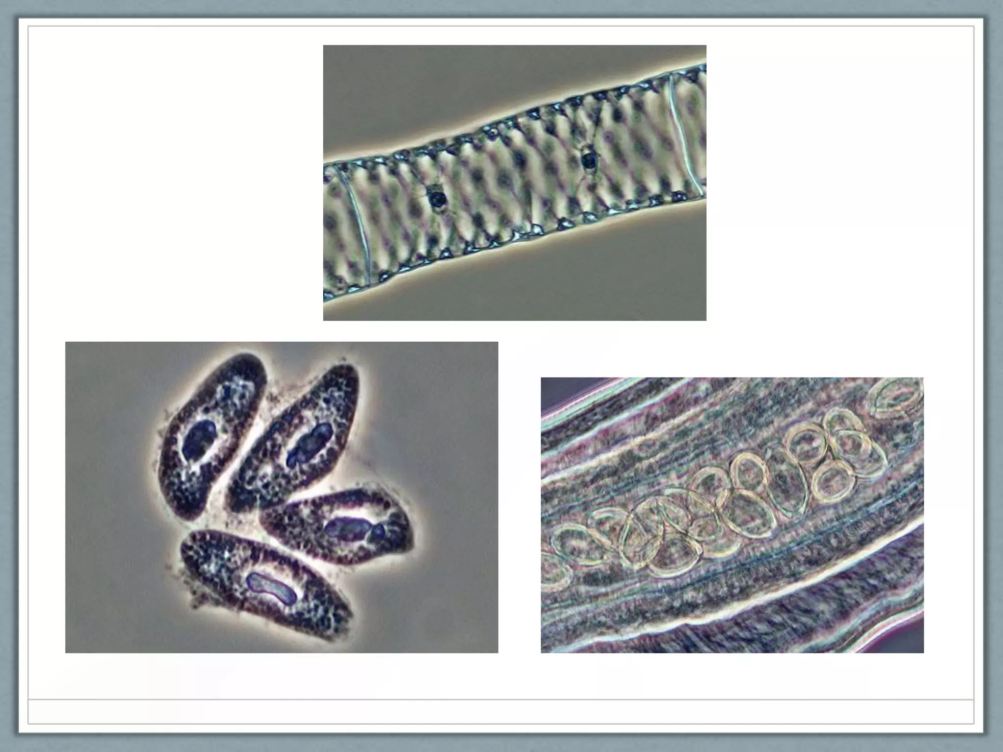
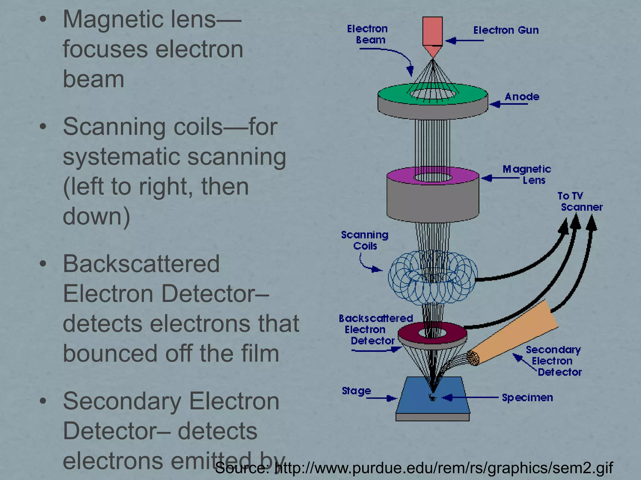
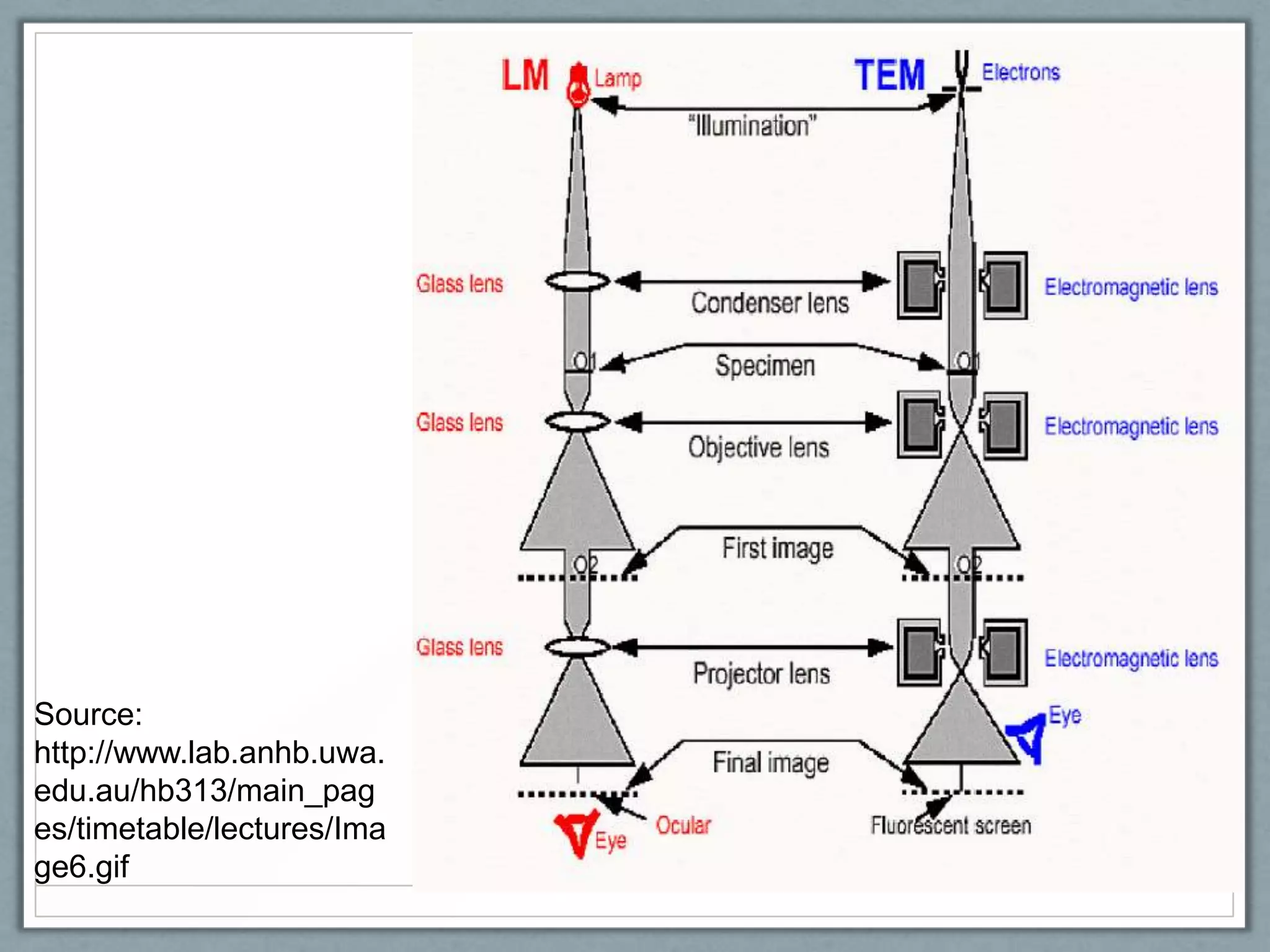
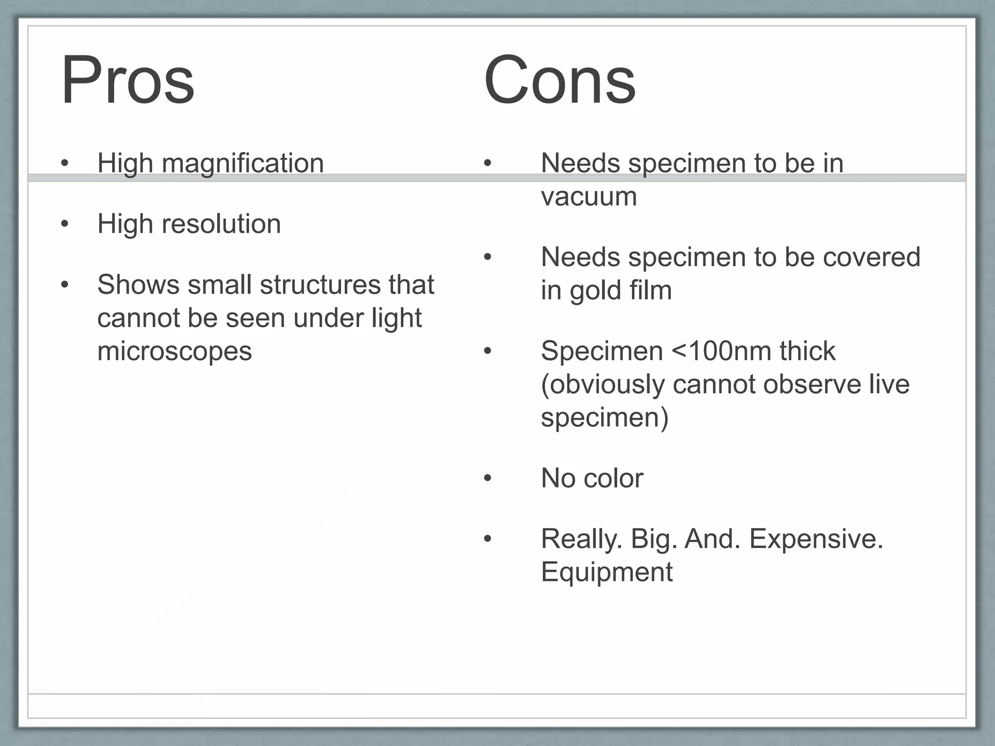
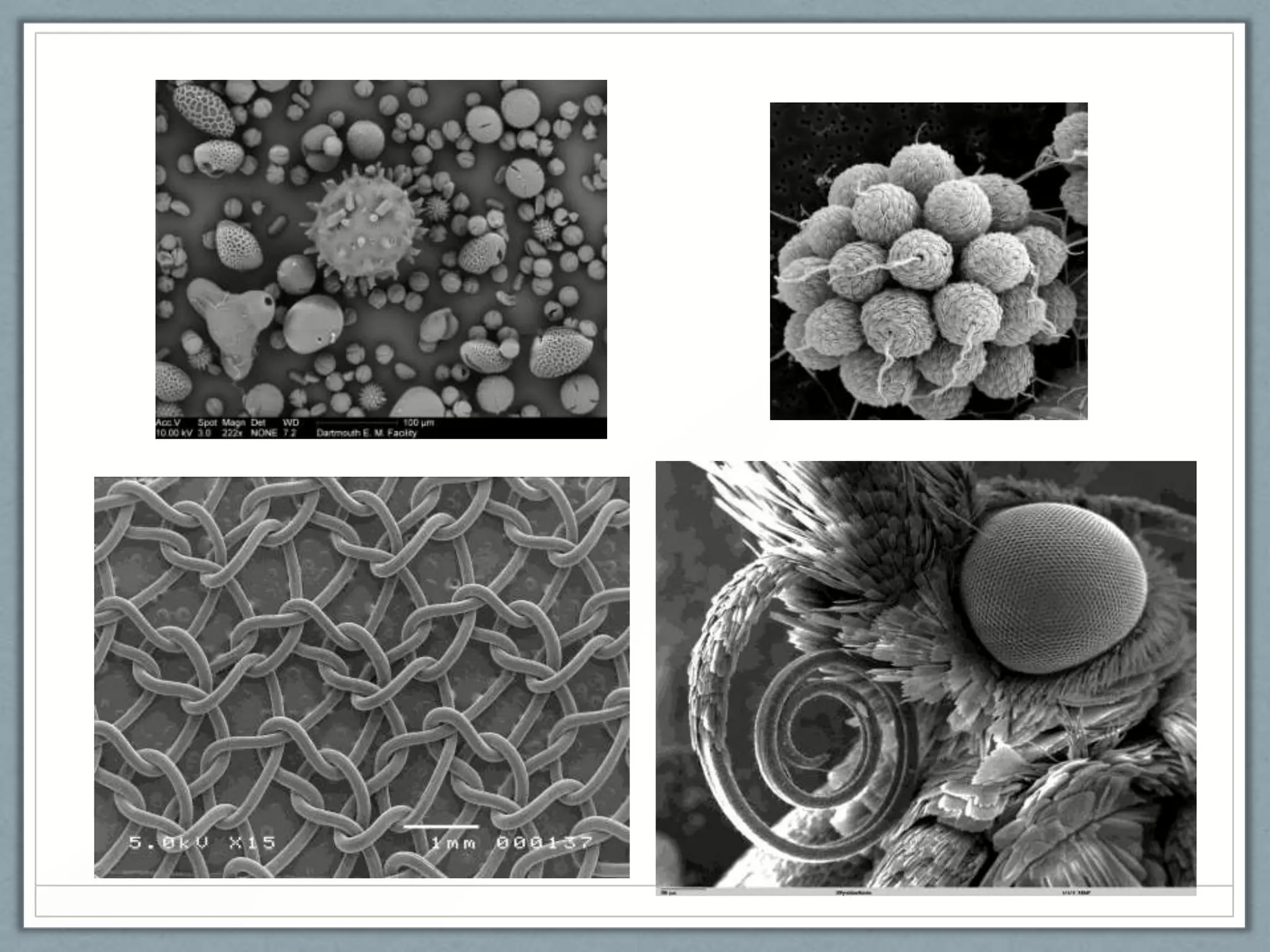
Microscopy
Group 2
Cabatit, Mendoza, Ramos, Rodriguez, Tan
Compound Light Microscope
• A device that transmits
light through several
lenses to produce an
enlarged image of a
microscopic specimen.
• Modern compound light
microscopes, under
optimal conditions, can
magnify an object from
1000X to 2000X (times)
the specimens original
diameter.
Bright Field Microscopy
•

Simplest optical microscopy illumination
technique

•

Uses visible light as source of illumination
•

> the shorter the wavelength, the greater the
resolution (blue is the best)

•

Contrast comes from absorbance of light in the
sample, or from staining.

•

When the diaphragm is wide open the image is
brighter and contrast is low.
Bright Field Microscopy
•

Sources of illumination: Lamp on the base

•

Types of image produced: Relatively large
internal structures and outline can be seen

•

Total Magnification: (if 10x ocular magnification is
used)
Range: 10x-1000x

•

Resolution: Up to 200nm (white light)
Source:
http://www.austincc.edu/biocr/1406/labm/ex3/prelab_3_8.htm
Dark Field Microscopy
• Type of microscopy which is the exact opposite of
a bright field microscope
• Dark background/field with the specimen being
the only one illuminated.
• Used in observing unstained specimens
• Most microscopes have the potential to do dark
field microscopy such as compound or
stereomicroscopes.
Dark Field Microscopy
• Light source: Light bulb from a microscope
• Condenser type: Specialized to block most light
from the source; contains an annular/patch stop
which disperses the light in various directions,
resulting to a ―cone of light‖
• Image formed: Dark background with illuminated
specimen; may be inverted or not depending on
microscope used
Dark Field Microscopy
• Total Magnification: Can range from those of
compound microscopes (10x to 1000x)
Pros and Cons of Dark Field
Microscopy
Advantages

Disadvantages

• Used to view unstained
specimens more clearly.

•

Can be inaccurate
compared to other methods.

• Can be used to study
various live bacteria,
protists, algae, fungi, and
many other cultures.

•

Special care if more needed
for this type of microscopy
to prevent aberrations.

•

Needs intense amount of
light which can hurt the
eyes and cause glare.

•

Air bubbles in the slide can
cause problems.

• Can examine the external
of the specimen with detail
Phase Contrast
Microscopy
Phase-contrast
microscope
Phase-contrast
microscope
• Type of light microscope
• Enhances contrast in micrographs by converting
phase shifts of light waves into brightness
• Offers more contrast than brightfield microscopy

• Does not require the use of staining procedures
which usually kill cells
• Especially useful for examining living, unpigmented
cells
Source of illumination
• Visible light from an illuminator

TOTAL MAGNIFICATION
• Phase contrast objective lenses (Nikon)
come in 4x, 10x, 20x, and 40x powers, so
the total magnification for phase contrast
microscopes range from 40x to 400x
Principle
• Differences in density (Campbell et. al., 1999) or
refractive index (Tortora et. al., 2007) within the
specimen or cell causes light waves to be
diffracted at different degrees
• Diffraction of light waves implies a change in the
phase of their wavelength
Principle
• A unique part of the phase-contrast microscope,
called the phase-plate, amplifies this change in
phase to one-half wavelength
• When both the direct (undiffracted) and reflected
(diffracted) types of light waves converge at the
ocular lens, constructive and destructive
interference occurs
Principle
Principle
Principle
• Constructive interference corresponds to bright
spots in the field of view
• Destructive interference corresponds to dark spots
Type of image produced
• The end result is a magnified and highly
contrasted view of a living, unstained, normally
transparent specimen
Electron Microscopy
Scanning Electron
Microscope
• Illumination: electrons
• Magnification: ~100,000x
• How it works: Detect electrons back-scattered by
the sample.
• Image: Monotone (but may be color enhanced), 3D surface of specimen
• Magnetic lens—
focuses electron
beam
• Scanning coils—for
systematic scanning
(left to right, then
down)
• Backscattered
Electron Detector–
detects electrons that
bounced off the film

• Secondary Electron
Detector– detects
electrons emitted by
Source: http://www.purdue.edu/rem/rs/graphics/sem2.gif
Pros

Cons

• High magnification

•

• High resolution

•
• Shows the surface of specimen

Needs specimen to be in
vacuum
Needs living cells and tissues
and whole, soft-bodied
organisms to be treated, usu.
coated w/ gold film

•

No color

•

Cannot examine live specimen

•

Really. Big. And Expensive.
Equipment.
Transmission Electron
Microscope
• Illumination: electrons
• Magnification: ~100,000x
• How it works: Detect electrons scattered as they
move through the sample.
• Image: Monotone (but may be color enhanced), 2D structure of specimen
Source:
http://www.lab.anhb.uwa.
edu.au/hb313/main_pag
es/timetable/lectures/Ima
ge6.gif
Pros

Cons

• High magnification

•

Needs specimen to be in
vacuum

•

Needs specimen to be covered
in gold film

•

Specimen <100nm thick
(obviously cannot observe live
specimen)

•

No color

•

Really. Big. And. Expensive.
Equipment

• High resolution

• Shows small structures that
cannot be seen under light
microscopes
End

Microscopy 121125060708-phpapp01

  • 1.
  • 2.
    Compound Light Microscope •A device that transmits light through several lenses to produce an enlarged image of a microscopic specimen. • Modern compound light microscopes, under optimal conditions, can magnify an object from 1000X to 2000X (times) the specimens original diameter.
  • 3.
  • 4.
    Bright Field Microscopy • Simplestoptical microscopy illumination technique • Uses visible light as source of illumination • > the shorter the wavelength, the greater the resolution (blue is the best) • Contrast comes from absorbance of light in the sample, or from staining. • When the diaphragm is wide open the image is brighter and contrast is low.
  • 5.
    Bright Field Microscopy • Sourcesof illumination: Lamp on the base • Types of image produced: Relatively large internal structures and outline can be seen • Total Magnification: (if 10x ocular magnification is used) Range: 10x-1000x • Resolution: Up to 200nm (white light)
  • 6.
  • 8.
  • 9.
    Dark Field Microscopy •Type of microscopy which is the exact opposite of a bright field microscope • Dark background/field with the specimen being the only one illuminated. • Used in observing unstained specimens • Most microscopes have the potential to do dark field microscopy such as compound or stereomicroscopes.
  • 10.
    Dark Field Microscopy •Light source: Light bulb from a microscope • Condenser type: Specialized to block most light from the source; contains an annular/patch stop which disperses the light in various directions, resulting to a ―cone of light‖ • Image formed: Dark background with illuminated specimen; may be inverted or not depending on microscope used
  • 11.
    Dark Field Microscopy •Total Magnification: Can range from those of compound microscopes (10x to 1000x)
  • 12.
    Pros and Consof Dark Field Microscopy Advantages Disadvantages • Used to view unstained specimens more clearly. • Can be inaccurate compared to other methods. • Can be used to study various live bacteria, protists, algae, fungi, and many other cultures. • Special care if more needed for this type of microscopy to prevent aberrations. • Needs intense amount of light which can hurt the eyes and cause glare. • Air bubbles in the slide can cause problems. • Can examine the external of the specimen with detail
  • 14.
  • 15.
  • 16.
    Phase-contrast microscope • Type oflight microscope • Enhances contrast in micrographs by converting phase shifts of light waves into brightness • Offers more contrast than brightfield microscopy • Does not require the use of staining procedures which usually kill cells • Especially useful for examining living, unpigmented cells
  • 17.
    Source of illumination •Visible light from an illuminator TOTAL MAGNIFICATION • Phase contrast objective lenses (Nikon) come in 4x, 10x, 20x, and 40x powers, so the total magnification for phase contrast microscopes range from 40x to 400x
  • 18.
    Principle • Differences indensity (Campbell et. al., 1999) or refractive index (Tortora et. al., 2007) within the specimen or cell causes light waves to be diffracted at different degrees • Diffraction of light waves implies a change in the phase of their wavelength
  • 19.
    Principle • A uniquepart of the phase-contrast microscope, called the phase-plate, amplifies this change in phase to one-half wavelength • When both the direct (undiffracted) and reflected (diffracted) types of light waves converge at the ocular lens, constructive and destructive interference occurs
  • 20.
  • 21.
  • 22.
    Principle • Constructive interferencecorresponds to bright spots in the field of view • Destructive interference corresponds to dark spots
  • 23.
    Type of imageproduced • The end result is a magnified and highly contrasted view of a living, unstained, normally transparent specimen
  • 26.
  • 27.
    Scanning Electron Microscope • Illumination:electrons • Magnification: ~100,000x • How it works: Detect electrons back-scattered by the sample. • Image: Monotone (but may be color enhanced), 3D surface of specimen
  • 28.
    • Magnetic lens— focuseselectron beam • Scanning coils—for systematic scanning (left to right, then down) • Backscattered Electron Detector– detects electrons that bounced off the film • Secondary Electron Detector– detects electrons emitted by Source: http://www.purdue.edu/rem/rs/graphics/sem2.gif
  • 29.
    Pros Cons • High magnification • •High resolution • • Shows the surface of specimen Needs specimen to be in vacuum Needs living cells and tissues and whole, soft-bodied organisms to be treated, usu. coated w/ gold film • No color • Cannot examine live specimen • Really. Big. And Expensive. Equipment.
  • 30.
    Transmission Electron Microscope • Illumination:electrons • Magnification: ~100,000x • How it works: Detect electrons scattered as they move through the sample. • Image: Monotone (but may be color enhanced), 2D structure of specimen
  • 31.
  • 32.
    Pros Cons • High magnification • Needsspecimen to be in vacuum • Needs specimen to be covered in gold film • Specimen <100nm thick (obviously cannot observe live specimen) • No color • Really. Big. And. Expensive. Equipment • High resolution • Shows small structures that cannot be seen under light microscopes
  • 34.

Editor's Notes

  • #6 Hence, best suited to viewing stained or naturally pigmented specimens such as stained prepared slides of tissue sections or living photosynthetic organisms