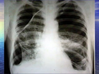
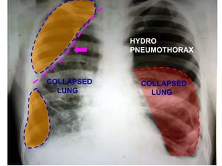
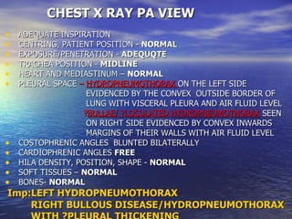
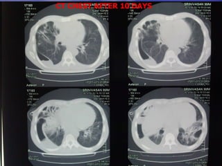
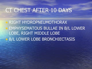
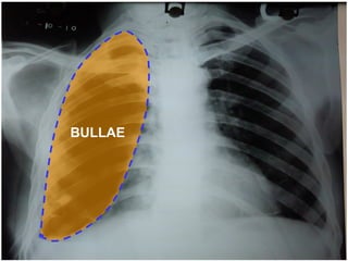
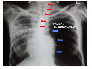
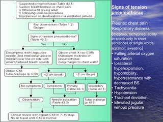
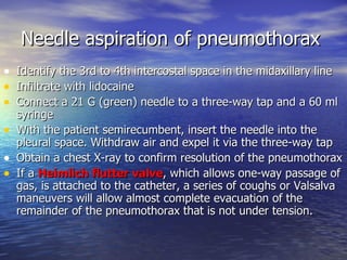
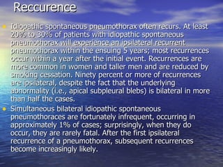
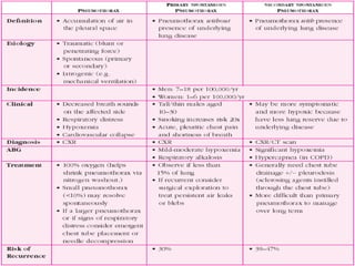
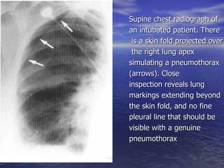
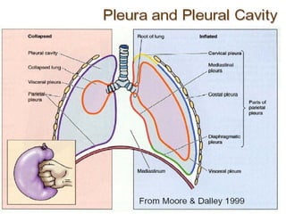
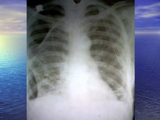
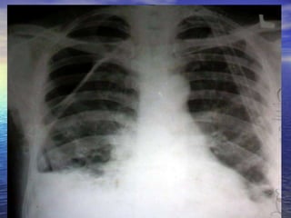
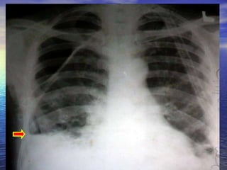
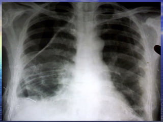
Prof .Dr.K.H.NOORUL AMEEN’S unit M6 Dr.G ARUN KUMAR IMAGE OF THE WEEK
30 YEAR OLD MALE CAME TO OPD WITH COMPLAINTS OF BREATHLESSNESS 1 MONTH DURATION. NOW INCREASED FOR 2 DAYS H/O COUGH WITH EXPECTORATION 1 MONTH CHRONIC ALCOHOLIC AND SMOKER 15 YEARS
 
COLLAPSED  LUNG COLLAPSED  LUNG HYDRO PNEUMOTHORAX
CHEST X RAY PA VIEW ADEQUATE INSPIRATION CENTRING, PATIENT POSITION -  NORMAL EXPOSURE/PENETRATION -  ADEQUQTE TRACHEA POSITION -  MIDLINE HEART AND MEDIASTINUM –  NORMAL PLEURAL SPACE  –  HYDROPNEUMOTHORAX  ON THE LEFT SIDE  EVIDENCED BY THE CONVEX  OUTSIDE BORDER OF  LUNG WITH VISCERAL PLEURA AND AIR FLUID LEVEL ? BULLAE/ ?LOCULATED HYDROPNEUMOTHORAX  SEEN  ON RIGHT SIDE EVIDENCED BY CONVEX INWARDS  MARGINS OF THEIR WALLS WITH AIR FLUID LEVEL COSTOPHRENIC ANGLES  BLUNTED BILATERALLY  CARDIOPHRENIC ANGLES  FREE HILA DENSITY, POSITION, SHAPE -  NORMAL SOFT TISSUES –  NORMAL BONES-  NORMAL Imp:LEFT HYDROPNEUMOTHORAX  RIGHT BULLOUS DISEASE/HYDROPNEUMOTHORAX WITH ?PLEURAL THICKENING
CT CHEST AFTER 10 DAYS
CT CHEST AFTER 10 DAYS RIGHT HYDROPNEUMOTHORAX EMPHYSEMATOUS BULLAE IN B/L LOWER LOBE, RIGHT MIDDLE LOBE B/L LOWER LOBE BRONCHIECTASIS
BULLAE
TENSION PNEUMOTHORAX
Pneumothorax Pneumothorax is the presence of air in the pleural cavity . Spontaneous pneumothorax Primary Secondary   Airways disease (COPD, cystic fibrosis, acute severe asthma) Infectious lung disease Interstitial lung disease (e.g. sarcoidosis) Connective tissue disease (e.g. rheumatoid arthritis, Marfan  Malignancy (bronchial carcinoma or sarcoma) Thoracic endometriosis Traumatic pneumothorax Iatrogenic pneumothorax   Transthoracic needle aspiration Subclavian vein puncture Thoracentesis and pleural biopsy Pericardiocentesis Barotrauma related to mech  ventilation
Because the normal pleural space contains a small volume of fluid , blunting of the costophrenic angle by a short fluid level is commonly seen in a pneumothorax In a small pneumothorax this fluid level may be the most obvious radiological sign. A larger fluid collection usually signifies a complication and represents exudate, pus or blood, depending an the aetiology of the pneumothorax.  Pleural thickening may cause blunting of the costophrenic angle, but is distinguished from pleural fluid by the fact that it occurs as a linear shadow ascending vertically and clinging to the ribs.
Peripheral shadowing on the right  Loss of right lung volume  Shadowing over the whole right lung due to circumferential pleural thickening
 
 
Pleural thickening Diffuse pleural thickening due to acute pleuritis :      Pneumonia      Tuberculosis      Empyema      Connective tissue disease      Drugs (eg. practolol, methysergide)      Fibrosing pleuritis      Post radiotherapy      Post-traumatic diffuse pleural thickening eg. haemothorax      Post-surgery (particularly coronary artery bypass grafting Other diagnoses that may resemble diffuse pleural thickening : Pleural plaques, Mesothelioma ,Other pleural- based tumours  Essentially all common causes of  nodular pleural thickening  are malignant and include:  mesothelioma ,  Lymphoma  ,  invasive thymoma  ,metastatic pleural disease particularly from adenocarcinomas
Pleural thickening is best seen at the lung edges where the pleura runs tangentially to the x-ray beam.  Visible pleural edge & Lung markings not visible beyond this edge Localized  pleural  thickening often occurs at the lung apices with increasing age, forming an  apical cap . This may be uni- or bilateral and is usually of homogeneous, soft tissue density, usually less than 5 mm thick, with a well-defined inferior margin. It should be distinguished from a  superior sulcus neoplasm The most useful signs in predicting the presence of malignancy are   1. Circumferential thickening 2. Nodularity 3. Thickening of greater than 1cm  4. Involvement of the mediastinal  pleura.
Signs of tension pneumothorax Pleuritic chest pain Respiratory distress  ( dyspnea, tachypnea, ability to speak only in short sentences or single words, agitation, sweating ) •  Falling arterial oxygen  saturation •  Ipsilateral  hyperexpansion,  hypomobility,  hyperresonance with  decreased BS  •  Tachycardia •  Hypotension  •  Tracheal deviation  •  Elevated jugular  venous pressure
Needle aspiration of pneumothorax Identify the 3rd to 4th intercostal space in the midaxillary line Infiltrate with lidocaine Connect a 21 G (green) needle to a three-way tap and a 60 ml syringe With the patient semirecumbent, insert the needle into the pleural space. Withdraw air and expel it via the three-way tap Obtain a chest X-ray to confirm resolution of the pneumothorax If a  Heimlich flutter valve , which allows one-way passage of gas, is attached to the catheter, a series of coughs or Valsalva maneuvers will allow almost complete evacuation of the remainder of the pneumothorax that is not under tension.
Reccurence Idiopathic spontaneous pneumothorax often recurs. At least 20% to 30% of patients with idiopathic spontaneous pneumothorax will experience an ipsilateral recurrent pneumothorax within the ensuing 5 years; most recurrences occur within a year after the initial event. Recurrences are more common in women and taller men and are reduced by smoking cessation. Ninety percent or more of recurrences are ipsilateral, despite the fact that the underlying abnormality (i.e., apical subpleural blebs) is bilateral in more than half the cases. Simultaneous bilateral idiopathic spontaneous pneumothoraces are fortunately infrequent, occurring in approximately 1% of cases; surprisingly, when they do occur, they are rarely fatal. After the first ipsilateral recurrence of a pneumothorax, subsequent recurrences become increasingly likely.
THANK YOU


Supine chest radiograph of  an intubated patient. There is a skin fold projected over the right lung apex  simulating a pneumothorax  (arrows). Close  inspection reveals lung  markings extending beyond  the skin fold, and no fine  pleural line that should be  visible with a genuine  pneumothorax


CXR: Pneumothorax / Pleural Thickening

  • 1.
    Prof .Dr.K.H.NOORUL AMEEN’Sunit M6 Dr.G ARUN KUMAR IMAGE OF THE WEEK
  • 2.
    30 YEAR OLDMALE CAME TO OPD WITH COMPLAINTS OF BREATHLESSNESS 1 MONTH DURATION. NOW INCREASED FOR 2 DAYS H/O COUGH WITH EXPECTORATION 1 MONTH CHRONIC ALCOHOLIC AND SMOKER 15 YEARS
  • 3.
  • 4.
    COLLAPSED LUNGCOLLAPSED LUNG HYDRO PNEUMOTHORAX
  • 5.
    CHEST X RAYPA VIEW ADEQUATE INSPIRATION CENTRING, PATIENT POSITION - NORMAL EXPOSURE/PENETRATION - ADEQUQTE TRACHEA POSITION - MIDLINE HEART AND MEDIASTINUM – NORMAL PLEURAL SPACE – HYDROPNEUMOTHORAX ON THE LEFT SIDE EVIDENCED BY THE CONVEX OUTSIDE BORDER OF LUNG WITH VISCERAL PLEURA AND AIR FLUID LEVEL ? BULLAE/ ?LOCULATED HYDROPNEUMOTHORAX SEEN ON RIGHT SIDE EVIDENCED BY CONVEX INWARDS MARGINS OF THEIR WALLS WITH AIR FLUID LEVEL COSTOPHRENIC ANGLES BLUNTED BILATERALLY CARDIOPHRENIC ANGLES FREE HILA DENSITY, POSITION, SHAPE - NORMAL SOFT TISSUES – NORMAL BONES- NORMAL Imp:LEFT HYDROPNEUMOTHORAX RIGHT BULLOUS DISEASE/HYDROPNEUMOTHORAX WITH ?PLEURAL THICKENING
  • 6.
  • 7.
    CT CHEST AFTER10 DAYS RIGHT HYDROPNEUMOTHORAX EMPHYSEMATOUS BULLAE IN B/L LOWER LOBE, RIGHT MIDDLE LOBE B/L LOWER LOBE BRONCHIECTASIS
  • 8.
  • 9.
  • 10.
    Pneumothorax Pneumothorax isthe presence of air in the pleural cavity . Spontaneous pneumothorax Primary Secondary Airways disease (COPD, cystic fibrosis, acute severe asthma) Infectious lung disease Interstitial lung disease (e.g. sarcoidosis) Connective tissue disease (e.g. rheumatoid arthritis, Marfan Malignancy (bronchial carcinoma or sarcoma) Thoracic endometriosis Traumatic pneumothorax Iatrogenic pneumothorax Transthoracic needle aspiration Subclavian vein puncture Thoracentesis and pleural biopsy Pericardiocentesis Barotrauma related to mech ventilation
  • 11.
    Because the normalpleural space contains a small volume of fluid , blunting of the costophrenic angle by a short fluid level is commonly seen in a pneumothorax In a small pneumothorax this fluid level may be the most obvious radiological sign. A larger fluid collection usually signifies a complication and represents exudate, pus or blood, depending an the aetiology of the pneumothorax. Pleural thickening may cause blunting of the costophrenic angle, but is distinguished from pleural fluid by the fact that it occurs as a linear shadow ascending vertically and clinging to the ribs.
  • 12.
    Peripheral shadowing onthe right Loss of right lung volume Shadowing over the whole right lung due to circumferential pleural thickening
  • 13.
  • 14.
  • 15.
    Pleural thickening Diffusepleural thickening due to acute pleuritis :      Pneumonia      Tuberculosis      Empyema      Connective tissue disease      Drugs (eg. practolol, methysergide)      Fibrosing pleuritis      Post radiotherapy      Post-traumatic diffuse pleural thickening eg. haemothorax      Post-surgery (particularly coronary artery bypass grafting Other diagnoses that may resemble diffuse pleural thickening : Pleural plaques, Mesothelioma ,Other pleural- based tumours Essentially all common causes of nodular pleural thickening are malignant and include: mesothelioma , Lymphoma , invasive thymoma ,metastatic pleural disease particularly from adenocarcinomas
  • 16.
    Pleural thickening isbest seen at the lung edges where the pleura runs tangentially to the x-ray beam. Visible pleural edge & Lung markings not visible beyond this edge Localized pleural thickening often occurs at the lung apices with increasing age, forming an apical cap . This may be uni- or bilateral and is usually of homogeneous, soft tissue density, usually less than 5 mm thick, with a well-defined inferior margin. It should be distinguished from a superior sulcus neoplasm The most useful signs in predicting the presence of malignancy are 1. Circumferential thickening 2. Nodularity 3. Thickening of greater than 1cm 4. Involvement of the mediastinal pleura.
  • 17.
    Signs of tensionpneumothorax Pleuritic chest pain Respiratory distress ( dyspnea, tachypnea, ability to speak only in short sentences or single words, agitation, sweating ) • Falling arterial oxygen saturation • Ipsilateral hyperexpansion, hypomobility, hyperresonance with decreased BS • Tachycardia • Hypotension • Tracheal deviation • Elevated jugular venous pressure
  • 18.
    Needle aspiration ofpneumothorax Identify the 3rd to 4th intercostal space in the midaxillary line Infiltrate with lidocaine Connect a 21 G (green) needle to a three-way tap and a 60 ml syringe With the patient semirecumbent, insert the needle into the pleural space. Withdraw air and expel it via the three-way tap Obtain a chest X-ray to confirm resolution of the pneumothorax If a Heimlich flutter valve , which allows one-way passage of gas, is attached to the catheter, a series of coughs or Valsalva maneuvers will allow almost complete evacuation of the remainder of the pneumothorax that is not under tension.
  • 19.
    Reccurence Idiopathic spontaneouspneumothorax often recurs. At least 20% to 30% of patients with idiopathic spontaneous pneumothorax will experience an ipsilateral recurrent pneumothorax within the ensuing 5 years; most recurrences occur within a year after the initial event. Recurrences are more common in women and taller men and are reduced by smoking cessation. Ninety percent or more of recurrences are ipsilateral, despite the fact that the underlying abnormality (i.e., apical subpleural blebs) is bilateral in more than half the cases. Simultaneous bilateral idiopathic spontaneous pneumothoraces are fortunately infrequent, occurring in approximately 1% of cases; surprisingly, when they do occur, they are rarely fatal. After the first ipsilateral recurrence of a pneumothorax, subsequent recurrences become increasingly likely.
  • 20.
  • 21.
  • 22.
  • 23.
  • 24.
  • 25.
  • 26.
    Supine chest radiographof an intubated patient. There is a skin fold projected over the right lung apex simulating a pneumothorax (arrows). Close inspection reveals lung markings extending beyond the skin fold, and no fine pleural line that should be visible with a genuine pneumothorax
  • 27.
  • 28.
  • 29.
  • 30.
  • 31.
  • 32.