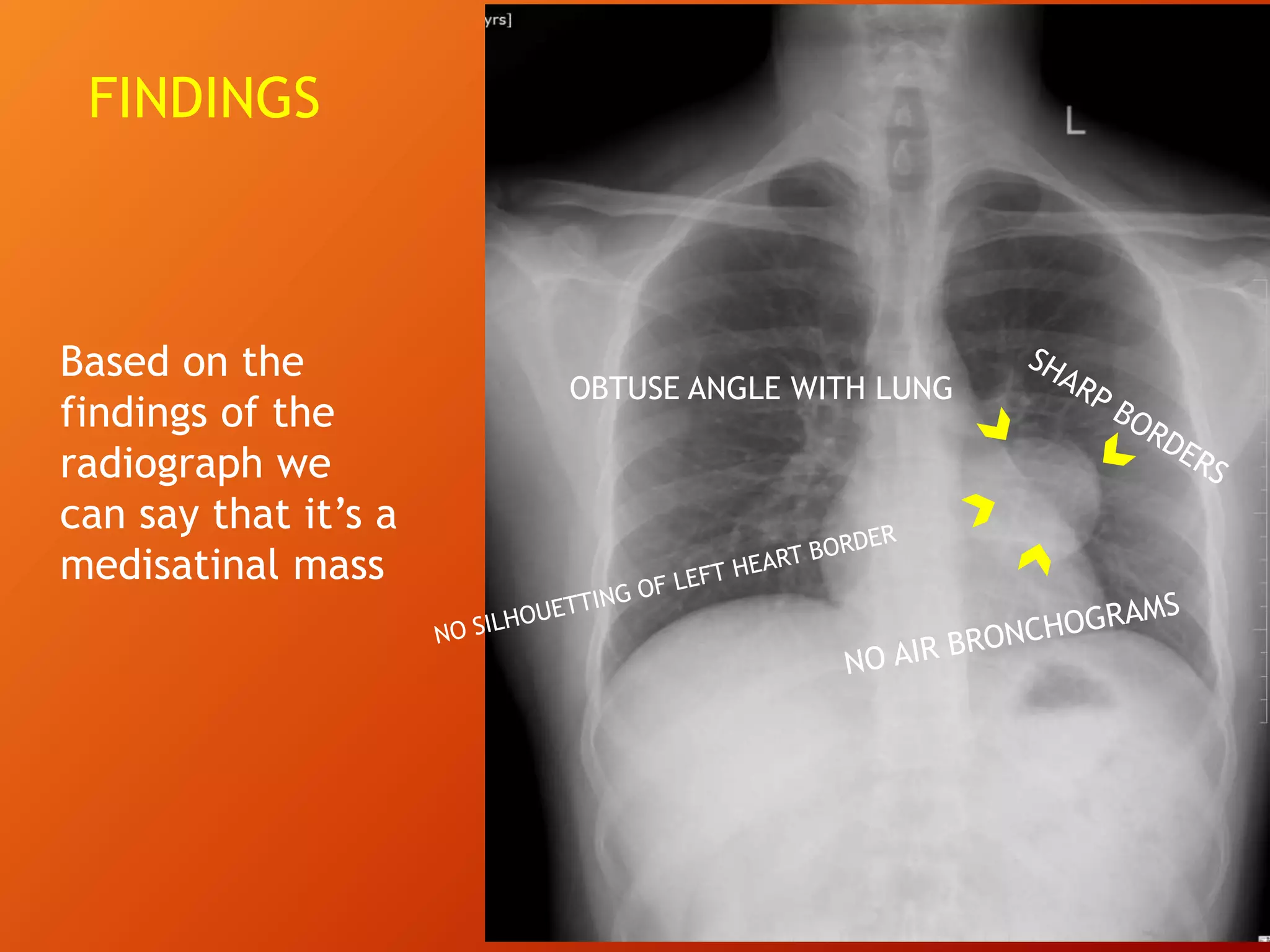
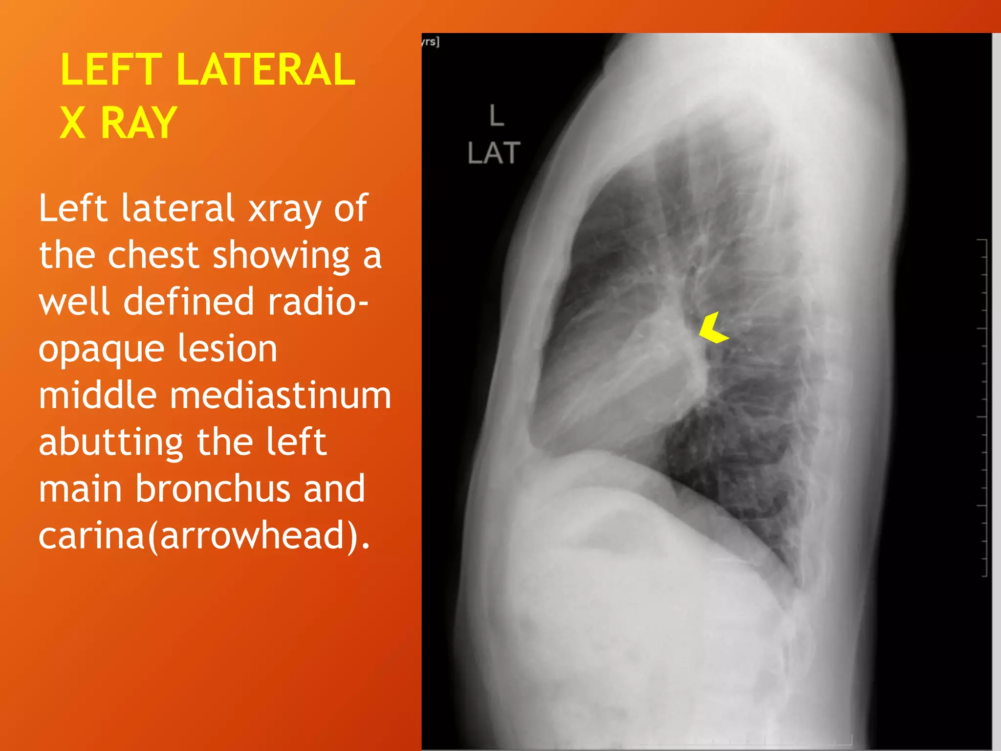
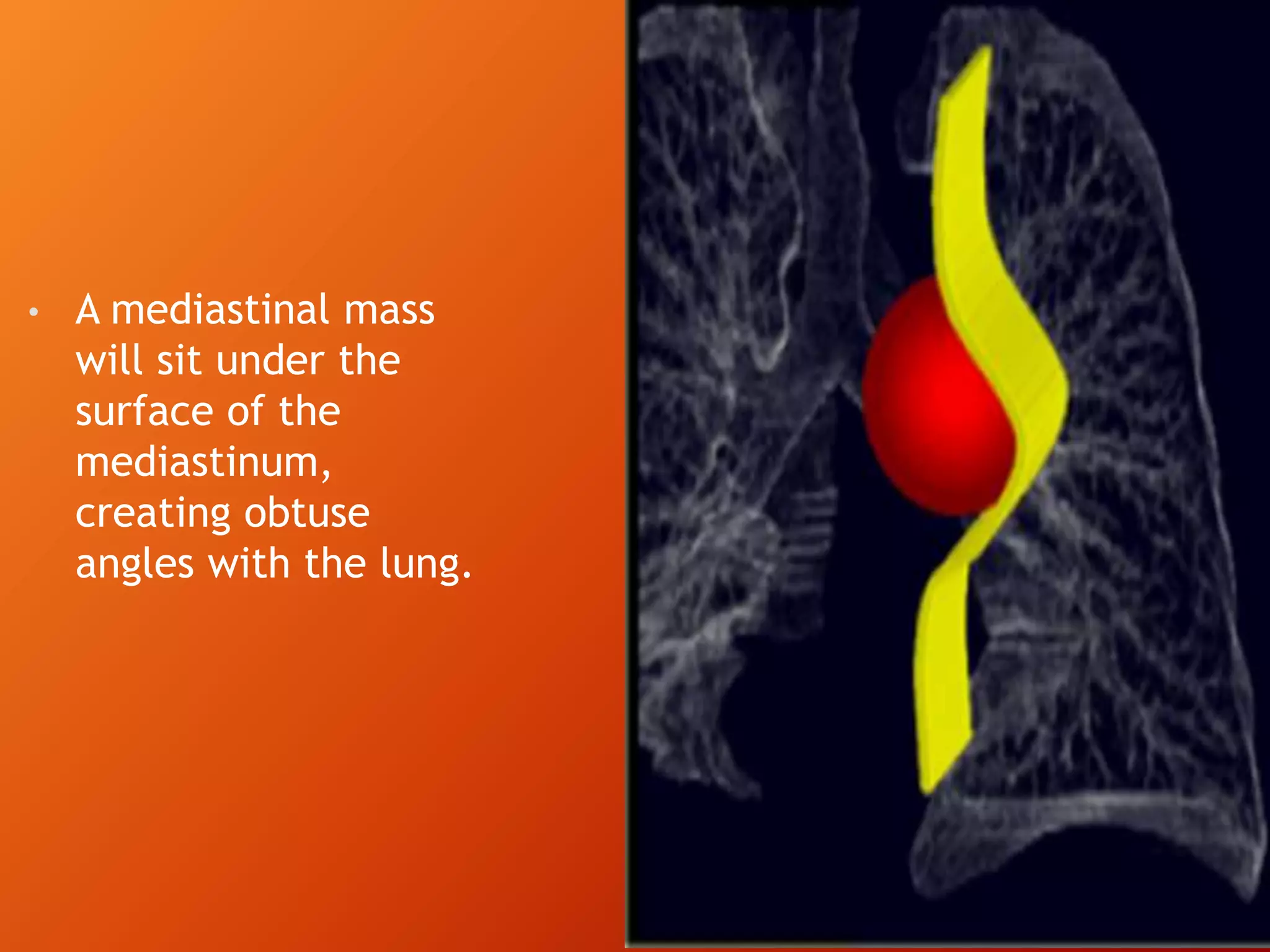
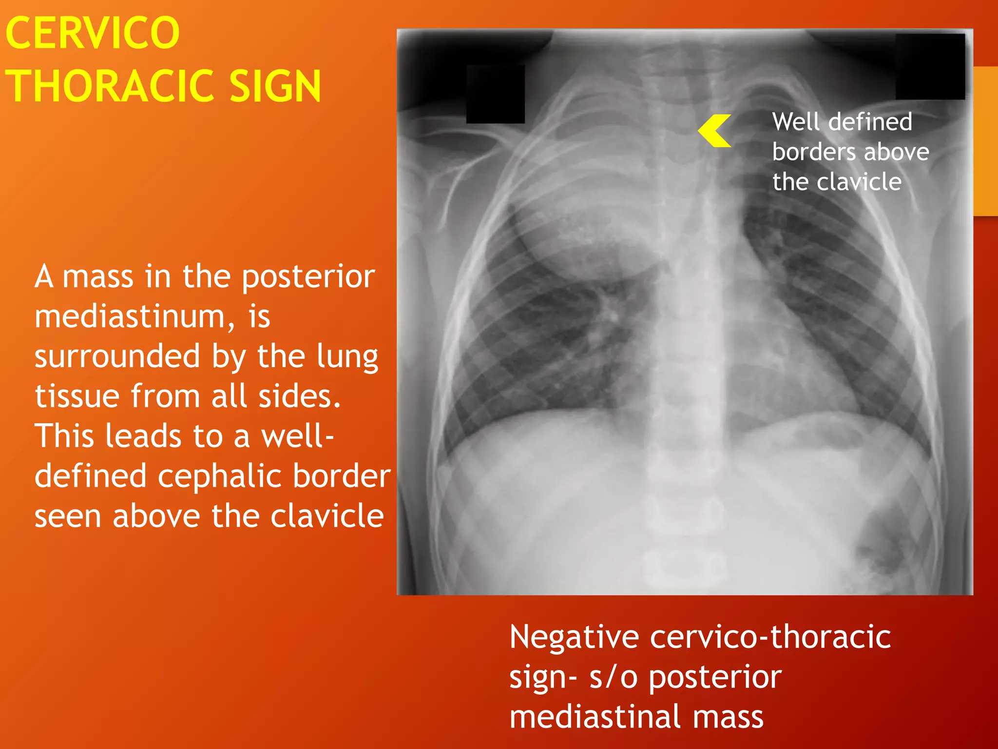
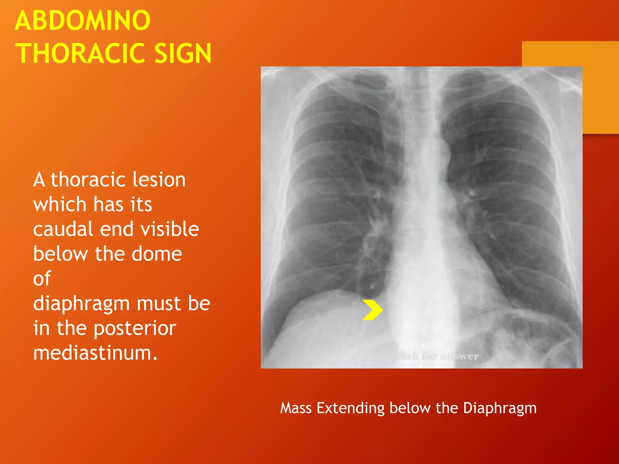
The document describes a case of a 27-year-old man presenting with chronic dry cough and referred for chest imaging. Chest x-ray revealed a well-defined round radio-opaque lesion in the left perihilar region. Further imaging found the mass to be arising from the left main bronchus in the middle mediastinum. Differential diagnoses included bronchogenic cysts and esophageal duplication cysts. Based on features of a sharply demarcated mass arising from the bronchus, the final diagnosis was determined to be a bronchogenic cyst, a congenital malformation of the bronchial tree.
Imaging plays an important role in diagnosis and formulating differential diagnosis in case of Solitary pulmonary nodule. It helps in differentiating and predicting benign and malignant nodules.
Artifact is defined as any opacity on the radiograph which does not correspond to an actual anatomic structure. Any misinterpretation of an actual anatomic structure. Anything decreasing radiographic quality.
PATOLOGÍA PLEURAL EN TOMOGRAFÍA COMPUTADANadia Rojas
Descripción de las principales enfermedades de la pleura y su presentación en tomografía computada. Derrame pleural. Neumotorax. Engrosamiento pleural. Fibrotorax. Tumores pleurales.
Concise overview of all the information that a Medico must know for his knowledge as well as to appear for entrance exams as well as for physicians for their routine practice.
Basic Radiology for Third Year Medical students BenjiH
Definitive guide to Third year radiology course featuring X-ray, CT, MRI features for basic pathologies, for medical students - third year Plovdiv medical university
Artficial Intelligence in Health universities(JSS) Vikram Patil
Artificial intelligence (AI) is a growing phenomenon
Will soon facilitate wide-scale changes in Health education and Healthcare delivery.
Students and Educators need to have the knowledge of AI in relation to learning and research
They need to know extent to which it will impact on education and healthcare delivery
The ppt is useful to understand the role of Universities in the era of AI
Lung Cancer: Artificial Intelligence, Synergetics, Complex System Analysis, S...Oleg Kshivets
RESULTS: Overall life span (LS) was 2252.1±1742.5 days and cumulative 5-year survival (5YS) reached 73.2%, 10 years – 64.8%, 20 years – 42.5%. 513 LCP lived more than 5 years (LS=3124.6±1525.6 days), 148 LCP – more than 10 years (LS=5054.4±1504.1 days).199 LCP died because of LC (LS=562.7±374.5 days). 5YS of LCP after bi/lobectomies was significantly superior in comparison with LCP after pneumonectomies (78.1% vs.63.7%, P=0.00001 by log-rank test). AT significantly improved 5YS (66.3% vs. 34.8%) (P=0.00000 by log-rank test) only for LCP with N1-2. Cox modeling displayed that 5YS of LCP significantly depended on: phase transition (PT) early-invasive LC in terms of synergetics, PT N0—N12, cell ratio factors (ratio between cancer cells- CC and blood cells subpopulations), G1-3, histology, glucose, AT, blood cell circuit, prothrombin index, heparin tolerance, recalcification time (P=0.000-0.038). Neural networks, genetic algorithm selection and bootstrap simulation revealed relationships between 5YS and PT early-invasive LC (rank=1), PT N0—N12 (rank=2), thrombocytes/CC (3), erythrocytes/CC (4), eosinophils/CC (5), healthy cells/CC (6), lymphocytes/CC (7), segmented neutrophils/CC (8), stick neutrophils/CC (9), monocytes/CC (10); leucocytes/CC (11). Correct prediction of 5YS was 100% by neural networks computing (area under ROC curve=1.0; error=0.0).
CONCLUSIONS: 5YS of LCP after radical procedures significantly depended on: 1) PT early-invasive cancer; 2) PT N0--N12; 3) cell ratio factors; 4) blood cell circuit; 5) biochemical factors; 6) hemostasis system; 7) AT; 8) LC characteristics; 9) LC cell dynamics; 10) surgery type: lobectomy/pneumonectomy; 11) anthropometric data. Optimal diagnosis and treatment strategies for LC are: 1) screening and early detection of LC; 2) availability of experienced thoracic surgeons because of complexity of radical procedures; 3) aggressive en block surgery and adequate lymph node dissection for completeness; 4) precise prediction; 5) adjuvant chemoimmunoradiotherapy for LCP with unfavorable prognosis.
Prix Galien International 2024 Forum ProgramLevi Shapiro
June 20, 2024, Prix Galien International and Jerusalem Ethics Forum in ROME. Detailed agenda including panels:
- ADVANCES IN CARDIOLOGY: A NEW PARADIGM IS COMING
- WOMEN’S HEALTH: FERTILITY PRESERVATION
- WHAT’S NEW IN THE TREATMENT OF INFECTIOUS,
ONCOLOGICAL AND INFLAMMATORY SKIN DISEASES?
- ARTIFICIAL INTELLIGENCE AND ETHICS
- GENE THERAPY
- BEYOND BORDERS: GLOBAL INITIATIVES FOR DEMOCRATIZING LIFE SCIENCE TECHNOLOGIES AND PROMOTING ACCESS TO HEALTHCARE
- ETHICAL CHALLENGES IN LIFE SCIENCES
- Prix Galien International Awards Ceremony
Pulmonary Thromboembolism - etilogy, types, medical- Surgical and nursing man...VarunMahajani
Disruption of blood supply to lung alveoli due to blockage of one or more pulmonary blood vessels is called as Pulmonary thromboembolism. In this presentation we will discuss its causes, types and its management in depth.
TEST BANK for Operations Management, 14th Edition by William J. Stevenson, Ve...kevinkariuki227
TEST BANK for Operations Management, 14th Edition by William J. Stevenson, Verified Chapters 1 - 19, Complete Newest Version.pdf
Anti ulcer drugs and their Advance pharmacology ||
Anti-ulcer drugs are medications used to prevent and treat ulcers in the stomach and upper part of the small intestine (duodenal ulcers). These ulcers are often caused by an imbalance between stomach acid and the mucosal lining, which protects the stomach lining.
||Scope: Overview of various classes of anti-ulcer drugs, their mechanisms of action, indications, side effects, and clinical considerations.
- Video recording of this lecture in English language: https://youtu.be/lK81BzxMqdo
- Video recording of this lecture in Arabic language: https://youtu.be/Ve4P0COk9OI
- Link to download the book free: https://nephrotube.blogspot.com/p/nephrotube-nephrology-books.html
- Link to NephroTube website: www.NephroTube.com
- Link to NephroTube social media accounts: https://nephrotube.blogspot.com/p/join-nephrotube-on-social-media.html
Flu Vaccine Alert in Bangalore Karnatakaaddon Scans
As flu season approaches, health officials in Bangalore, Karnataka, are urging residents to get their flu vaccinations. The seasonal flu, while common, can lead to severe health complications, particularly for vulnerable populations such as young children, the elderly, and those with underlying health conditions.
Dr. Vidisha Kumari, a leading epidemiologist in Bangalore, emphasizes the importance of getting vaccinated. "The flu vaccine is our best defense against the influenza virus. It not only protects individuals but also helps prevent the spread of the virus in our communities," he says.
This year, the flu season is expected to coincide with a potential increase in other respiratory illnesses. The Karnataka Health Department has launched an awareness campaign highlighting the significance of flu vaccinations. They have set up multiple vaccination centers across Bangalore, making it convenient for residents to receive their shots.
To encourage widespread vaccination, the government is also collaborating with local schools, workplaces, and community centers to facilitate vaccination drives. Special attention is being given to ensuring that the vaccine is accessible to all, including marginalized communities who may have limited access to healthcare.
Residents are reminded that the flu vaccine is safe and effective. Common side effects are mild and may include soreness at the injection site, mild fever, or muscle aches. These side effects are generally short-lived and far less severe than the flu itself.
Healthcare providers are also stressing the importance of continuing COVID-19 precautions. Wearing masks, practicing good hand hygiene, and maintaining social distancing are still crucial, especially in crowded places.
Protect yourself and your loved ones by getting vaccinated. Together, we can help keep Bangalore healthy and safe this flu season. For more information on vaccination centers and schedules, residents can visit the Karnataka Health Department’s official website or follow their social media pages.
Stay informed, stay safe, and get your flu shot today!
These lecture slides, by Dr Sidra Arshad, offer a quick overview of physiological basis of a normal electrocardiogram.
Learning objectives:
1. Define an electrocardiogram (ECG) and electrocardiography
2. Describe how dipoles generated by the heart produce the waveforms of the ECG
3. Describe the components of a normal electrocardiogram of a typical bipolar leads (limb II)
4. Differentiate between intervals and segments
5. Enlist some common indications for obtaining an ECG
Study Resources:
1. Chapter 11, Guyton and Hall Textbook of Medical Physiology, 14th edition
2. Chapter 9, Human Physiology - From Cells to Systems, Lauralee Sherwood, 9th edition
3. Chapter 29, Ganong’s Review of Medical Physiology, 26th edition
4. Electrocardiogram, StatPearls - https://www.ncbi.nlm.nih.gov/books/NBK549803/
5. ECG in Medical Practice by ABM Abdullah, 4th edition
6. ECG Basics, http://www.nataliescasebook.com/tag/e-c-g-basics
MANAGEMENT OF ATRIOVENTRICULAR CONDUCTION BLOCK.pdfJim Jacob Roy
Cardiac conduction defects can occur due to various causes.
Atrioventricular conduction blocks ( AV blocks ) are classified into 3 types.
This document describes the acute management of AV block.
Report Back from SGO 2024: What’s the Latest in Cervical Cancer?bkling
Are you curious about what’s new in cervical cancer research or unsure what the findings mean? Join Dr. Emily Ko, a gynecologic oncologist at Penn Medicine, to learn about the latest updates from the Society of Gynecologic Oncology (SGO) 2024 Annual Meeting on Women’s Cancer. Dr. Ko will discuss what the research presented at the conference means for you and answer your questions about the new developments.
ARTIFICIAL INTELLIGENCE IN HEALTHCARE.pdfAnujkumaranit
Artificial intelligence (AI) refers to the simulation of human intelligence processes by machines, especially computer systems. It encompasses tasks such as learning, reasoning, problem-solving, perception, and language understanding. AI technologies are revolutionizing various fields, from healthcare to finance, by enabling machines to perform tasks that typically require human intelligence.
The prostate is an exocrine gland of the male mammalian reproductive system
It is a walnut-sized gland that forms part of the male reproductive system and is located in front of the rectum and just below the urinary bladder
Function is to store and secrete a clear, slightly alkaline fluid that constitutes 10-30% of the volume of the seminal fluid that along with the spermatozoa, constitutes semen
A healthy human prostate measures (4cm-vertical, by 3cm-horizontal, 2cm ant-post ).
It surrounds the urethra just below the urinary bladder. It has anterior, median, posterior and two lateral lobes
It’s work is regulated by androgens which are responsible for male sex characteristics
Generalised disease of the prostate due to hormonal derangement which leads to non malignant enlargement of the gland (increase in the number of epithelial cells and stromal tissue)to cause compression of the urethra leading to symptoms (LUTS
1. JSS Medical College, Mysuru
CASE OF THE WEEK
DR KAVITHA K.
DR SHIKHAR GARG
(Post Graduate Residents-Radiology)
2. PRESENTING COMPLAINT
27 year old man came with history of chronic
dry cough. No h/o fever, hemoptysis, loss of
weight.
He was referred to the radiology department for
further evaluation.
9. LEFT LATERAL
X RAY
Left lateral xray of
the chest showing a
well defined radio-
opaque lesion
middle mediastinum
abutting the left
main bronchus and
carina(arrowhead).
10. SO HOW DO WE DIFFERENTIATE
MEDIASTINAL MASS FROM PARENCHYMAL
MASS?
11. MEDIASTINAL VS PARENCHYMAL MASS
• Unlike lung lesions, a mediastinal mass will not
contain air bronchograms.
• Margins with the lung will be obtuse.
• Will not move with resipration on fluoroscopy.
• Pencil sharp borders.
• Broad based towards the mediastinum.
12. A lung mass abutts the
mediastinal surface
and creates acute
angle with the lung.
13. • A mediastinal mass
will sit under the
surface of the
mediastinum,
creating obtuse
angles with the lung.
16. Anterior
mediastinum: Anterior to
the pericardium
Middle
mediastinum: Within the
pericardium
Posterior
mediastinum: Posterior
to the pericardium
DIVISIONS OF INFERIOR
MEDIASTINUM
17. Anterior mediastinum
Thymus, lymph nodes
and
retrosternal thyroid
CONTENTS OF INFERIOR
MEDIASTINUM
Middle mediastinum
The heart,
Pericardium, Great
vessels,
Tracheal bifurcation
and both main
bronchi.
Posterior mediastinum
Descending aorta,
Oesophagus, Thoracic
duct, Azygous &
hemiazygous venous
systems.
18. LETS LEARN THE SIGNS WHICH HELP US
TO LOCALISE A MEDIASTINAL MASS ON A
FRONTAL RADIOGRAPH
19. The differential
attenuation of x-ray
photons by two
adjacent structures
defines the
silhouette
SILHOUETTE
SIGN
Loss of right cardiac silhouette due tor right lung middle lobe pneumonia
20. When a mass arises from
the hilum, the pulmonary
vessels are in contact with
the mass and their
silhouette is obliterated.
Visible vessles implies that
the mass is not contacting
the hilum, and is either
anterior or posterior to it.
HILUM OVERLAY
SIGN
21. Helps to distinguish a
bulky hilum due to
pulmonary artery
dilatation from a
mass.
Vessels can be seen to
converge and join a
dilated pulmonary
artery.
HILUM
CONVERGENCE
SIGN
A CASE OF PULMONARY ARTERY
HYPERTENSION
22. A mass in the posterior
mediastinum, is
surrounded by the lung
tissue from all sides.
This leads to a well-
defined cephalic border
seen above the clavicle
CERVICO
THORACIC SIGN
Negative cervico-thoracic
sign- s/o posterior
mediastinal mass
Well defined
borders above
the clavicle
23. ABDOMINO
THORACIC SIGN
A thoracic lesion
which has its
caudal end visible
below the dome
of
diaphragm must be
in the posterior
mediastinum.
Mass Extending below the Diaphragm
26. CT AND MRI WILL HELP US TO LOCALIZE,
FURTHER CHARACTERISE VARIOUS
MEDISTINAL MASS AND ALSO HELPS IN
EVALUTING INVASION INTO ADJACENT
STRUCTURES.
FURTHER IMAGING
27. Final conclusion: Well demarcated radio-opaque mass in
the middle mediastinum arising from the left main
bronchus.
28. BASED ON THE XRAY FINDINGS
DIFFERENTIALS –
Oesophageal duplication cysts - Thick walled cysts
found adjacent to the oesophagus
Bronchogenic Duplication cysts - Sharply demarcated
round/ oval thin walled mass filled with proteinacious
fluid usually in the medial 1/3 of lungs arising from the
bronchus.
30. BRONCHOGENIC CYST
Bronchogenic cysts are congenital malformations of the bronchial tree.
They can present as a mediastinal mass that may enlarge and cause local
compression.
It is also considered the commonest of foregut duplication cysts.
31. Bronchogenic cysts are asymptomatic and are found
incidentally.
When large, mass effect may result in bronchial
obstruction leading to air trapping and respiratory
distress.
CLINICAL PRESENTATION
32. Sharply demarcated round mass in the medial 1/3 of
lungs.
They do not communicate with the bronchial tree, and
are therefore not air filled.
They contain fluid ,variable amounts of proteinaceous
material, blood products, and calcium oxalate .
It is the latter three components that result in
increased attenuation mimicking solid lesions.
FEATURES
33. CT findings
Well circumscribed
spherical mass of
variable
attenuation with
variable fluid
composition explaining
the different CT
attenuations observed.
The degree of CT
attenuation often
depends on the amount
of internal proteinaceous
content .
FURTHER IMAGING