Downloaded 1,089 times


![Tissue Doppler of mitral annulus
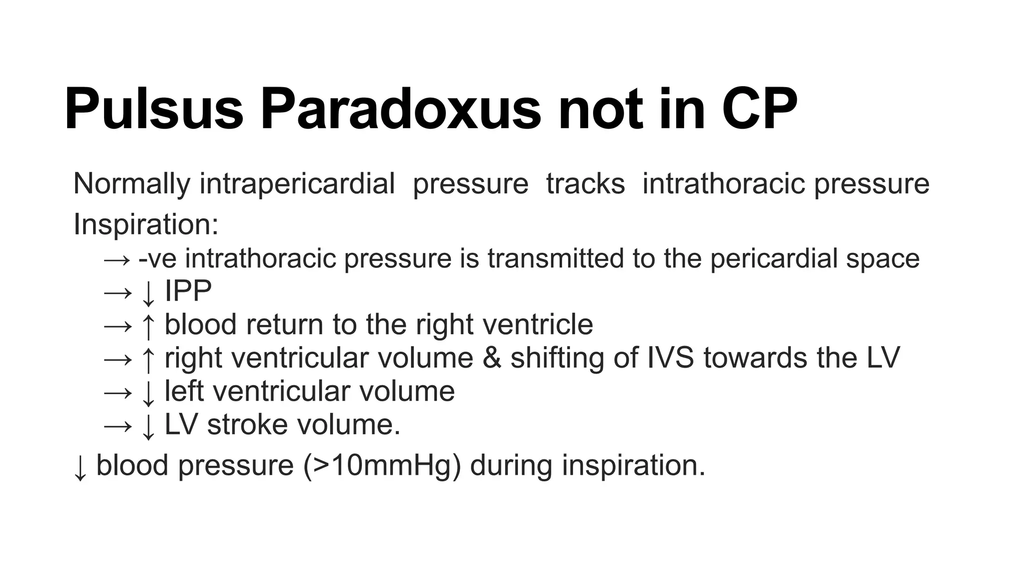
Constrictictive pericarditis:
Annular paradox:
E’ increases as severity of CP increase[as increased filing pressure]
Peak E’ ≥ 8 cm/s
89% senstive for constriction
100% specific.
RCM:
E’ decreases as severity ↑
E’< 8 cm/s.](https://image.slidesharecdn.com/constrictivepericarditis-inprocess-140508025746-phpapp01/75/Constrictive-pericarditis-24-2048.jpg)


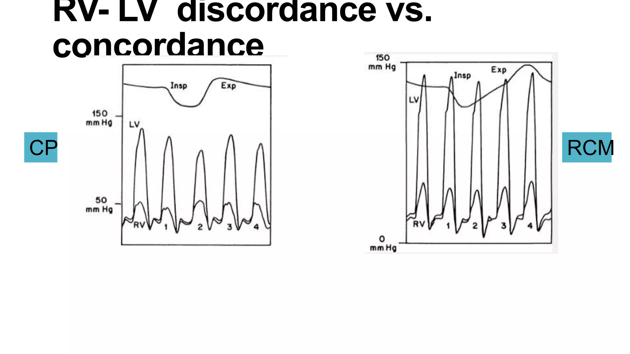


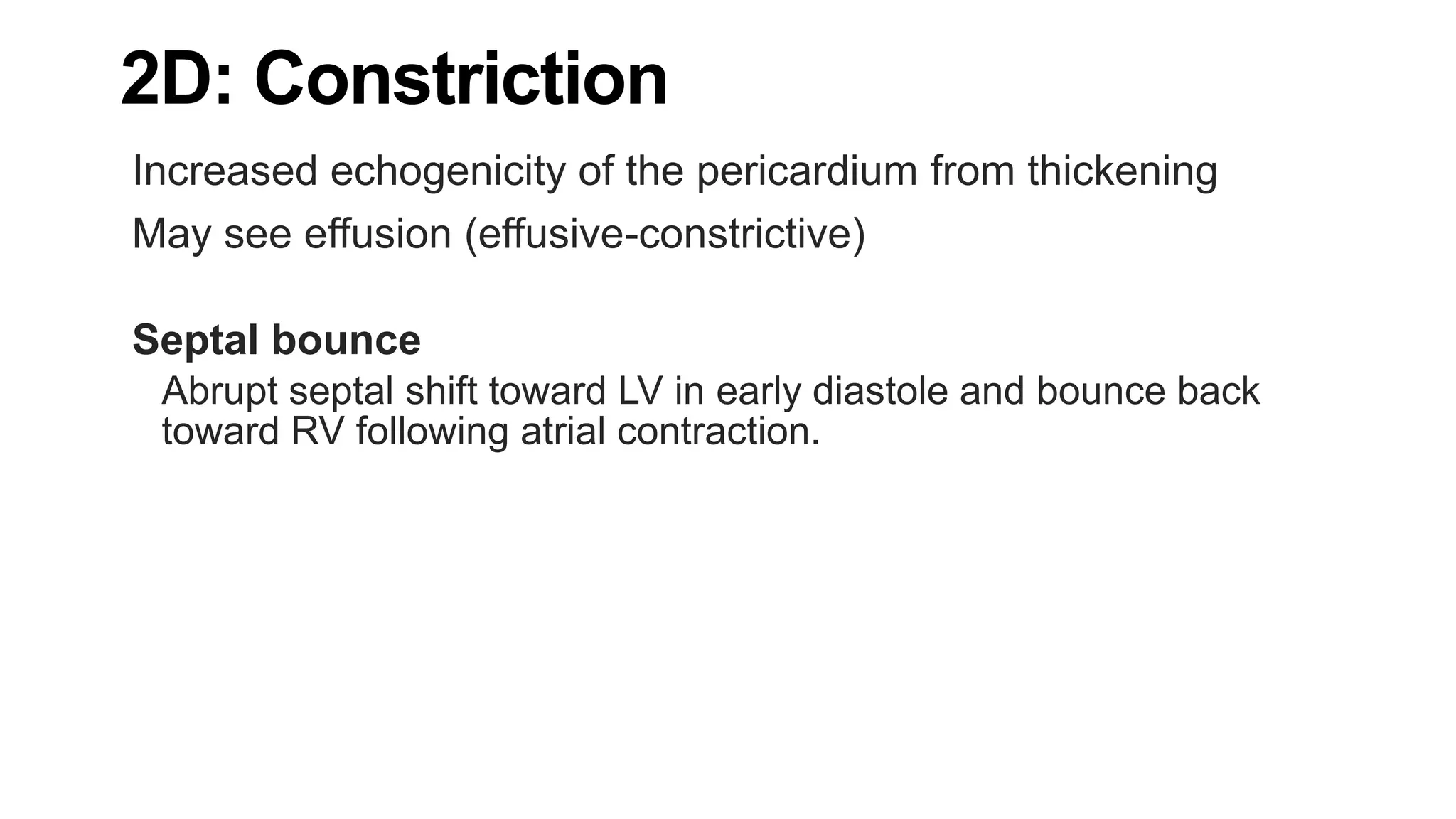
The document discusses constrictive pericarditis, providing details on: 1) The pathology of constrictive pericarditis which involves thickening and scarring of the pericardium leading to loss of elasticity. 2) The pathophysiology of constrictive pericarditis where the inelastic pericardium constrains cardiac filling and prevents adaptation to volume changes. 3) Key diagnostic features of constrictive pericarditis seen on echocardiogram include septal bounce, rapid early diastolic mitral inflow, and increased mitral annular velocities that rise with inspiration.


