Download to read offline
![Review began 02/22/2023
Review ended 03/06/2023
Published 03/09/2023
© Copyright 2023
Barik et al. This is an open access article
distributed under the terms of the Creative
Commons Attribution License CC-BY 4.0.,
which permits unrestricted use, distribution,
and reproduction in any medium, provided
the original author and source are credited.
Pacemaker Pocket Infection After Splenectomy
Ramachandra Barik Sr. , Pranjit Deb , Abhinav Kumar , Rudrapratap Mahapatra
1. Cardiology, All India Institute of Medical Sciences, Bhubaneswar, IND 2. Cardiothoracic Surgery, All India Institute
of Medical Sciences, Bhubaneswar, IND
Corresponding author: Pranjit Deb, pranjeetdeb@gmail.com
Abstract
A post-splenectomy patient suffers from frequent infections due to capsulated bacteria like Streptococcus
pneumoniae, Hemophilus influenzae, and Neisseria meningitidis despite vaccination because of a lack of
memory B lymphocytes. Pacemaker implantation after splenectomy is less common. Our patient underwent
splenectomy for splenic rupture after a road traffic accident. He developed a complete heart block after
seven years, during which a dual-chamber pacemaker was implanted. However, he was operated on seven
times to treat the complication related to that pacemaker over a period of one year because of various
reasons, which have been shared in this case report. The clinical translation of this interesting observation
is that, though the pacemaker implantation procedure is a well-established procedure, the procedural
outcome is influenced by patient factors like the absence of a spleen, procedural factors like septic measures,
and device factors like the reuse of an already-used pacemaker or leads.
Categories: Cardiology, General Surgery, Infectious Disease
Keywords: pneumococcal vaccine, reuse of pacemaker, abandoned leads, pacemaker pocket infection, splenectomy
Introduction
Pacemaker pocket infection or infective endocarditis due to vegetation over the pacemaker leads has several
causes, like patient factors, procedural technique, and the type of pacemaker and its leads. When it occurs, it
is associated with high treatment costs, prolonged hospital stays, frequent outpatient visits, morbidity, and
in a few cases, may even result in death [1], which adds to the cost [2]. Asplenia or post-splenectomy status
causes significant bloodstream infection by capsulated bacteria due to the absence of memory B cells [3].
Pacemaker pocket infection, especially after splenectomy, is less common. We came across a patient who
had undergone a splenectomy for a spleen rupture in a road traffic accident in 2014. This patient underwent
dual chamber pacemaker implantation after seven years to treat complete heart failure due to sick sinus
syndrome. However, the pacemaker pocket had repeated infections, most probably due to impaired
immunity because of its post-splenectomy status. The case report is very interesting because the patient was
operated on seven times over a period of one year for a pacemaker pocket infection.
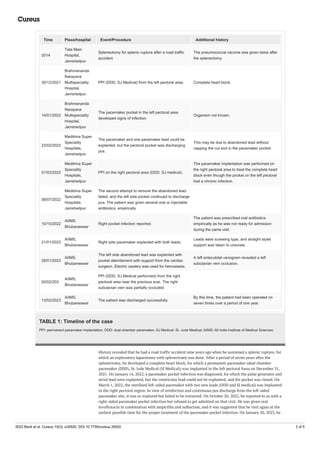
Case Presentation
A 56-year-old man presented with a permanent pacemaker pocket infection in the right pectoral area for a
month. The detailed case history with respect to the timeline is mentioned in Table 1.
1 1 1 2
Open Access Case
Report DOI: 10.7759/cureus.35920
How to cite this article
Barik R, Deb P, Kumar A, et al. (March 09, 2023) Pacemaker Pocket Infection After Splenectomy. Cureus 15(3): e35920. DOI
10.7759/cureus.35920](https://image.slidesharecdn.com/cureus-0015-00000035920-230319111819-aa465822/85/Pacemaker-Pocket-Infection-After-Splenectomy-1-320.jpg)
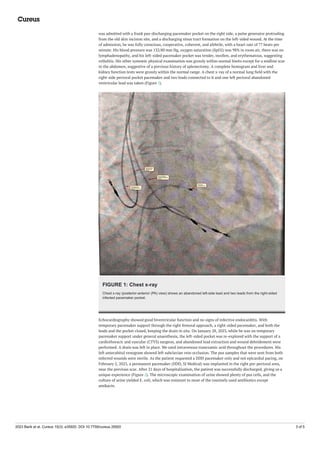
![FIGURE 2: Anterior chest wall
The anterior chest wall shows two healed pacemaker implantation sites on the right side and one healed
pacemaker site on the left side.
Discussion
Pacemaker pocket infection is defined as the presence of local signs of inflammation at the site of the
pacemaker pocket [4]. It may be associated with vegetation over the implanted pacemaker leads and heart
valves, pulmonary thromboembolism, and other systemic infections due to bacteremia. By the time a patient
goes to another hospital for proper care, the pacemaker is usually out of pocket, as in our case. The rate of
pocket infection is significantly higher after reimplantation procedures [5]. Nearly 15% of the patients with
pacemaker pocket infections may have systemic infections.
Risk factors for pacemaker pocket infection can be divided into three categories: patient-related, procedure-
related, and device-related. Some of these are modifiable factors. Identification of modifiable risk factors is
important because that may help to take preventive measures to reduce the risk of recurrent infections. Our
patient had three apparent risk factors, namely post-splenectomy status, an abandoned pacemaker lead in
the left pectoral pocket, and the reuse of a sterilised pacemaker (Table 1). Recurrent infection of the
pacemaker pocket, as in our case, has not been reported earlier. It may be due to the repeated use of several
empiric antibiotic regimens one after another that our patient did not grow bacteria on a blood or pus
culture from the infected pocket, or the organism may be atypical. Our patient received the pneumococcal
vaccination twice. In our case, the infection related to the abandoned lead on the left and the reuse of a
sterilised pacemaker could have been prevented to reduce recurring infection [6,7].
Chronic kidney disease, diabetes mellitus, chronic obstructive pulmonary disease, corticosteroid use, a
history of previous device infection, malignancy, heart failure, pre-procedural infection in the body,
anticoagulant or antiplatelet use, and skin disorders are important patient-related risk factors. The
procedure-related factors are lack of proper hand washing, an inadequate infection control programme in the
hospital, no standardised sterilisation protocol, pocket hematoma, poor hemostasis due to the lack of all the
layers being stitched systematically, early lead repositioning, and inexperienced operators [8]. The device-
related factors are few but have a significant impact on the incidence rate of pacemaker pocket infections.
The abdominal pocket site, a heavier device, a greater number of leads (including epicardial leads), and the
reuse of sterile pacemakers or leads significantly increase pacemaker pocket infection and infective
endocarditis, as in our case [4,9]. Cultures from pocket sites, lead tips, and blood samples may become
positive in 60 to 70% of the patients, but these were negative in our case because of repeated use of
empirical antibiotics or because the patient had an atypical bacterial infection [10]. As our patient had
undergone splenectomy earlier, the multiple blood cultures or pus from both the pacemaker site were sent
for aerobic, anaerobic, and fungal cultures, but none had growth of any organism. The reasons for repeated
pacemaker infections in our case may be related to the post-splenectomy status, the re-use of an explanted
pacemaker, abandoned leads without capping the cut end of them in the pacemaker packet, and the
treatment of the patient in multiple centres. However, we feel the post-splenectomy status is the main
reason for repeated infections. Even though there is no evidence of encapsulated bacterial growth in the
blood or pus culture in our case, the repeated use of antibiotics can be explained. As we notice in everyday
practice, all the risk factors except the splenectomy are usual risk factors, and they do not pose great
challenges, i.e., seven operations by different experts over a period of one year.
2023 Barik et al. Cureus 15(3): e35920. DOI 10.7759/cureus.35920 4 of 5](https://image.slidesharecdn.com/cureus-0015-00000035920-230319111819-aa465822/85/Pacemaker-Pocket-Infection-After-Splenectomy-4-320.jpg)

This case report describes a patient who underwent seven operations over one year to treat recurrent pacemaker pocket infections. The patient had undergone a splenectomy seven years prior due to a splenic rupture from a traffic accident. This left the patient immunocompromised and at higher risk for infection. The patient later required a pacemaker implantation for complete heart block. The pacemaker pocket developed repeated infections, likely due to the patient's asplenic state impairing immunity. The infections were difficult to treat due to multiple complicating factors, including an abandoned pacemaker lead and reuse of a sterilized pacemaker. This highlights the influence of patient factors like asplenia on procedural outcomes like pacemaker implantation.


