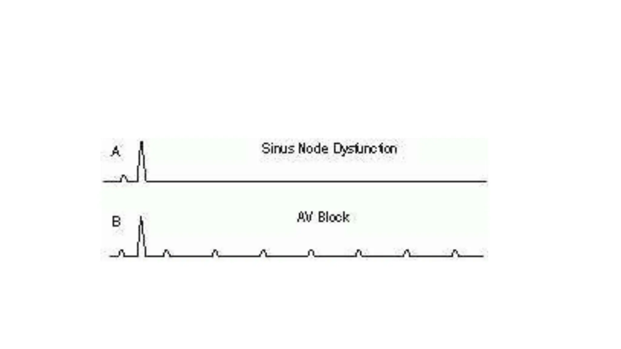
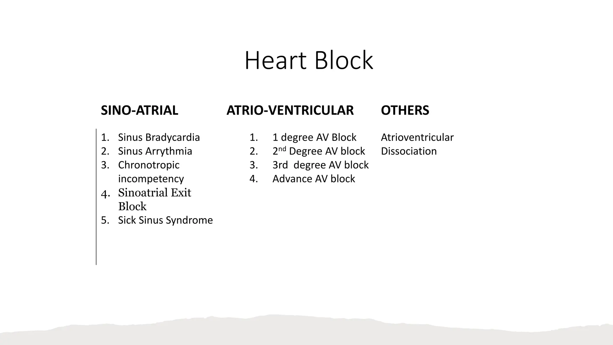
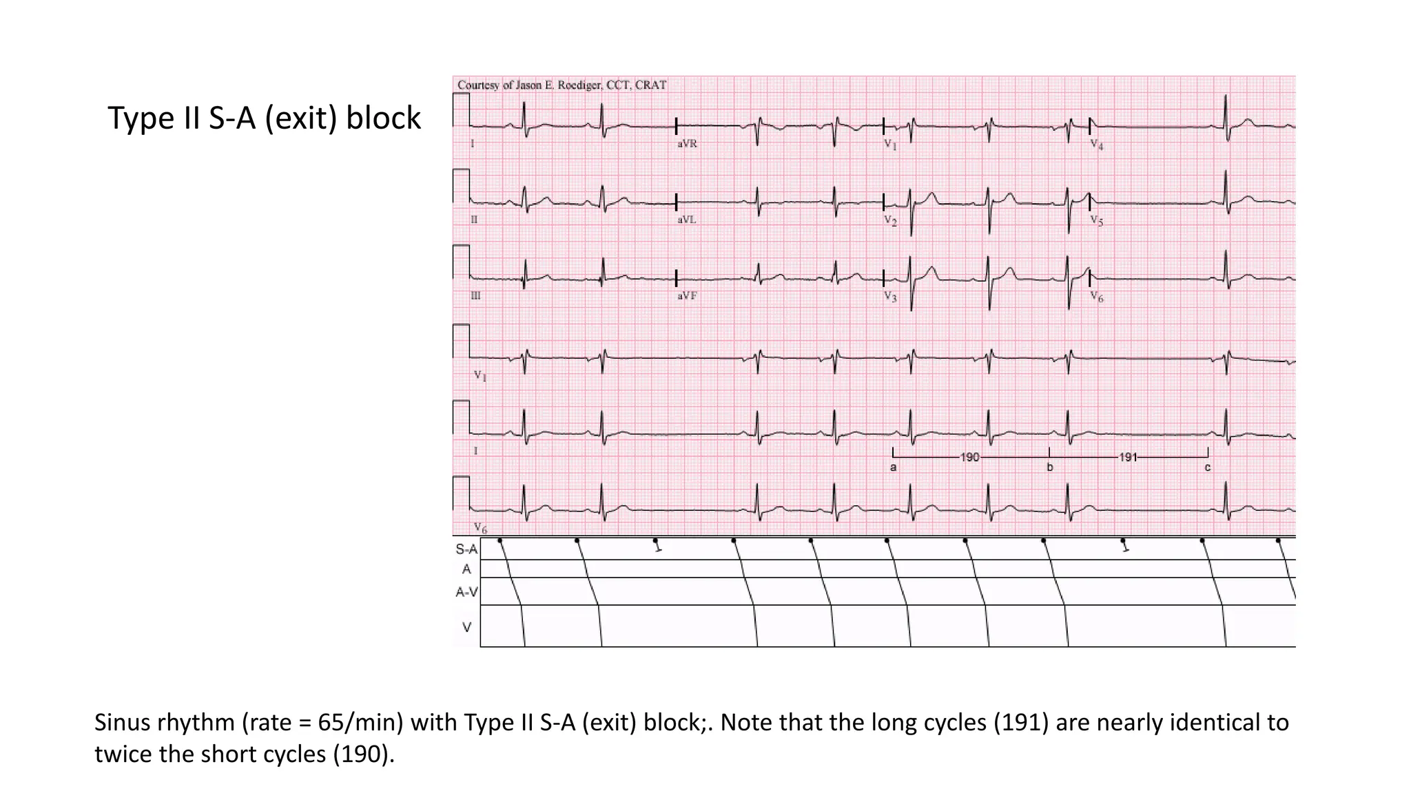
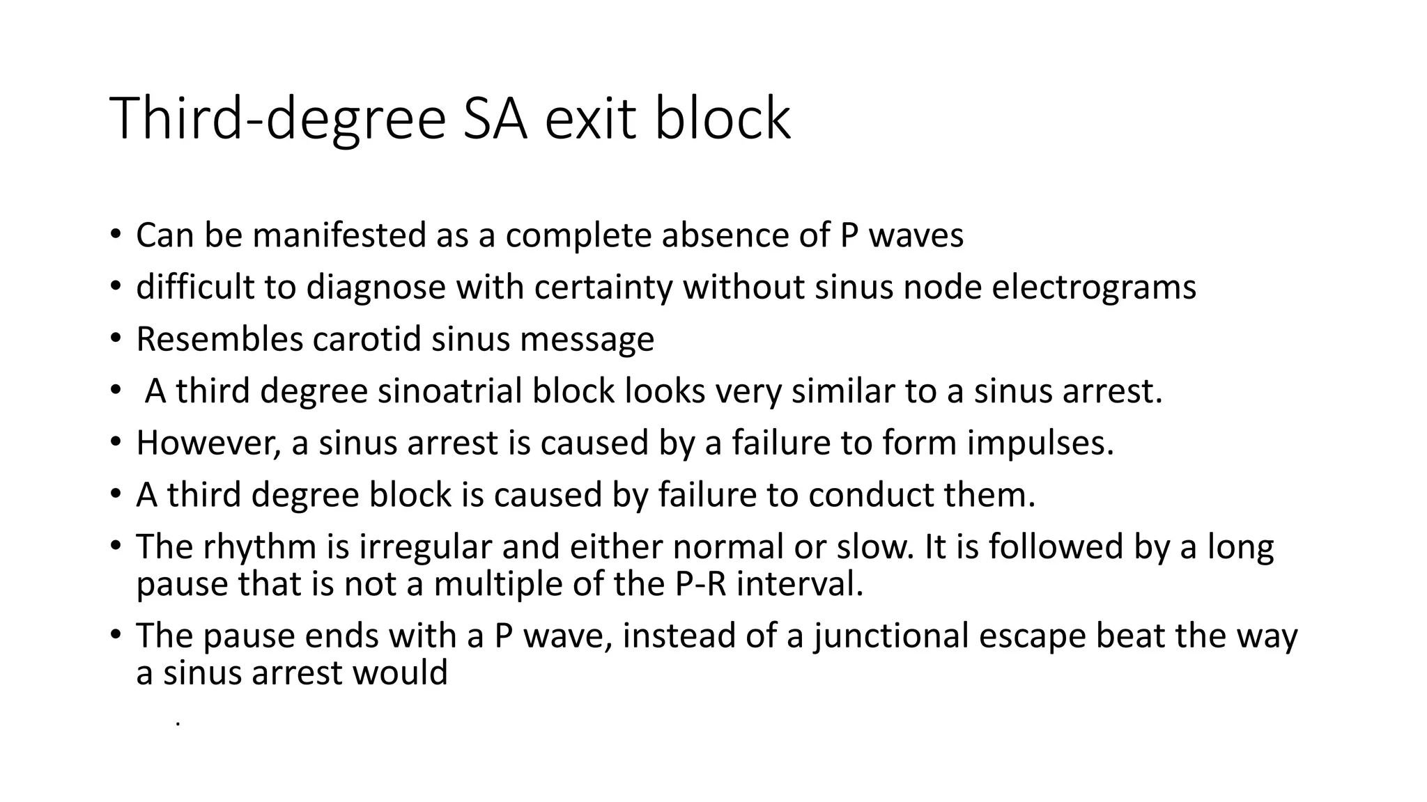
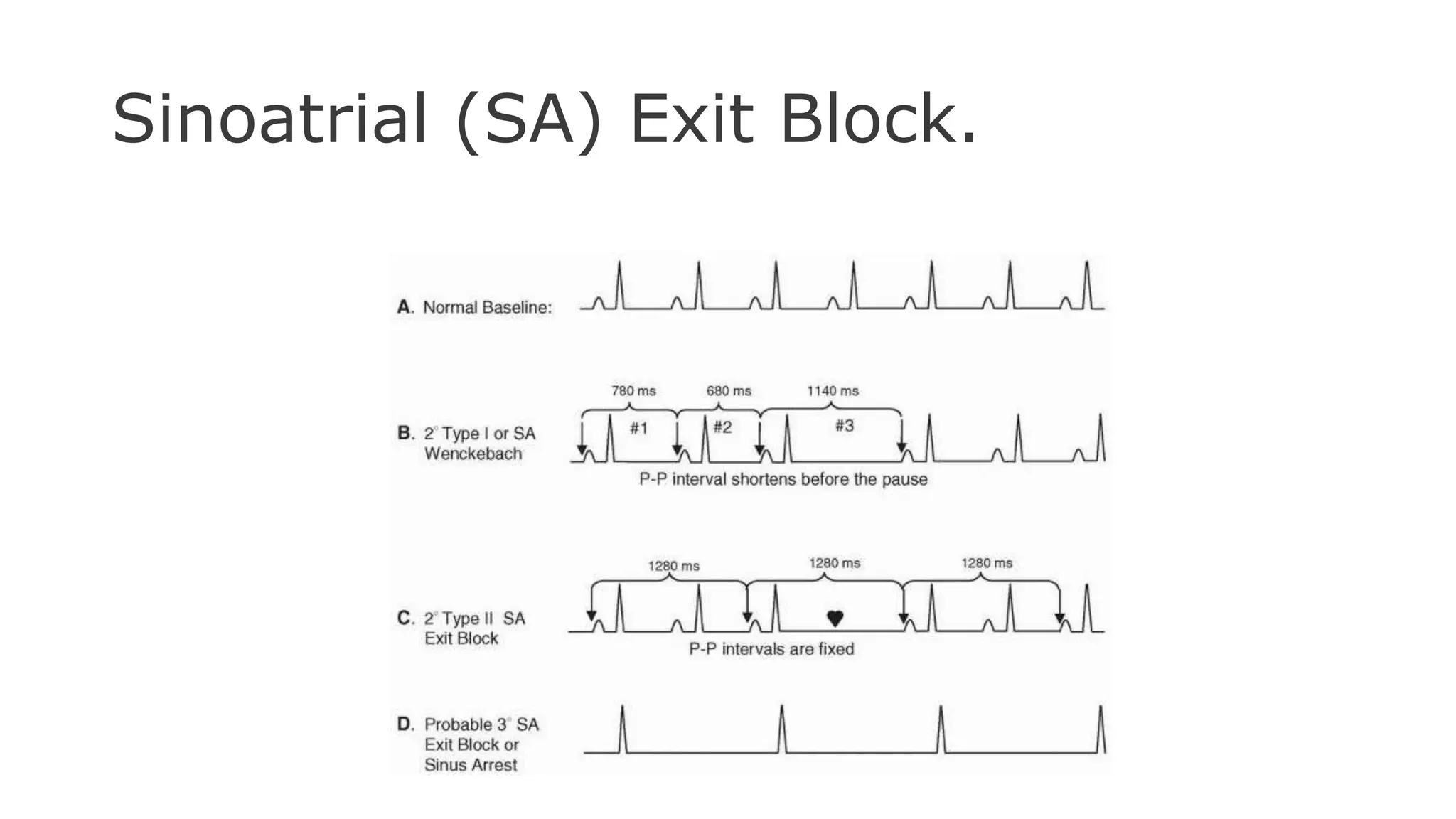
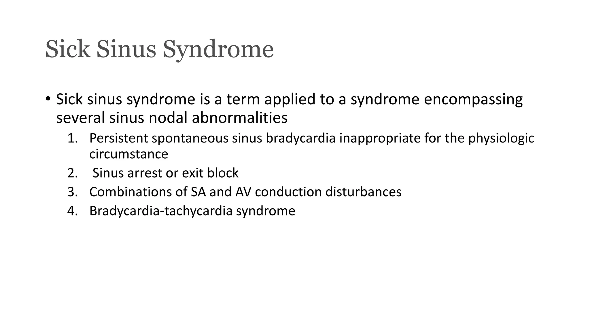
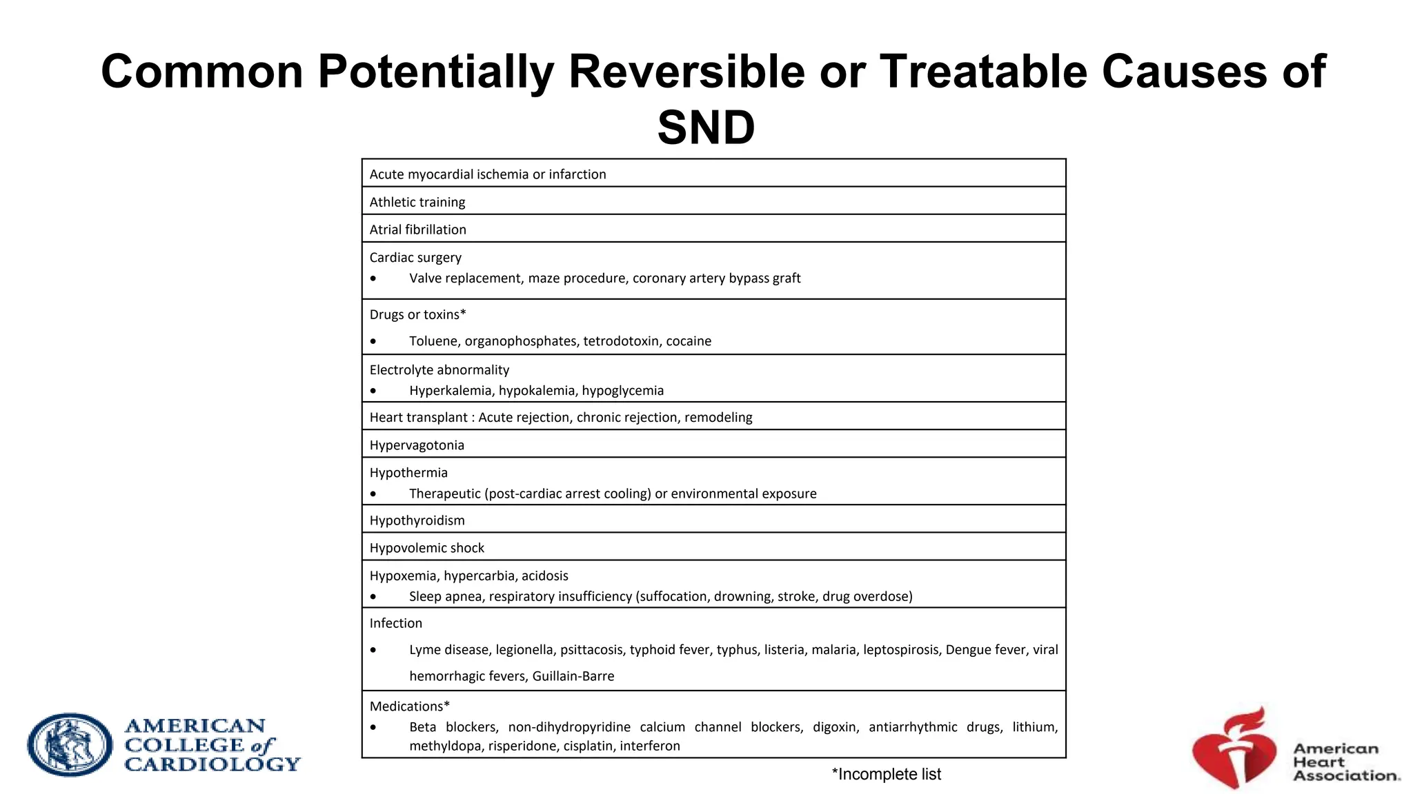
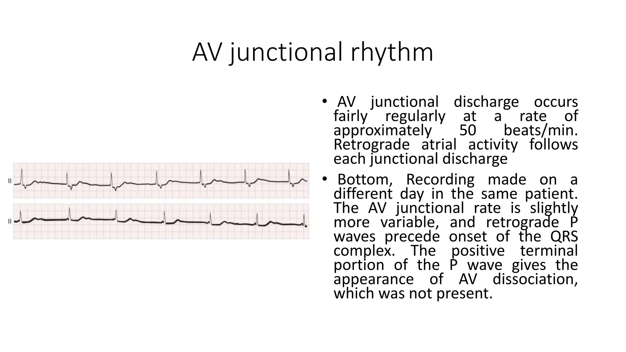
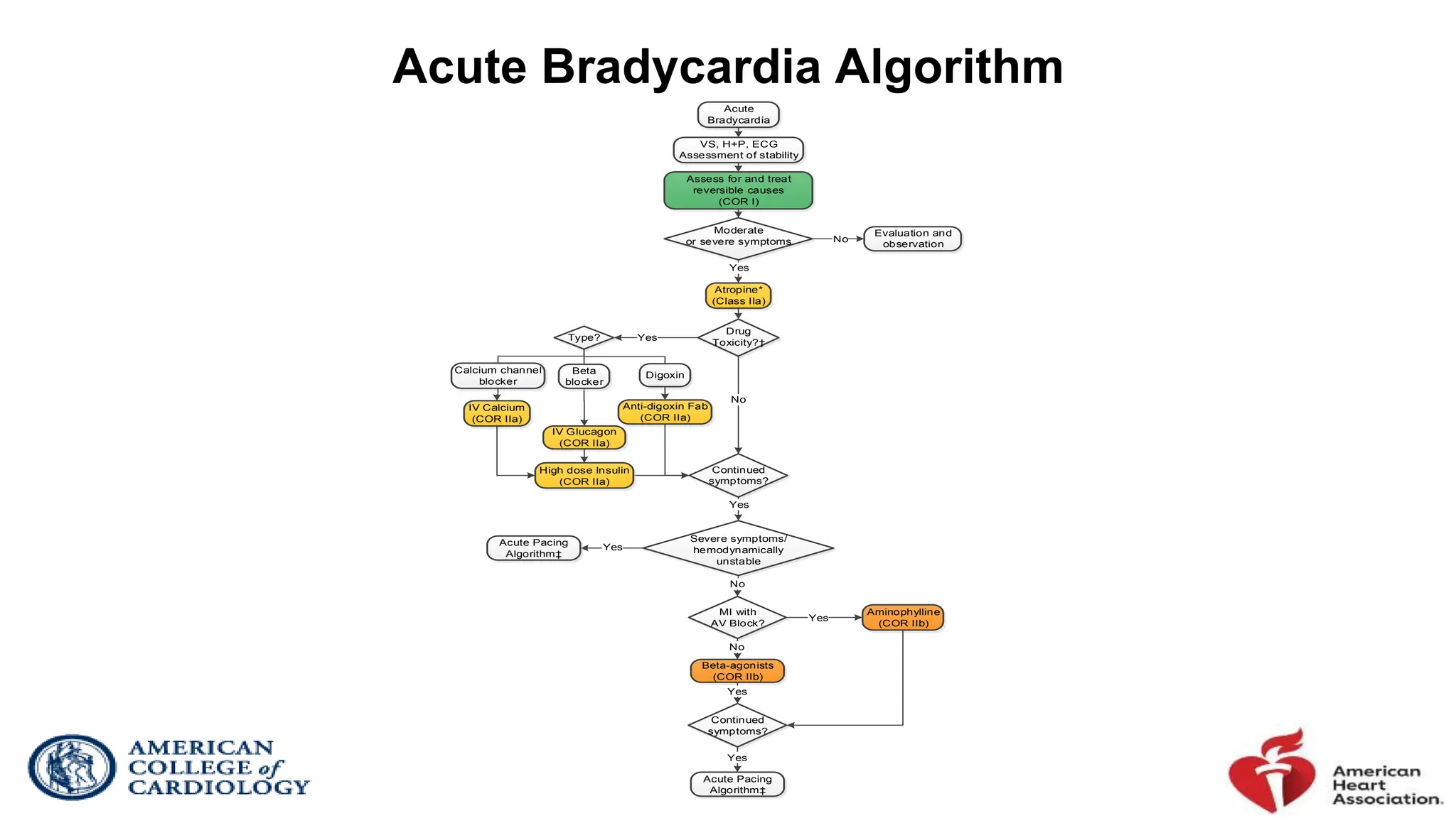
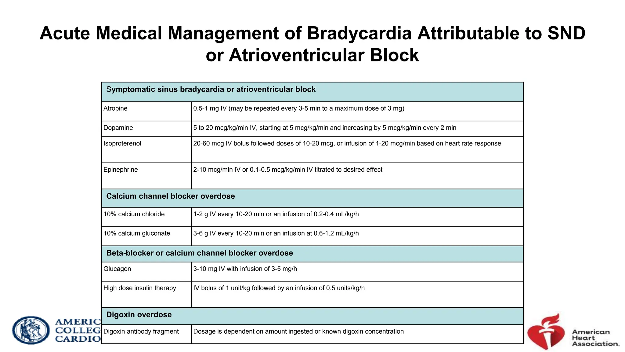
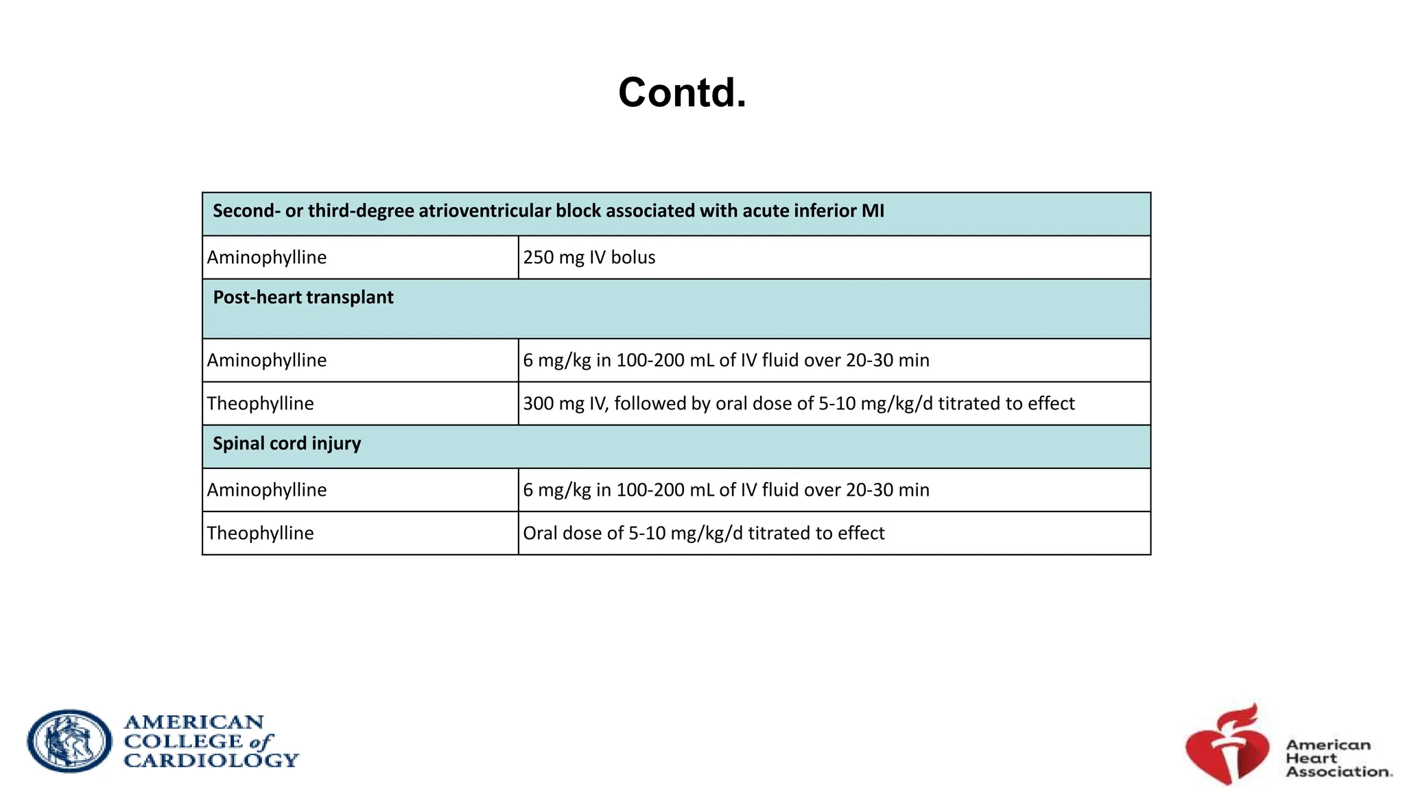
Brady arrythmia part-II
Heart Block
Introduction
• Natural pacemaker of the heart fails to generate electrical impulse in
normal rate or conduction fiber fails to conduct normally
• Sometimes either issues may be there
• Subsidiary pacemaker takes but heart beats at a slower rate
Heart Block
SINO-ATRIAL ATRIO-VENTRICULAR OTHERS
1. Sinus Bradycardia
2. Sinus Arrythmia
3. Chronotropic
incompetency
4. Sinoatrial Exit
Block
5. Sick Sinus Syndrome
1. 1 degree AV Block
2. 2nd Degree AV block
3. 3rd degree AV block
4. Advance AV block
Atrioventricular
Dissociation
Sinoatrial Exit Block
• Absence of the normally expected P wave
• The duration of the pause is a multiple of the basic P-P interval
• SA exit block is caused by a conduction disturbance during which an
impulse formed within the sinus node fails to depolarize the atria or
does so with delay
First-degree SA exit block
• First degree sinoatrial block, there is a lag between the time that the
SA node fires and actual depolarization of the atria.
• This rhythm is not easily detectable , not recognizable on an ECG strip
because an ECG strip does not denote when the SA node fires.
• It can be detected only during an electrophysiology study
Second degree block
• Type I (Wenckebach) second-degree SA exit block: the P-P
interval progressively shortens before the pause, and the
duration of the pause is less than two P-P cycles
• Type II second-degree SA exit block : An interval without P
waves that equals approximately two, three, or four times
the normal P-P cycle characterizes
Sinus nodal exit block
• Type I : The P-P interval shortens from
the first to the second cycle in each
grouping, followed by a pause. The
duration of the pause is less than
twice the shortest cycle length, and
the cycle after the pause exceeds the
cycle before the pause. The PR
interval is normal and constant.
• TYPE II: The P-P interval varies slightly
because of sinus arrhythmia. The two
pauses in sinus nodal activity equal
twice the basic P-P interval and are
consistent with a type II 2 : 1 SA nodal
exit block. The PR interval is normal
and constant. Lead III recording is
shown.
Type II S-A (exit) block
Sinus rhythm (rate = 65/min) with Type II S-A (exit) block;. Note that the long cycles (191) are nearly identical to
twice the short cycles (190).
Third-degree SA exit block
• Can be manifested as a complete absence of P waves
• difficult to diagnose with certainty without sinus node electrograms
• Resembles carotid sinus message
• A third degree sinoatrial block looks very similar to a sinus arrest.
• However, a sinus arrest is caused by a failure to form impulses.
• A third degree block is caused by failure to conduct them.
• The rhythm is irregular and either normal or slow. It is followed by a long
pause that is not a multiple of the P-R interval.
• The pause ends with a P wave, instead of a junctional escape beat the way
a sinus arrest would
.
Sinoatrial (SA) Exit Block.
Sick Sinus Syndrome
• Sick sinus syndrome is a term applied to a syndrome encompassing
several sinus nodal abnormalities
1. Persistent spontaneous sinus bradycardia inappropriate for the physiologic
circumstance
2. Sinus arrest or exit block
3. Combinations of SA and AV conduction disturbances
4. Bradycardia-tachycardia syndrome
Anatomic basis of sick sinus syndrome
• Can involve total or subtotal destruction of the sinus node, areas of nodal-atrial
discontinuity, inflammatory or degenerative changes in the nerves and ganglia
surrounding the node, and pathologic changes in the atrial wall
• Fibrosis and fatty infiltration occur, and the sclerodegenerative processes
generally involve the sinus node and the AV node or the bundle of His and its
branches or distal subdivisions.
• Occlusion of the sinus node artery can cause sinus node dysfunction.
SINUS ARREST
• The patient had a long-term
electrocardiographic recorder
connected when he died suddenly
of cardiac standstill. The rhythms
demonstrate progressive sinus
bradycardia and sinus arrest at
8:41 am . The rhythm then
becomes a ventricular escape
rhythm, which progressively slows
and finally ceases at 8:47 am . The
paired electrocardiographic strips
are continuous recordings.
SA exit block vs Sinus arrest
Tachycardia-Bradycardia Syndrome
• Typically, atrial fibrillation or atrial flutter terminates, with a resultant
excessive post-conversion pause.
• It typically occurs as a result of treatment with beta blockers or
calcium channel blockers
• Pacing for bradycardia, combined with drug therapy to treat the
tachycardia, is required in those with bradycardia-tachycardia
syndrome
Sick sinus syndrome with bradycardia-
tachycardia
• Sick sinus syndrome with
bradycardia-tachycardia. Top,
Intermittent sinus arrest is
apparent with junctional escape
beats at irregular intervals ( red
circles ). Bottom, In this continuous
monitor lead recording, a short
episode of atrial flutter is followed
by almost 5 seconds of asystole
before a junctional escape rhythm
resumes. The patient became
presyncopal at this point.
Wandering atrial pacemaker
• As the heart rate slows, the P
waves become inverted and then
gradually revert toward normal
when the heart rate speeds up
again. The PR interval shortens to
0.14 second with the inverted P
wave and is 0.16 second with the
upright P wave. This phasic
variation in cycle length with
varying P wave contour suggests a
shift in pacemaker site and is
characteristic of a wandering atrial
pacemaker
Common Potentially Reversible or Treatable Causes of
SND
Acute myocardial ischemia or infarction
Athletic training
Atrial fibrillation
Cardiac surgery
 Valve replacement, maze procedure, coronary artery bypass graft
Drugs or toxins*
 Toluene, organophosphates, tetrodotoxin, cocaine
Electrolyte abnormality
 Hyperkalemia, hypokalemia, hypoglycemia
Heart transplant : Acute rejection, chronic rejection, remodeling
Hypervagotonia
Hypothermia
 Therapeutic (post-cardiac arrest cooling) or environmental exposure
Hypothyroidism
Hypovolemic shock
Hypoxemia, hypercarbia, acidosis
 Sleep apnea, respiratory insufficiency (suffocation, drowning, stroke, drug overdose)
Infection
 Lyme disease, legionella, psittacosis, typhoid fever, typhus, listeria, malaria, leptospirosis, Dengue fever, viral
hemorrhagic fevers, Guillain-Barre
Medications*
 Beta blockers, non-dihydropyridine calcium channel blockers, digoxin, antiarrhythmic drugs, lithium,
methyldopa, risperidone, cisplatin, interferon
*Incomplete list
AV junctional rhythm
• AV junctional discharge occurs
fairly regularly at a rate of
approximately 50 beats/min.
Retrograde atrial activity follows
each junctional discharge
• Bottom, Recording made on a
different day in the same patient.
The AV junctional rate is slightly
more variable, and retrograde P
waves precede onset of the QRS
complex. The positive terminal
portion of the P wave gives the
appearance of AV dissociation,
which was not present.
Atrioventricular Block (Heart Block)
• Heart block is a disturbance of impulse conduction that can be permanent
or transient, depending on the anatomic or functional impairment
• Interference or block can occur at any site where impulses are conducted,
but they are recognized most often between the sinus node and atrium (SA
block), between the atria and ventricles (AV block), within the atria (intra-
atrial block), or within the ventricles
• AV block exists if the atrial impulse is conducted with delay or is not
conducted at all to the ventricle when the AV junction is not physiologically
refractory
• The site of block can be in the AV node, His bundle, or bundle branches
AV block is classification
• first-degree heart block, conduction time is prolonged but all
impulses are conducted
• Second-degree heart block
• Mobitz type I (Wenckebach) : characterized by progressive lengthening of the
conduction time until an impulse is not conducted
• Type II heart block : Occasional or repetitive sudden block of conduction of
an impulse, without prior measurable lengthening of conduction time
• Third degree block : no impulses are conducted
• Advanced or high-grade heart block : blockage of two or more
consecutive impulses.
Etiology of Atrioventricular Block
Congenital/genetic Vagotonic-associated with increased vagal tone
 Congenital AV block (associated with maternal systemic
lupus erythematosus)
 Congenital heart defects (e.g., L-TGA)
 Genetic (e.g., SCN5A mutations)
 Sleep, obstructive sleep apnea
 High-level athletic conditioning
 Neurocardiogenic
Infectious Metabolic/endocrine
 Lyme carditis
 Bacterial endocarditis with perivalvar abscess
 Acute rheumatic fever
 Chagas disease
 Toxoplasmosis
 Acid-base disorders
 Poisoning/overdose (e.g., mercury, cyanide, carbon
monoxide, mad honey)
 Thyroid disease (both hypothyroidism and
hyperthyroidism)
 Adrenal disease (e.g., pheochromocytoma,
hypoaldosteronism)
Inflammatory/infiltrative Other diseases
 Myocarditis
 Amyloidosis
 Cardiac sarcoidosis
 Rheumatologic disease: Systemic sclerosis, SLE, RA, reactive
arthritis (Reiter’s syndrome)
 Other cardiomyopathy-idiopathic, valvular
 Neuromuscular diseases (e.g., myotonic dystrophy, Kearns-
Sayre syndrome, Erb’s dystrophy)
 Lymphoma
Ischemic Iatrogenic
 Acute MI
 Coronary ischemia without infarction—unstable angina,
variant angina
 Chronic ischemic cardiomyopathy
 Medication related
o Beta blockers, verapamil, diltiazem, digoxin
o Antiarrhythmic drugs
o Neutraceuticals
 Catheter ablation
 Cardiac surgery, especially valve surgery
 TAVR, alcohol septal ablation
Degenerative
 Lev’s and Lenegre’s diseases
FIRST DEGREE
• Every atrial impulse is conducted to the ventricles
• Regular ventricular rate is produced
• PR interval exceeds 0.20 second in adults
• PR interval prolongation can result from a conduction delay in the AV node
(A-H interval), in the His-Purkinje system (H-V interval), or at both sites
• If the QRS complex on the ECG is normal in contour and duration, the AV
delay almost always resides in the AV node and rarely within the His bundle
• If the QRS complex shows a BBB pattern, the conduction delay may be within
the AV node or the His-Purkinje system .
• In the latter case, a His bundle electrogram is necessary to localize the site of
conduction delay.
First-degree AV block
• Left panel: PR interval measured 370 milliseconds
(PA = 25 msec; A-H = 310 msec; H-V = 35 msec)
during a right bundle branch block. Conduction
delay in the AV node causes the first-degree AV
block
• Right panel: PR interval is 230 milliseconds (PA =
39 msec; A-H = 100 msec; H-V = 95 msec) during a
left bundle branch block. The conduction delay in
the His-Purkinje system is causing the first-degree
AV block.
Diagnosis ?
Second-Degree Atrioventricular Block
• Blocking of some atrial impulses conducted to the ventricle at a time when
physiologic interference is not involved defines second-degree AV block
• The non-conducted P wave can be intermittent or frequent, can occur at regular
or irregular intervals, and may be preceded by fixed or lengthening PR intervals
• A distinguishing feature is that conducted P waves relate to the QRS complex
with recurring PR intervals; that is, the association of P with QRS is not random
• typical type I second-degree AV block is characterized by progressive PR
prolongation culminating in a non-conducted P wave
• type II second-degree AV block, the PR interval remains constant before the
blocked P
• Frequently, the eponyms Mobitz type I and Mobitz type II are applied to the two
types of block, whereas Wenckebach block refers to type I block only
Second-Degree Atrioventricular Block
• Typical 4 : 3 Wenckebach cycle
• P waves occur at a cycle length of 1000 milliseconds
• PR interval (AV tier) is 200 milliseconds for the first beat and
generates a ventricular response
• PR interval increases by 100 milliseconds in the next
• The increment in the PR interval is only 50 milliseconds for
the third cycle, and the PR interval becomes 350 milliseconds
• R-R interval shortens to 1050 milliseconds (1000 + 50)
• The next P wave is blocked, and an R-R interval is created that
is less than twice the P-P interval by an amount equal to the
increments in the PR interval
• If the increment in the PR interval of the last conducted
complex increased rather than decreased (e.g., 150 msec
rather than 50 msec), the last R-R interval before the block
would increase (1150 msec) rather than decrease and thus
become an example of an atypical Wenckebach cycle
Diagnosis ?
Type II AV block
• A :His-Purkinje block
• SR and LBBB
• A-H and H-V intervals constant
• PR interval is normal
• B : Wenckebach AV block
• SR and RBBB
• Block in the His-Purkinje system
• Second QRS complex in the 3 : 2
conduction
• H-V interval increases from 70 to
280 milliseconds
• Block distal to the His bundle results.
Diagnosis?
THIRD DEGREE AV BLOCK
• No atrial activity is conducted to the ventricles
• atria and ventricles are controlled by independent pacemakers
• complete AV dissociation
• The atrial pacemaker can be sinus or ectopic (tachycardia, flutter, or fibrillation) or can result
from an AV junctional focus occurring above the level of block with retrograde atrial conduction
• The ventricular focus is usually located just below the region of the block, which can be above or
below the His bundle bifurcation
• Sites of ventricular pacemaker activity that are in or closer to the His bundle appear to be more
stable and can produce a faster escape rate than those located more distally in the ventricular
conduction system
• The ventricular rate in acquired complete heart block is less than 40 beats/min but can be faster
with congenital complete AV block
• The ventricular rhythm, usually regular, can vary in response to PVCs, a shift in the pacemaker
site, an irregularly discharging pacemaker focus, or autonomic influences.
Third-Degree (Complete) Atrioventricular
Block
• The ventricular rate in acquired
complete heart block is less than
40 beats/min
• Can be faster with congenital
complete AV block
Diagnosis ?
Diagnosis ?
High-Grade Atrioventricular Block
• An intermittent relationship between atrial and ventricular activity,
yet conduction that is more impaired than in second-degree AV block
• The ventricular rhythm will not be regular since the diagnosis of high-
grade AV block requires demonstration of intermittent AV conduction
• Commonly, two or more consecutive non-conducted P waves are
noted on ECG
• Acute coronary syndromes, rheumatic heart disease, autoimmune
disorders, myocarditis, and infiltrative cardiomyopathies
• The clinical presentation, symptoms, and outcomes are
indistinguishable from third-degree AV block.
Atrioventricular Dissociation
1. Slowing of the dominant pacemaker
2. Acceleration of a latent pacemaker capture
3. A block, generally at the AV junction
4. A combination of causes, as when excess digitalis results in the
production of nonparoxysmal AV junctional tachycardia associated
with SA or AV block.
Acute Bradycardia Algorithm
Acute
Bradycardia
Assess for and treat
reversible causes
(COR I)
Drug
Toxicity?†
Yes
Anti-digoxin Fab
(COR IIa)
Yes
Aminophylline
(COR IIb)
Beta-agonists
(COR IIb)
IV Glucagon
(COR IIa)
IV Calcium
(COR IIa)
VS, H+P, ECG
Assessment of stability
No
Evaluation and
observation
No
MI with
AV Block?
No
Continued
symptoms?
Acute Pacing
Algorithm‡
Yes
High dose Insulin
(COR IIa)
Type?
Calcium channel
blocker
Beta
blocker
Digoxin
No
Yes
Atropine*
(Class IIa)
Yes
Acute Pacing
Algorithm‡
Continued
symptoms?
Yes
Severe symptoms/
hemodynamically
unstable
Yes
Moderate
or severe symptoms
Acute Medical Management of Bradycardia Attributable to SND
or Atrioventricular Block
Symptomatic sinus bradycardia or atrioventricular block
Atropine 0.5-1 mg IV (may be repeated every 3-5 min to a maximum dose of 3 mg)
Dopamine 5 to 20 mcg/kg/min IV, starting at 5 mcg/kg/min and increasing by 5 mcg/kg/min every 2 min
Isoproterenol 20-60 mcg IV bolus followed doses of 10-20 mcg, or infusion of 1-20 mcg/min based on heart rate response
Epinephrine 2-10 mcg/min IV or 0.1-0.5 mcg/kg/min IV titrated to desired effect
Calcium channel blocker overdose
10% calcium chloride 1-2 g IV every 10-20 min or an infusion of 0.2-0.4 mL/kg/h
10% calcium gluconate 3-6 g IV every 10-20 min or an infusion at 0.6-1.2 mL/kg/h
Beta-blocker or calcium channel blocker overdose
Glucagon 3-10 mg IV with infusion of 3-5 mg/h
High dose insulin therapy IV bolus of 1 unit/kg followed by an infusion of 0.5 units/kg/h
Digoxin overdose
Digoxin antibody fragment Dosage is dependent on amount ingested or known digoxin concentration
Second- or third-degree atrioventricular block associated with acute inferior MI
Aminophylline 250 mg IV bolus
Post-heart transplant
Aminophylline 6 mg/kg in 100-200 mL of IV fluid over 20-30 min
Theophylline 300 mg IV, followed by oral dose of 5-10 mg/kg/d titrated to effect
Spinal cord injury
Aminophylline 6 mg/kg in 100-200 mL of IV fluid over 20-30 min
Theophylline Oral dose of 5-10 mg/kg/d titrated to effect
Contd.
Thank you

COMPLETE ATRIOVENTRICULAR HEART BLOCK.pptx

  • 1.
  • 2.
    Introduction • Natural pacemakerof the heart fails to generate electrical impulse in normal rate or conduction fiber fails to conduct normally • Sometimes either issues may be there • Subsidiary pacemaker takes but heart beats at a slower rate
  • 4.
    Heart Block SINO-ATRIAL ATRIO-VENTRICULAROTHERS 1. Sinus Bradycardia 2. Sinus Arrythmia 3. Chronotropic incompetency 4. Sinoatrial Exit Block 5. Sick Sinus Syndrome 1. 1 degree AV Block 2. 2nd Degree AV block 3. 3rd degree AV block 4. Advance AV block Atrioventricular Dissociation
  • 5.
    Sinoatrial Exit Block •Absence of the normally expected P wave • The duration of the pause is a multiple of the basic P-P interval • SA exit block is caused by a conduction disturbance during which an impulse formed within the sinus node fails to depolarize the atria or does so with delay
  • 6.
    First-degree SA exitblock • First degree sinoatrial block, there is a lag between the time that the SA node fires and actual depolarization of the atria. • This rhythm is not easily detectable , not recognizable on an ECG strip because an ECG strip does not denote when the SA node fires. • It can be detected only during an electrophysiology study
  • 7.
    Second degree block •Type I (Wenckebach) second-degree SA exit block: the P-P interval progressively shortens before the pause, and the duration of the pause is less than two P-P cycles • Type II second-degree SA exit block : An interval without P waves that equals approximately two, three, or four times the normal P-P cycle characterizes
  • 8.
    Sinus nodal exitblock • Type I : The P-P interval shortens from the first to the second cycle in each grouping, followed by a pause. The duration of the pause is less than twice the shortest cycle length, and the cycle after the pause exceeds the cycle before the pause. The PR interval is normal and constant. • TYPE II: The P-P interval varies slightly because of sinus arrhythmia. The two pauses in sinus nodal activity equal twice the basic P-P interval and are consistent with a type II 2 : 1 SA nodal exit block. The PR interval is normal and constant. Lead III recording is shown.
  • 9.
    Type II S-A(exit) block Sinus rhythm (rate = 65/min) with Type II S-A (exit) block;. Note that the long cycles (191) are nearly identical to twice the short cycles (190).
  • 10.
    Third-degree SA exitblock • Can be manifested as a complete absence of P waves • difficult to diagnose with certainty without sinus node electrograms • Resembles carotid sinus message • A third degree sinoatrial block looks very similar to a sinus arrest. • However, a sinus arrest is caused by a failure to form impulses. • A third degree block is caused by failure to conduct them. • The rhythm is irregular and either normal or slow. It is followed by a long pause that is not a multiple of the P-R interval. • The pause ends with a P wave, instead of a junctional escape beat the way a sinus arrest would .
  • 11.
  • 12.
    Sick Sinus Syndrome •Sick sinus syndrome is a term applied to a syndrome encompassing several sinus nodal abnormalities 1. Persistent spontaneous sinus bradycardia inappropriate for the physiologic circumstance 2. Sinus arrest or exit block 3. Combinations of SA and AV conduction disturbances 4. Bradycardia-tachycardia syndrome
  • 13.
    Anatomic basis ofsick sinus syndrome • Can involve total or subtotal destruction of the sinus node, areas of nodal-atrial discontinuity, inflammatory or degenerative changes in the nerves and ganglia surrounding the node, and pathologic changes in the atrial wall • Fibrosis and fatty infiltration occur, and the sclerodegenerative processes generally involve the sinus node and the AV node or the bundle of His and its branches or distal subdivisions. • Occlusion of the sinus node artery can cause sinus node dysfunction.
  • 14.
    SINUS ARREST • Thepatient had a long-term electrocardiographic recorder connected when he died suddenly of cardiac standstill. The rhythms demonstrate progressive sinus bradycardia and sinus arrest at 8:41 am . The rhythm then becomes a ventricular escape rhythm, which progressively slows and finally ceases at 8:47 am . The paired electrocardiographic strips are continuous recordings.
  • 15.
    SA exit blockvs Sinus arrest
  • 16.
    Tachycardia-Bradycardia Syndrome • Typically,atrial fibrillation or atrial flutter terminates, with a resultant excessive post-conversion pause. • It typically occurs as a result of treatment with beta blockers or calcium channel blockers • Pacing for bradycardia, combined with drug therapy to treat the tachycardia, is required in those with bradycardia-tachycardia syndrome
  • 17.
    Sick sinus syndromewith bradycardia- tachycardia • Sick sinus syndrome with bradycardia-tachycardia. Top, Intermittent sinus arrest is apparent with junctional escape beats at irregular intervals ( red circles ). Bottom, In this continuous monitor lead recording, a short episode of atrial flutter is followed by almost 5 seconds of asystole before a junctional escape rhythm resumes. The patient became presyncopal at this point.
  • 18.
    Wandering atrial pacemaker •As the heart rate slows, the P waves become inverted and then gradually revert toward normal when the heart rate speeds up again. The PR interval shortens to 0.14 second with the inverted P wave and is 0.16 second with the upright P wave. This phasic variation in cycle length with varying P wave contour suggests a shift in pacemaker site and is characteristic of a wandering atrial pacemaker
  • 19.
    Common Potentially Reversibleor Treatable Causes of SND Acute myocardial ischemia or infarction Athletic training Atrial fibrillation Cardiac surgery  Valve replacement, maze procedure, coronary artery bypass graft Drugs or toxins*  Toluene, organophosphates, tetrodotoxin, cocaine Electrolyte abnormality  Hyperkalemia, hypokalemia, hypoglycemia Heart transplant : Acute rejection, chronic rejection, remodeling Hypervagotonia Hypothermia  Therapeutic (post-cardiac arrest cooling) or environmental exposure Hypothyroidism Hypovolemic shock Hypoxemia, hypercarbia, acidosis  Sleep apnea, respiratory insufficiency (suffocation, drowning, stroke, drug overdose) Infection  Lyme disease, legionella, psittacosis, typhoid fever, typhus, listeria, malaria, leptospirosis, Dengue fever, viral hemorrhagic fevers, Guillain-Barre Medications*  Beta blockers, non-dihydropyridine calcium channel blockers, digoxin, antiarrhythmic drugs, lithium, methyldopa, risperidone, cisplatin, interferon *Incomplete list
  • 20.
    AV junctional rhythm •AV junctional discharge occurs fairly regularly at a rate of approximately 50 beats/min. Retrograde atrial activity follows each junctional discharge • Bottom, Recording made on a different day in the same patient. The AV junctional rate is slightly more variable, and retrograde P waves precede onset of the QRS complex. The positive terminal portion of the P wave gives the appearance of AV dissociation, which was not present.
  • 21.
    Atrioventricular Block (HeartBlock) • Heart block is a disturbance of impulse conduction that can be permanent or transient, depending on the anatomic or functional impairment • Interference or block can occur at any site where impulses are conducted, but they are recognized most often between the sinus node and atrium (SA block), between the atria and ventricles (AV block), within the atria (intra- atrial block), or within the ventricles • AV block exists if the atrial impulse is conducted with delay or is not conducted at all to the ventricle when the AV junction is not physiologically refractory • The site of block can be in the AV node, His bundle, or bundle branches
  • 22.
    AV block isclassification • first-degree heart block, conduction time is prolonged but all impulses are conducted • Second-degree heart block • Mobitz type I (Wenckebach) : characterized by progressive lengthening of the conduction time until an impulse is not conducted • Type II heart block : Occasional or repetitive sudden block of conduction of an impulse, without prior measurable lengthening of conduction time • Third degree block : no impulses are conducted • Advanced or high-grade heart block : blockage of two or more consecutive impulses.
  • 23.
    Etiology of AtrioventricularBlock Congenital/genetic Vagotonic-associated with increased vagal tone  Congenital AV block (associated with maternal systemic lupus erythematosus)  Congenital heart defects (e.g., L-TGA)  Genetic (e.g., SCN5A mutations)  Sleep, obstructive sleep apnea  High-level athletic conditioning  Neurocardiogenic Infectious Metabolic/endocrine  Lyme carditis  Bacterial endocarditis with perivalvar abscess  Acute rheumatic fever  Chagas disease  Toxoplasmosis  Acid-base disorders  Poisoning/overdose (e.g., mercury, cyanide, carbon monoxide, mad honey)  Thyroid disease (both hypothyroidism and hyperthyroidism)  Adrenal disease (e.g., pheochromocytoma, hypoaldosteronism) Inflammatory/infiltrative Other diseases  Myocarditis  Amyloidosis  Cardiac sarcoidosis  Rheumatologic disease: Systemic sclerosis, SLE, RA, reactive arthritis (Reiter’s syndrome)  Other cardiomyopathy-idiopathic, valvular  Neuromuscular diseases (e.g., myotonic dystrophy, Kearns- Sayre syndrome, Erb’s dystrophy)  Lymphoma Ischemic Iatrogenic  Acute MI  Coronary ischemia without infarction—unstable angina, variant angina  Chronic ischemic cardiomyopathy  Medication related o Beta blockers, verapamil, diltiazem, digoxin o Antiarrhythmic drugs o Neutraceuticals  Catheter ablation  Cardiac surgery, especially valve surgery  TAVR, alcohol septal ablation Degenerative  Lev’s and Lenegre’s diseases
  • 24.
    FIRST DEGREE • Everyatrial impulse is conducted to the ventricles • Regular ventricular rate is produced • PR interval exceeds 0.20 second in adults • PR interval prolongation can result from a conduction delay in the AV node (A-H interval), in the His-Purkinje system (H-V interval), or at both sites • If the QRS complex on the ECG is normal in contour and duration, the AV delay almost always resides in the AV node and rarely within the His bundle • If the QRS complex shows a BBB pattern, the conduction delay may be within the AV node or the His-Purkinje system . • In the latter case, a His bundle electrogram is necessary to localize the site of conduction delay.
  • 25.
    First-degree AV block •Left panel: PR interval measured 370 milliseconds (PA = 25 msec; A-H = 310 msec; H-V = 35 msec) during a right bundle branch block. Conduction delay in the AV node causes the first-degree AV block • Right panel: PR interval is 230 milliseconds (PA = 39 msec; A-H = 100 msec; H-V = 95 msec) during a left bundle branch block. The conduction delay in the His-Purkinje system is causing the first-degree AV block.
  • 26.
  • 27.
    Second-Degree Atrioventricular Block •Blocking of some atrial impulses conducted to the ventricle at a time when physiologic interference is not involved defines second-degree AV block • The non-conducted P wave can be intermittent or frequent, can occur at regular or irregular intervals, and may be preceded by fixed or lengthening PR intervals • A distinguishing feature is that conducted P waves relate to the QRS complex with recurring PR intervals; that is, the association of P with QRS is not random • typical type I second-degree AV block is characterized by progressive PR prolongation culminating in a non-conducted P wave • type II second-degree AV block, the PR interval remains constant before the blocked P • Frequently, the eponyms Mobitz type I and Mobitz type II are applied to the two types of block, whereas Wenckebach block refers to type I block only
  • 28.
    Second-Degree Atrioventricular Block •Typical 4 : 3 Wenckebach cycle • P waves occur at a cycle length of 1000 milliseconds • PR interval (AV tier) is 200 milliseconds for the first beat and generates a ventricular response • PR interval increases by 100 milliseconds in the next • The increment in the PR interval is only 50 milliseconds for the third cycle, and the PR interval becomes 350 milliseconds • R-R interval shortens to 1050 milliseconds (1000 + 50) • The next P wave is blocked, and an R-R interval is created that is less than twice the P-P interval by an amount equal to the increments in the PR interval • If the increment in the PR interval of the last conducted complex increased rather than decreased (e.g., 150 msec rather than 50 msec), the last R-R interval before the block would increase (1150 msec) rather than decrease and thus become an example of an atypical Wenckebach cycle
  • 29.
  • 30.
    Type II AVblock • A :His-Purkinje block • SR and LBBB • A-H and H-V intervals constant • PR interval is normal • B : Wenckebach AV block • SR and RBBB • Block in the His-Purkinje system • Second QRS complex in the 3 : 2 conduction • H-V interval increases from 70 to 280 milliseconds • Block distal to the His bundle results.
  • 31.
  • 32.
    THIRD DEGREE AVBLOCK • No atrial activity is conducted to the ventricles • atria and ventricles are controlled by independent pacemakers • complete AV dissociation • The atrial pacemaker can be sinus or ectopic (tachycardia, flutter, or fibrillation) or can result from an AV junctional focus occurring above the level of block with retrograde atrial conduction • The ventricular focus is usually located just below the region of the block, which can be above or below the His bundle bifurcation • Sites of ventricular pacemaker activity that are in or closer to the His bundle appear to be more stable and can produce a faster escape rate than those located more distally in the ventricular conduction system • The ventricular rate in acquired complete heart block is less than 40 beats/min but can be faster with congenital complete AV block • The ventricular rhythm, usually regular, can vary in response to PVCs, a shift in the pacemaker site, an irregularly discharging pacemaker focus, or autonomic influences.
  • 33.
    Third-Degree (Complete) Atrioventricular Block •The ventricular rate in acquired complete heart block is less than 40 beats/min • Can be faster with congenital complete AV block
  • 34.
  • 35.
  • 36.
    High-Grade Atrioventricular Block •An intermittent relationship between atrial and ventricular activity, yet conduction that is more impaired than in second-degree AV block • The ventricular rhythm will not be regular since the diagnosis of high- grade AV block requires demonstration of intermittent AV conduction • Commonly, two or more consecutive non-conducted P waves are noted on ECG • Acute coronary syndromes, rheumatic heart disease, autoimmune disorders, myocarditis, and infiltrative cardiomyopathies • The clinical presentation, symptoms, and outcomes are indistinguishable from third-degree AV block.
  • 37.
    Atrioventricular Dissociation 1. Slowingof the dominant pacemaker 2. Acceleration of a latent pacemaker capture 3. A block, generally at the AV junction 4. A combination of causes, as when excess digitalis results in the production of nonparoxysmal AV junctional tachycardia associated with SA or AV block.
  • 38.
    Acute Bradycardia Algorithm Acute Bradycardia Assessfor and treat reversible causes (COR I) Drug Toxicity?† Yes Anti-digoxin Fab (COR IIa) Yes Aminophylline (COR IIb) Beta-agonists (COR IIb) IV Glucagon (COR IIa) IV Calcium (COR IIa) VS, H+P, ECG Assessment of stability No Evaluation and observation No MI with AV Block? No Continued symptoms? Acute Pacing Algorithm‡ Yes High dose Insulin (COR IIa) Type? Calcium channel blocker Beta blocker Digoxin No Yes Atropine* (Class IIa) Yes Acute Pacing Algorithm‡ Continued symptoms? Yes Severe symptoms/ hemodynamically unstable Yes Moderate or severe symptoms
  • 39.
    Acute Medical Managementof Bradycardia Attributable to SND or Atrioventricular Block Symptomatic sinus bradycardia or atrioventricular block Atropine 0.5-1 mg IV (may be repeated every 3-5 min to a maximum dose of 3 mg) Dopamine 5 to 20 mcg/kg/min IV, starting at 5 mcg/kg/min and increasing by 5 mcg/kg/min every 2 min Isoproterenol 20-60 mcg IV bolus followed doses of 10-20 mcg, or infusion of 1-20 mcg/min based on heart rate response Epinephrine 2-10 mcg/min IV or 0.1-0.5 mcg/kg/min IV titrated to desired effect Calcium channel blocker overdose 10% calcium chloride 1-2 g IV every 10-20 min or an infusion of 0.2-0.4 mL/kg/h 10% calcium gluconate 3-6 g IV every 10-20 min or an infusion at 0.6-1.2 mL/kg/h Beta-blocker or calcium channel blocker overdose Glucagon 3-10 mg IV with infusion of 3-5 mg/h High dose insulin therapy IV bolus of 1 unit/kg followed by an infusion of 0.5 units/kg/h Digoxin overdose Digoxin antibody fragment Dosage is dependent on amount ingested or known digoxin concentration
  • 40.
    Second- or third-degreeatrioventricular block associated with acute inferior MI Aminophylline 250 mg IV bolus Post-heart transplant Aminophylline 6 mg/kg in 100-200 mL of IV fluid over 20-30 min Theophylline 300 mg IV, followed by oral dose of 5-10 mg/kg/d titrated to effect Spinal cord injury Aminophylline 6 mg/kg in 100-200 mL of IV fluid over 20-30 min Theophylline Oral dose of 5-10 mg/kg/d titrated to effect Contd.
  • 41.

Editor's Notes

  • #20 *Partial list.
  • #24 RA indicates rheumatoid arthritis; MI, myocardial infarction; SLE, systemic lupus erythematosus; and TAVR, transcatheter aortic valve replacement.
  • #39 Colors correspond to Class of Recommendation in Table 1. See Sections 5.3. and 6.3. for discussion. *Atropine should not be given in patients after heart transplant. †In patients with drug toxicity and severe symptoms, preparation for pacing should proceed simultaneously with pharmacologic treatment of drug toxicity. ‡Refer to Section 5.3.3., Figure 5. AADs indicates antiarrhythmic drugs; AV, atrioventricular; BB, beta blocker; CCB, calcium channel blocker; COR, Class of Recommendation; ECG, electrocardiographic; H+P, history and physical examination; IMI, inferior myocardial infarction; IV, intravenous; PM, pacemaker; S/P, status post; and VS, vital signs.