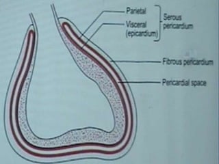
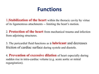
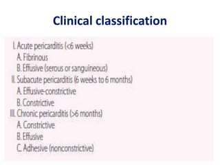
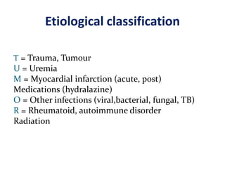
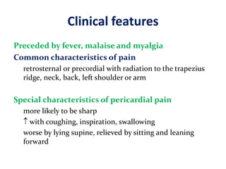
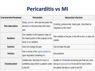
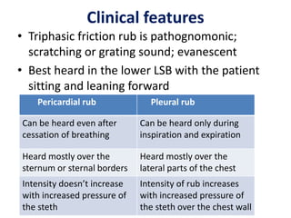
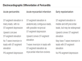
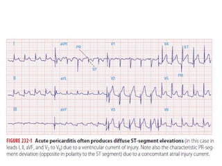
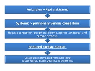
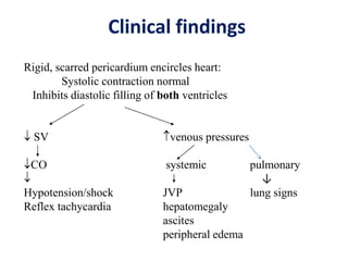
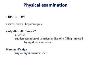
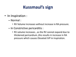
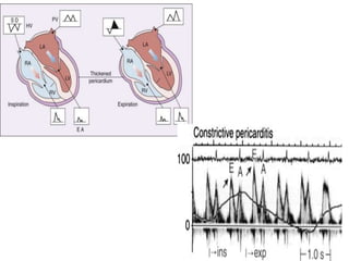
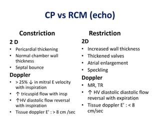
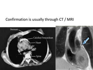
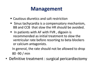
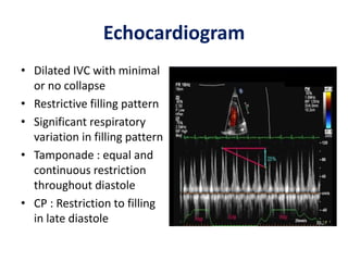
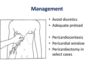
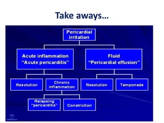
Pericardial diseases
Overview
• Introduction
• Acute pericarditis
• Constrictive pericarditis
• Pericardial effusion
• Cardiac tamponade
Pericardium is the sac that surrounds the heart
Made up of
- outer fibrous pericardium
- inner serous pericardium (parietal & visceral)
• Pericardial fluid :
– up to 50 ml of clear plasma ultrafiltrate between
the two layers of the serous pericardium
Functions
1.Stabilization of the heart within the thoracic cavity by virtue
of its ligamentous attachments -- limiting the heart’s motion.
2. Protection of the heart from mechanical trauma and infection
from adjoining structures.
3. The pericardial fluid functions as a lubricant and decreases
friction of cardiac surface during systole and diastole.
4. Prevention of excessive dilation of heart especially during
sudden rise in intra-cardiac volume (e.g. acute aortic or mitral
regurgitation).
Acute pericarditis
Acute pericarditis
• Most common pathologic process involving
the pericardium.
• Classification Of Pericarditis :
 Clinical
 Etiological
Clinical classification
Etiological classification
T = Trauma, Tumour
U = Uremia
M = Myocardial infarction (acute, post)
Medications (hydralazine)
O = Other infections (viral,bacterial, fungal, TB)
R = Rheumatoid, autoimmune disorder
Radiation
Clinical features
Preceded by fever, malaise and myalgia
Common characteristics of pain
retrosternal or precordial with radiation to the trapezius
ridge, neck, back, left shoulder or arm
Special characteristics of pericardial pain
more likely to be sharp
 with coughing, inspiration, swallowing
worse by lying supine, relieved by sitting and leaning
forward
Pericarditis vs MI
Clinical features
• Triphasic friction rub is pathognomonic;
scratching or grating sound; evanescent
• Best heard in the lower LSB with the patient
sitting and leaning forward
Pericardial rub Pleural rub
Can be heard even after
cessation of breathing
Can be heard only during
inspiration and expiration
Heard mostly over the
sternum or sternal borders
Heard mostly over the
lateral parts of the chest
Intensity doesn’t increase
with increased pressure of
the steth
Intensity of rub increases
with increased pressure of
the steth over the chest wall
Acute Pericarditis
Management
• Treat underlying cause
• Analgesic agents
– codeine 15-30 mg q 4-6 hrs
• Anti-inflammatory agents
– Aspirin
– NSAID (indomethacin 25-50 mg qid)
– Corticosteroids are symptomatically effective , but
preferably avoided
16
Pericarditis after AMI
Early
 Occurs - 1 to 3 days (no more
than a week
 due to transmural necrosis
with pericardial inflammation
 40% of patients with large, Q-
wave MIs have pericarditis
 Benign
 aspirin doses (650 mg orally
three or four times per day
for 2 to 5 days) or
acetaminophen is usually
effective
Late (Dresslers Syndrome)
 Occurs - 1 week to a few
months after AMI .
 autoimmune etiology
 3% to 4%.
 Polyserositis with pericardial
or pleural effusions
 Aspirin , Colchicine .
 Prednisone, 40 to 60 mg /d
with a 7- to 10-day taper(If
not responding to treatment
or for recurrent symptoms)
Constrictive pericarditis
Etiology
• Idiopathic or viral — 42 to 49 %
• Post cardiac surgery — 11 to 37 %
• Post radiation therapy — 9 to 31 %
• Connective tissue disorder — 3 to 7 %
• Postinfectious (tuberculous or purulent
pericarditis) — 3 to 6 %
• Miscellaneous causes (malignancy, trauma,
drug-induced, asbestosis, sarcoidosis, uremic
pericarditis) — 1 to 10 %
Pathophysiology
• A thickened, fibrotic pericardium forms a non-
compliant shell around the heart (“constricts
it”).
• Ventricles are like a fixed cavity; encased by
the thick pericardium
• This shell prevents the heart from expanding
when blood enters it.
• So it interferes with ventricular filling
Pathophysiology
• In early diastole the ventricle relaxes (expands) to
a certain extent and stops abruptly because it
cannot expand any more
• Almost all of the ventricular filling occurs in
early diastole; very little filling in late diastole
because the ventricle cannot expand any more
• CP  restriction of filling in late diastole
• Filling of one ventricle occurs at the expense of
the other ventricle
Consequence of impaired ventricular filling
causes fatigue, muscle wasting, and weight loss
Reduced cardiac output :
Hepatic congestion, peripheral edema, ascites , anasarca, and
cardiac cirrhosis.
Systemic > pulmonary venous congestion
Pericardium – Rigid and Scarred
Clinical findings
Rigid, scarred pericardium encircles heart:
Systolic contraction normal
Inhibits diastolic filling of both ventricles
 SV venous pressures
CO systemic pulmonary
 ↓
Hypotension/shock JVP lung signs
Reflex tachycardia hepatomegaly
ascites
peripheral edema
Physical examination
BP, HR  JVP
ascites, edema, hepatomegaly
early diastolic “knock”
after S2
sudden cessation of ventricular diastolic filling imposed
by rigid pericardial sac
Kussmaul’s sign
inspiratory increase in JVP
Kussmaul’s sign
• In Inspiration :
– Normal :
• RV Volume increases without increase in RA pressure.
– In Constrictive pericarditis :
• RV volume increases , as the RV cannot expand due to
thickened pericardium ,this results in increase in RA
pressure which causes Elevated JVP in inspiration.
Diagnosis
• Clinical suspicion followed by confirmation
with certain diagnostic tests
( many patients are initially seen for abdominal
symptoms)
• ECG : AF in 1/3rd of patients
flattened or inverted T waves
Chest X ray
Pericardial calcification
• Echocardiogram
- pericardial thickening
- septal bounce : abrupt displacement of IVS
during early diastole
- restrictive filling pattern
- >25% increase in mitral E velocity during expiration
compared with inspiration
CP vs RCM (echo)
Constriction
2 D
• Pericardial thickening
• Normal chamber wall
thickness
• Septal bounce
Doppler
• > 25% ↓ in mitral E velocity
with inspiration
• ↑ tricuspid flow with insp
• ↑HV diastolic flow reversal
with inspiration
• Tissue doppler E’ : > 8 cm /sec
Restriction
2D
• Increased wall thickness
• Thickened valves
• Atrial enlargement
• Speckling
Doppler
• MR, TR
• ↑ HV diastolic diastolic flow
reversal with expiration
• Tissue doppler E’ : < 8
cm/sec
Confirmation is usually through CT / MRI
Management
 Cautious diuretics and salt restriction
 Sinus tachycardia is a compensatory mechanism,
BB and CCB that slow the HR should be avoided.
 In patients with AF with FVR , digoxin is
recommended as initial treatment to slow the
ventricular rate before resorting to beta blockers
or calcium antagonists.
In general, the rate should not be allowed to drop
80 -90 / min
• Definitive treatment : surgical pericardiectomy
Pericardial effusion
• Accumulation of fluid between the visceral and
parietal layers of serous pericardium
• Serous
– Transudative – CHF , Renal failure
• Suppurative
– Pyogenic infection
• Hemorrhagic
– occurs with any type of pericarditis
– especially with infections and malignancies
Etiology
1. Inflammation from infection, immunologic process.
2. Trauma causing bleeding in pericardial space.
3. Noninfectious conditions such as:
a. increase in hydrostatic pressure e.g. congestive
heart failure.
b. increase in capillary permeability e.g. hypothyroidism
c. decrease in plasma oncotic pressure e.g. cirrhosis.
4. Decreased drainage of pericardial fluid due to obstruction of
thoracic duct as a result of malignancy or damage during surgery.
Quantification
• Trivial : 50 – 100 cc
• Small : 100 cc
• Moderate : 500 cc
• Large : 1000 cc
Clinical features
• Usually asymptomatic
• Can have signs of compression
- dyspnoea, dysphagia, hoarseness of voice,
hiccoughs, nausea
• Signs : muffled heart sounds
paradoxically reduced intensity of rub
Chest x ray
– usually requires >
200 ml of fluid
– cannot distinguish
between pericardial
effusion and
cardiomegaly
Electrocardiogram
Low voltage complexes
Echocardiogram
Management
• Depends on the etiology , presence of
hemodynamic compromise and the volume of
fluid.
• No role for diuretics
• Pericardiocentesis is not always necessary.
• Pericardiocentesis if
 Malignancy or Purulent pericarditis is suspected
 Hemodynamic compromise present
Cardiac tamponade
What is Tamponade ?
• Accumulation of fluid in the pericardial space
causing increase in pressure with subsequent
cardiac compression.
• Pericardial pressures > intracardiac pressures
• Most common causes :
– Malignancy
– Idiopathic pericarditis
– Renal failure.
– Bleeding following cardiac Sx and trauma , TB &
Hemopericardium
Pathophysiology
– Most critical point occurs when an effusion
reduces the volume of the cardiac chambers such
that cardiac output begins to decline
– Mainly by impeding right-sided heart filling, with
much of the effect on the left side of the heart
due to secondary under filling.
Pathophysiology
• A ) Modest amounts of
rapidly accumulating fluid
can have major effects on
cardiac function.
• B) Large, slowly
accumulating effusions
are often well tolerated,
presumably because of
chronic changes in the
pericardial pressure-
volume relation described
earlier.
Cardiac Tamponade -- Pathophysiology
Accumulation of fluid under high pressure:
compresses cardiac chambers & impairs
diastolic filling of both ventricles
 SV venous pressures
 CO systemic
Hypotension/shock JVP
Reflex tachycardia hepatomegaly
ascites
peripheral edema
Clinical features
• Symptoms
acute : confusion / agitation
• Signs ( Becks triad)
- hypotension
- elevated JVP
- muffled heart sounds
Pulsus paradoxus : insp drop in SBP > 10 mmhg
Pulsus paradoxus also seen in CP, COPD, asthma
Pulsus Paradoxus
• When severe, it may be detected by palpating
weakness or disappearance of the arterial pulse
during inspiration.
• Measured by noting the difference between the
systolic pressure at which the Korotkoff sounds
are first heard (during expiration) and the systolic
pressure at which the Korotkoff sounds are heard
with each beat, independent of respiratory phase
• Between these two pressures, the sounds are
heard only intermittently (during expiration).
• Since both ventricles share
a tight incompressible
covering, the inspiratory
enlargement of the RV
compresses and reduces LV
volume; leftward bulging
of the IVS further reduces
the LV cavity as the RV
enlarges during inspiration
• The normal inspiratory
augmentation of RV
volume causes an
exaggerated reciprocal
reduction in LV volume.
JVP
• Elevated JVP with prominent positive ways :
heart failure
• Elevated JVP with prominent negative ways :
pericardial disease
• Elevated JVP with absent pulsations : SVC
obstruction
ECG
Chest x ray
• Cardiac shadow
rounded ; Flask like
appearance
• Lungs appear oligemic
Echocardiogram
RA collapse RV collapse
Echocardiogram
• Dilated IVC with minimal
or no collapse
• Restrictive filling pattern
• Significant respiratory
variation in filling pattern
• Tamponade : equal and
continuous restriction
throughout diastole
• CP : Restriction to filling
in late diastole
Management
• Avoid diuretics
• Adequate preload
• Pericardiocentesis
• Pericardial window
• Pericardiectomy in
select cases
Take aways…
Take aways…
• Symptoms may be non cardiac
• CP and PE will mimic right heart failure
• In any RHF symptoms rule out pericardial disease
• Because treatment is completely different
(diuretics,digoxin)
• Clinical suspicion is essential for diagnosis
• Correct diagnosis is imperative
• Potential for permanent cure
ECG
ECG in MI
 ST elevations are convex,
 Restricted to arterial territory
 Reciprocal depression - More
prominent
 QRS changes occur( Q waves,
as well as notching and loss of
R-wave amplitude)
 T-wave inversions are usually
seen within hours before the
ST segments have become
isoelectric.
ECG in pericarditis
• ST Elevations are concave.
• Not restricted to arterial
territory.
• Reciprocal ST depression in
avR /V1
• T wave inversions after ST
segment becomes isoelectric.
• elevated ST segments return
to normal within hours

Pericardial diseases

  • 1.
  • 2.
    Overview • Introduction • Acutepericarditis • Constrictive pericarditis • Pericardial effusion • Cardiac tamponade
  • 3.
    Pericardium is thesac that surrounds the heart Made up of - outer fibrous pericardium - inner serous pericardium (parietal & visceral) • Pericardial fluid : – up to 50 ml of clear plasma ultrafiltrate between the two layers of the serous pericardium
  • 5.
    Functions 1.Stabilization of theheart within the thoracic cavity by virtue of its ligamentous attachments -- limiting the heart’s motion. 2. Protection of the heart from mechanical trauma and infection from adjoining structures. 3. The pericardial fluid functions as a lubricant and decreases friction of cardiac surface during systole and diastole. 4. Prevention of excessive dilation of heart especially during sudden rise in intra-cardiac volume (e.g. acute aortic or mitral regurgitation).
  • 6.
  • 7.
    Acute pericarditis • Mostcommon pathologic process involving the pericardium. • Classification Of Pericarditis :  Clinical  Etiological
  • 8.
  • 9.
    Etiological classification T =Trauma, Tumour U = Uremia M = Myocardial infarction (acute, post) Medications (hydralazine) O = Other infections (viral,bacterial, fungal, TB) R = Rheumatoid, autoimmune disorder Radiation
  • 10.
    Clinical features Preceded byfever, malaise and myalgia Common characteristics of pain retrosternal or precordial with radiation to the trapezius ridge, neck, back, left shoulder or arm Special characteristics of pericardial pain more likely to be sharp  with coughing, inspiration, swallowing worse by lying supine, relieved by sitting and leaning forward
  • 11.
  • 12.
    Clinical features • Triphasicfriction rub is pathognomonic; scratching or grating sound; evanescent • Best heard in the lower LSB with the patient sitting and leaning forward Pericardial rub Pleural rub Can be heard even after cessation of breathing Can be heard only during inspiration and expiration Heard mostly over the sternum or sternal borders Heard mostly over the lateral parts of the chest Intensity doesn’t increase with increased pressure of the steth Intensity of rub increases with increased pressure of the steth over the chest wall
  • 16.
    Acute Pericarditis Management • Treatunderlying cause • Analgesic agents – codeine 15-30 mg q 4-6 hrs • Anti-inflammatory agents – Aspirin – NSAID (indomethacin 25-50 mg qid) – Corticosteroids are symptomatically effective , but preferably avoided 16
  • 17.
    Pericarditis after AMI Early Occurs - 1 to 3 days (no more than a week  due to transmural necrosis with pericardial inflammation  40% of patients with large, Q- wave MIs have pericarditis  Benign  aspirin doses (650 mg orally three or four times per day for 2 to 5 days) or acetaminophen is usually effective Late (Dresslers Syndrome)  Occurs - 1 week to a few months after AMI .  autoimmune etiology  3% to 4%.  Polyserositis with pericardial or pleural effusions  Aspirin , Colchicine .  Prednisone, 40 to 60 mg /d with a 7- to 10-day taper(If not responding to treatment or for recurrent symptoms)
  • 18.
  • 19.
    Etiology • Idiopathic orviral — 42 to 49 % • Post cardiac surgery — 11 to 37 % • Post radiation therapy — 9 to 31 % • Connective tissue disorder — 3 to 7 % • Postinfectious (tuberculous or purulent pericarditis) — 3 to 6 % • Miscellaneous causes (malignancy, trauma, drug-induced, asbestosis, sarcoidosis, uremic pericarditis) — 1 to 10 %
  • 20.
    Pathophysiology • A thickened,fibrotic pericardium forms a non- compliant shell around the heart (“constricts it”). • Ventricles are like a fixed cavity; encased by the thick pericardium • This shell prevents the heart from expanding when blood enters it. • So it interferes with ventricular filling
  • 21.
    Pathophysiology • In earlydiastole the ventricle relaxes (expands) to a certain extent and stops abruptly because it cannot expand any more • Almost all of the ventricular filling occurs in early diastole; very little filling in late diastole because the ventricle cannot expand any more • CP  restriction of filling in late diastole • Filling of one ventricle occurs at the expense of the other ventricle
  • 24.
    Consequence of impairedventricular filling causes fatigue, muscle wasting, and weight loss Reduced cardiac output : Hepatic congestion, peripheral edema, ascites , anasarca, and cardiac cirrhosis. Systemic > pulmonary venous congestion Pericardium – Rigid and Scarred
  • 25.
    Clinical findings Rigid, scarredpericardium encircles heart: Systolic contraction normal Inhibits diastolic filling of both ventricles  SV venous pressures CO systemic pulmonary  ↓ Hypotension/shock JVP lung signs Reflex tachycardia hepatomegaly ascites peripheral edema
  • 26.
    Physical examination BP, HR JVP ascites, edema, hepatomegaly early diastolic “knock” after S2 sudden cessation of ventricular diastolic filling imposed by rigid pericardial sac Kussmaul’s sign inspiratory increase in JVP
  • 27.
    Kussmaul’s sign • InInspiration : – Normal : • RV Volume increases without increase in RA pressure. – In Constrictive pericarditis : • RV volume increases , as the RV cannot expand due to thickened pericardium ,this results in increase in RA pressure which causes Elevated JVP in inspiration.
  • 28.
    Diagnosis • Clinical suspicionfollowed by confirmation with certain diagnostic tests ( many patients are initially seen for abdominal symptoms) • ECG : AF in 1/3rd of patients flattened or inverted T waves
  • 29.
  • 30.
    • Echocardiogram - pericardialthickening - septal bounce : abrupt displacement of IVS during early diastole - restrictive filling pattern - >25% increase in mitral E velocity during expiration compared with inspiration
  • 33.
    CP vs RCM(echo) Constriction 2 D • Pericardial thickening • Normal chamber wall thickness • Septal bounce Doppler • > 25% ↓ in mitral E velocity with inspiration • ↑ tricuspid flow with insp • ↑HV diastolic flow reversal with inspiration • Tissue doppler E’ : > 8 cm /sec Restriction 2D • Increased wall thickness • Thickened valves • Atrial enlargement • Speckling Doppler • MR, TR • ↑ HV diastolic diastolic flow reversal with expiration • Tissue doppler E’ : < 8 cm/sec
  • 34.
    Confirmation is usuallythrough CT / MRI
  • 35.
    Management  Cautious diureticsand salt restriction  Sinus tachycardia is a compensatory mechanism, BB and CCB that slow the HR should be avoided.  In patients with AF with FVR , digoxin is recommended as initial treatment to slow the ventricular rate before resorting to beta blockers or calcium antagonists. In general, the rate should not be allowed to drop 80 -90 / min • Definitive treatment : surgical pericardiectomy
  • 36.
  • 37.
    • Accumulation offluid between the visceral and parietal layers of serous pericardium • Serous – Transudative – CHF , Renal failure • Suppurative – Pyogenic infection • Hemorrhagic – occurs with any type of pericarditis – especially with infections and malignancies
  • 38.
    Etiology 1. Inflammation frominfection, immunologic process. 2. Trauma causing bleeding in pericardial space. 3. Noninfectious conditions such as: a. increase in hydrostatic pressure e.g. congestive heart failure. b. increase in capillary permeability e.g. hypothyroidism c. decrease in plasma oncotic pressure e.g. cirrhosis. 4. Decreased drainage of pericardial fluid due to obstruction of thoracic duct as a result of malignancy or damage during surgery.
  • 39.
    Quantification • Trivial :50 – 100 cc • Small : 100 cc • Moderate : 500 cc • Large : 1000 cc
  • 40.
    Clinical features • Usuallyasymptomatic • Can have signs of compression - dyspnoea, dysphagia, hoarseness of voice, hiccoughs, nausea • Signs : muffled heart sounds paradoxically reduced intensity of rub
  • 41.
    Chest x ray –usually requires > 200 ml of fluid – cannot distinguish between pericardial effusion and cardiomegaly
  • 42.
  • 43.
  • 44.
    Management • Depends onthe etiology , presence of hemodynamic compromise and the volume of fluid. • No role for diuretics • Pericardiocentesis is not always necessary. • Pericardiocentesis if  Malignancy or Purulent pericarditis is suspected  Hemodynamic compromise present
  • 45.
  • 46.
    What is Tamponade? • Accumulation of fluid in the pericardial space causing increase in pressure with subsequent cardiac compression. • Pericardial pressures > intracardiac pressures • Most common causes : – Malignancy – Idiopathic pericarditis – Renal failure. – Bleeding following cardiac Sx and trauma , TB & Hemopericardium
  • 50.
    Pathophysiology – Most criticalpoint occurs when an effusion reduces the volume of the cardiac chambers such that cardiac output begins to decline – Mainly by impeding right-sided heart filling, with much of the effect on the left side of the heart due to secondary under filling.
  • 51.
    Pathophysiology • A )Modest amounts of rapidly accumulating fluid can have major effects on cardiac function. • B) Large, slowly accumulating effusions are often well tolerated, presumably because of chronic changes in the pericardial pressure- volume relation described earlier.
  • 52.
    Cardiac Tamponade --Pathophysiology Accumulation of fluid under high pressure: compresses cardiac chambers & impairs diastolic filling of both ventricles  SV venous pressures  CO systemic Hypotension/shock JVP Reflex tachycardia hepatomegaly ascites peripheral edema
  • 53.
    Clinical features • Symptoms acute: confusion / agitation • Signs ( Becks triad) - hypotension - elevated JVP - muffled heart sounds Pulsus paradoxus : insp drop in SBP > 10 mmhg Pulsus paradoxus also seen in CP, COPD, asthma
  • 54.
    Pulsus Paradoxus • Whensevere, it may be detected by palpating weakness or disappearance of the arterial pulse during inspiration. • Measured by noting the difference between the systolic pressure at which the Korotkoff sounds are first heard (during expiration) and the systolic pressure at which the Korotkoff sounds are heard with each beat, independent of respiratory phase • Between these two pressures, the sounds are heard only intermittently (during expiration).
  • 55.
    • Since bothventricles share a tight incompressible covering, the inspiratory enlargement of the RV compresses and reduces LV volume; leftward bulging of the IVS further reduces the LV cavity as the RV enlarges during inspiration • The normal inspiratory augmentation of RV volume causes an exaggerated reciprocal reduction in LV volume.
  • 56.
    JVP • Elevated JVPwith prominent positive ways : heart failure • Elevated JVP with prominent negative ways : pericardial disease • Elevated JVP with absent pulsations : SVC obstruction
  • 57.
  • 58.
    Chest x ray •Cardiac shadow rounded ; Flask like appearance • Lungs appear oligemic
  • 59.
  • 60.
    Echocardiogram • Dilated IVCwith minimal or no collapse • Restrictive filling pattern • Significant respiratory variation in filling pattern • Tamponade : equal and continuous restriction throughout diastole • CP : Restriction to filling in late diastole
  • 61.
    Management • Avoid diuretics •Adequate preload • Pericardiocentesis • Pericardial window • Pericardiectomy in select cases
  • 62.
  • 63.
    Take aways… • Symptomsmay be non cardiac • CP and PE will mimic right heart failure • In any RHF symptoms rule out pericardial disease • Because treatment is completely different (diuretics,digoxin) • Clinical suspicion is essential for diagnosis • Correct diagnosis is imperative • Potential for permanent cure
  • 65.
    ECG ECG in MI ST elevations are convex,  Restricted to arterial territory  Reciprocal depression - More prominent  QRS changes occur( Q waves, as well as notching and loss of R-wave amplitude)  T-wave inversions are usually seen within hours before the ST segments have become isoelectric. ECG in pericarditis • ST Elevations are concave. • Not restricted to arterial territory. • Reciprocal ST depression in avR /V1 • T wave inversions after ST segment becomes isoelectric. • elevated ST segments return to normal within hours