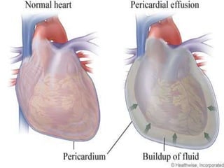
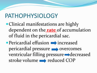
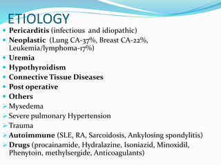
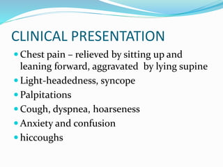
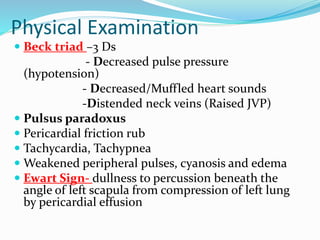
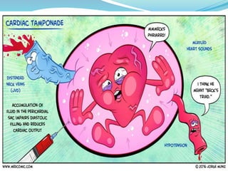
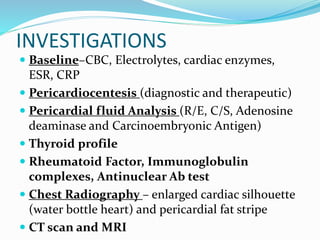
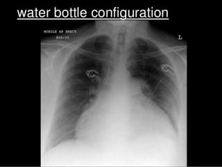
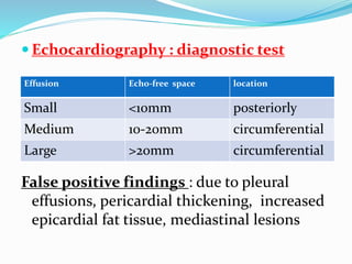
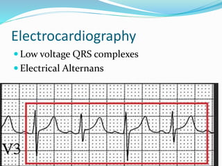
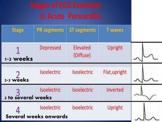
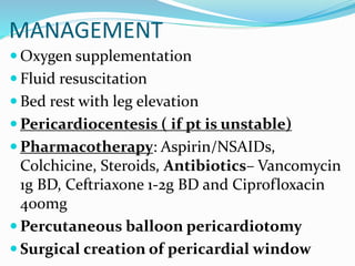
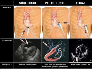
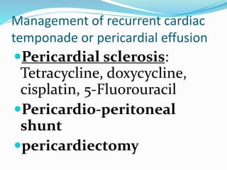
This document defines pericardial effusion and cardiac tamponade, discusses their pathophysiology, etiology, clinical presentation, investigations, and management. Pericardial effusion is an abnormal amount of fluid in the pericardial space, while cardiac tamponade is acute heart failure caused by compression of the heart from a large or rapidly developing effusion. Clinical manifestations depend on the rate of fluid accumulation and include chest pain, lightheadedness, and decreased pulse pressure. Investigations include echocardiography, electrocardiography, and pericardiocentesis. Management involves bed rest, medications, drainage procedures, and surgery in severe cases.
2. DEFINITION
Presence of an abnormal amount of fluid
and/or an abnormal character to fluid in the
pericardial space.
The pericardial space normally contains 15-
50 mL of fluid
Cardiac temponade is acute heart failure
due to compression of heart by a large or
rapidly developing effusion.
3.
4. PATHOPHYSIOLOGY
Clinical manifestations are highly
dependent on the rate of accumulation
of fluid in the pericardial sac.
Pericardial effusion increased
pericardial pressure overcomes
ventricular filling pressure decreased
stroke volume reduced COP
6. CLINICAL PRESENTATION
Chest pain – relieved by sitting up and
leaning forward, aggravated by lying supine
Light-headedness, syncope
Palpitations
Cough, dyspnea, hoarseness
Anxiety and confusion
hiccoughs
7. Physical Examination
Beck triad –3 Ds
- Decreased pulse pressure
(hypotension)
- Decreased/Muffled heart sounds
-Distended neck veins (Raised JVP)
Pulsus paradoxus
Pericardial friction rub
Tachycardia, Tachypnea
Weakened peripheral pulses, cyanosis and edema
Ewart Sign- dullness to percussion beneath the
angle of left scapula from compression of left lung
by pericardial effusion
8.
9. INVESTIGATIONS
Baseline–CBC, Electrolytes, cardiac enzymes,
ESR, CRP
Pericardiocentesis (diagnostic and therapeutic)
Pericardial fluid Analysis (R/E, C/S, Adenosine
deaminase and Carcinoembryonic Antigen)
Thyroid profile
Rheumatoid Factor, Immunoglobulin
complexes, Antinuclear Ab test
Chest Radiography – enlarged cardiac silhouette
(water bottle heart) and pericardial fat stripe
CT scan and MRI
10.
11. Echocardiography : diagnostic test
False positive findings : due to pleural
effusions, pericardial thickening, increased
epicardial fat tissue, mediastinal lesions
Effusion Echo-free space location
Small <10mm posteriorly
Medium 10-20mm circumferential
Large >20mm circumferential