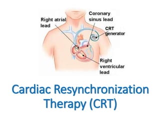
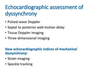
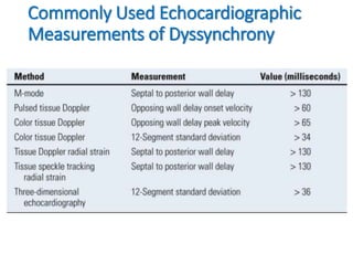
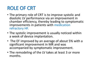
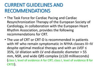
Welcome to 5 minutes
presentation
Dr. Sayeedur Rahman Khan Rumi
Dr.rumibd@gmail.com
MD (Cardiology) Final Part Student
National Heart Foundation Hospital and Research
Cardiac Resynchronization
Therapy (CRT)
• Typical LV myocardial activation occurs from the
apex to base, simultaneously in the septum and in
the LV free wall, and is described as synchronous.
• In the setting of conduction delay, the
electromechanical coupling of the heart is
disrupted, leading to dyssynchrony.
• Over time, electromechanical uncoupling leads to
impaired stroke volume, worsened mitral
insufficiency, prolonged LV isovolumetric events,
and impaired diastolic filling.
• These effects contribute to adverse remodeling in
the already impaired heart, creating a vicious cycle
that perpetuates this process into more advanced
HF.
TYPES OF DYSSYNCHRONY
• AV dyssynchrony
• Interventricular dyssynchrony
• Intraventricular dyssynchrony
Others types:
• Mechanical dyssynchrony
• Electrical dyssynchrony
ASSESSMENT OF DYSSYNCHRONY
• Approximately 30% of patients meeting current
implantation criteria fail to respond to CRT.
• Studies have revealed that up to 30% of patients
with a prolonged QRS duration do not have
mechanical dyssynchrony as assessed by MRI or
echocardiography.
• Whereas up to 30% of patients with a normal QRS
duration and symptomatic HF have evidence of
mechanical dyssynchrony on echo or MRI and could
potentially benefit from resynchronization therapy.
Echocardiographic assessment of
dyssynchrony
• Pulsed-wave Doppler
• Septal to posterior wall motion delay
• Tissue Doppler imaging
• Three-dimensional imaging
New echocardiographic indices of mechanical
dyssynchrony:
• Strain imaging
• Speckle tracking
Commonly Used Echocardiographic
Measurements of Dyssynchrony
ROLE OF CRT
• The primary role of CRT is to improve systolic and
diastolic LV performance via an improvement in
chamber efficiency, thereby leading to symptomatic
improvements in patients with medication
refractory HF.
• The systolic improvement is usually noticed within
a week of device implantation.
• The EF improved by an average of about 5% with a
significant improvement in MR and was
accompanied by symptomatic improvement.
• The remodeling of the LV takes at least 3 or more
months.
CURRENT GUIDELINES AND
RECOMMENDATIONS
• The Task Force for Cardiac Pacing and Cardiac
Resynchronization Therapy of the European Society of
Cardiology, in collaboration with the European Heart
Rhythm Association, provides the following
recommendations for CRT.
• The use of CRT or CRT-D is recommended in patients
with HF who remain symptomatic in NYHA classes III–IV
despite optimal medical therapy and with an LVEF ≤
35%, LV dilation with LV end-diastolic diameter > 55
mm, NSR, and wide QRS complex (≥120 milliseconds)
(class I, level of evidence A for CRT; class I, level of evidence B for
CRTD).
• The use of CRT is also recommended in patients with HF
with NYHA classes III–IV despite optimal medical therapy
and with an LVEF ≤ 35%, LV dilation, and a concomitant
indication for permanent pacing (first implant or upgrade of
conventional pacemaker) (class IIa, level of evidence C).
• The use of CRT is also recommended in patients with HF
who remain symptomatic in classes III–IV despite optimal
medical therapy and with an LVEF ≤ 35%, LV dilation,
permanent atrial fibrillation, and indication for AV junction
ablation (class IIa, level of evidence C).
• The use of CRT is also recommended in patients with HF
who remain symptomatic in classes III–IV despite optimal
medical therapy and with an LVEF ≤ 35%, QRS ≥ 130
milliseconds, permanent atrial fibrillation, and indication for
AV junction ablation (class IIa, level of evidence C).
IMPLANTATION PROCEDURE
• Unlike conventional
transvenous pacemaker or
ICD implantation that
requires lead placement in
the right atrium and/or the
right ventricle only, Bi-V
pacing requires LV lead
implantation.
• Initially, this was achieved
via a thoracotomy;
however, currently up to
98% of Bi-V devices are
placed via a transvenous
approach.
• Typically, a cephalic or axillary vein approach to
venous access is used.
• The right atrial and RV leads are implanted in a
fashion similar to a pacemaker or ICD implantation.
• The LV lead is placed through the CS into a CS
branch on the lateral free wall of the left ventricle.
• Performing an occlusive CS venogram may help
identify the appropriate vein.
Thank You

Cardiac resynchronization therapy

  • 1.
    Welcome to 5minutes presentation Dr. Sayeedur Rahman Khan Rumi Dr.rumibd@gmail.com MD (Cardiology) Final Part Student National Heart Foundation Hospital and Research
  • 2.
  • 3.
    • Typical LVmyocardial activation occurs from the apex to base, simultaneously in the septum and in the LV free wall, and is described as synchronous. • In the setting of conduction delay, the electromechanical coupling of the heart is disrupted, leading to dyssynchrony. • Over time, electromechanical uncoupling leads to impaired stroke volume, worsened mitral insufficiency, prolonged LV isovolumetric events, and impaired diastolic filling. • These effects contribute to adverse remodeling in the already impaired heart, creating a vicious cycle that perpetuates this process into more advanced HF.
  • 4.
    TYPES OF DYSSYNCHRONY •AV dyssynchrony • Interventricular dyssynchrony • Intraventricular dyssynchrony Others types: • Mechanical dyssynchrony • Electrical dyssynchrony
  • 5.
    ASSESSMENT OF DYSSYNCHRONY •Approximately 30% of patients meeting current implantation criteria fail to respond to CRT. • Studies have revealed that up to 30% of patients with a prolonged QRS duration do not have mechanical dyssynchrony as assessed by MRI or echocardiography. • Whereas up to 30% of patients with a normal QRS duration and symptomatic HF have evidence of mechanical dyssynchrony on echo or MRI and could potentially benefit from resynchronization therapy.
  • 6.
    Echocardiographic assessment of dyssynchrony •Pulsed-wave Doppler • Septal to posterior wall motion delay • Tissue Doppler imaging • Three-dimensional imaging New echocardiographic indices of mechanical dyssynchrony: • Strain imaging • Speckle tracking
  • 7.
  • 8.
    ROLE OF CRT •The primary role of CRT is to improve systolic and diastolic LV performance via an improvement in chamber efficiency, thereby leading to symptomatic improvements in patients with medication refractory HF. • The systolic improvement is usually noticed within a week of device implantation. • The EF improved by an average of about 5% with a significant improvement in MR and was accompanied by symptomatic improvement. • The remodeling of the LV takes at least 3 or more months.
  • 9.
    CURRENT GUIDELINES AND RECOMMENDATIONS •The Task Force for Cardiac Pacing and Cardiac Resynchronization Therapy of the European Society of Cardiology, in collaboration with the European Heart Rhythm Association, provides the following recommendations for CRT. • The use of CRT or CRT-D is recommended in patients with HF who remain symptomatic in NYHA classes III–IV despite optimal medical therapy and with an LVEF ≤ 35%, LV dilation with LV end-diastolic diameter > 55 mm, NSR, and wide QRS complex (≥120 milliseconds) (class I, level of evidence A for CRT; class I, level of evidence B for CRTD).
  • 10.
    • The useof CRT is also recommended in patients with HF with NYHA classes III–IV despite optimal medical therapy and with an LVEF ≤ 35%, LV dilation, and a concomitant indication for permanent pacing (first implant or upgrade of conventional pacemaker) (class IIa, level of evidence C). • The use of CRT is also recommended in patients with HF who remain symptomatic in classes III–IV despite optimal medical therapy and with an LVEF ≤ 35%, LV dilation, permanent atrial fibrillation, and indication for AV junction ablation (class IIa, level of evidence C). • The use of CRT is also recommended in patients with HF who remain symptomatic in classes III–IV despite optimal medical therapy and with an LVEF ≤ 35%, QRS ≥ 130 milliseconds, permanent atrial fibrillation, and indication for AV junction ablation (class IIa, level of evidence C).
  • 11.
    IMPLANTATION PROCEDURE • Unlikeconventional transvenous pacemaker or ICD implantation that requires lead placement in the right atrium and/or the right ventricle only, Bi-V pacing requires LV lead implantation. • Initially, this was achieved via a thoracotomy; however, currently up to 98% of Bi-V devices are placed via a transvenous approach.
  • 13.
    • Typically, acephalic or axillary vein approach to venous access is used. • The right atrial and RV leads are implanted in a fashion similar to a pacemaker or ICD implantation. • The LV lead is placed through the CS into a CS branch on the lateral free wall of the left ventricle. • Performing an occlusive CS venogram may help identify the appropriate vein.
  • 14.