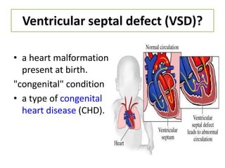
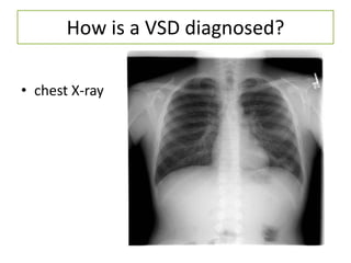
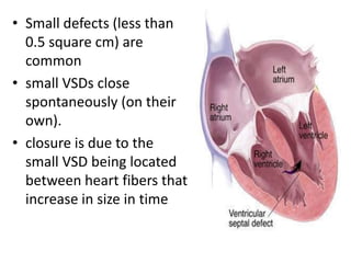
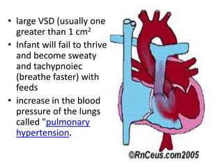
Ventricular Septal
   defect (VSD)


  Miljie
Tompong
Ventricular septal defect (VSD)?

• a heart malformation
  present at birth.
"congenital" condition
• a type of congenital
  heart disease (CHD).
Anatomy and physiology


•   The heart with a VSD has a hole in the wall (the septum) between its two lower
    chambers (the ventricles).
How do VSDs cause problems?
How is a VSD diagnosed?

• chest X-ray
• Echocardiogram
  (used to define
  the anatomy
  and evaluate
  the
  characteristics
  (amount and
  pressures) of
  the shunted
  blood)
Electrocardiogram
• is a noninvasive
  test that is used
  to reflect
  underlying
  heart
  conditions by
  measuring the
  electrical
  activity of the
  heart.
What are the symptoms of a VSD?

• A murmur is a sound
  generated by abnormally
  turbulent flow of blood
  through the heart.
• symptomless at birth
  usually develop until later
  in the first week of life.
• no signs of cyanosis.
• Small defects (less than
  0.5 square cm) are
  common
• small VSDs close
  spontaneously (on their
  own).
• closure is due to the
  small VSD being located
  between heart fibers that
  increase in size in time
• large VSD (usually one
  greater than 1 cm2
• Infant will fail to thrive
  and become sweaty
  and tachypnoiec
  (breathe faster) with
  feeds
• increase in the blood
  pressure of the lungs
  called "pulmonary
  hypertension.
Medical Management
• Digoxin (LANOXIN)
• works directly on the heart to increase the
  velocity of contraction and the force of the
  contraction.
• FUROSEMIDE (LASIX)
• Diuretics remove edema and act to lower
  blood pressure. This helps reduce the extra
  load on the pulmonary system.
Treatment
• PA Banding
  a palliative
  treatment
  particularly
  reserved for those
  patients who are
  unable to
  withstand an open
  heart procedure
  for total
  correction.
Surgical Treatment
• median sternotomy
• Surgical access to the VSD depends upon it's
  location and the presence of other cardiac
  defects.
• The right ventricle is often opened to gain
  access to perimembranous or inlet defects.
• The pulmonary artery may be used to close
  outlet septum VSDs
• Large defects
  may be repaired
  using a
  pericardium
  patch sutured
  into place to
  occlude the
  space.
Preoperative and postoperative
• prophylactic antibiotics are often required to
  prevent infectious endocarditis.

• The child should be assessed postoperatively
  for dysrhythmia, since edema in the septum
  may interfere with conduction.
Nursing Diagnosis

   “Decrease in cardiac output
      associated with heart
         malformations”
Objective: improve the heart Rainfall
Outcome criteria: signs of improvement in cardiac
  output
Nursing Intervention :
• Observation of the quality and strength of heart
  rate, peripheral pulse, skin color and warmth.
• Set the degree of cyanosis (mucous membranes,
  clubbing)
• Monitor signs of CHF (anxiety, tachycardia,
  tachipnea, cramped, tired while drinking milk,
  periorbital edema, and hepatomegaly Oliguria.
• Collaboration for the administration of drugs
  (diuretics, to reduce after load) as indicated.

Ventricular septal defect (vsd)

  • 1.
    Ventricular Septal defect (VSD) Miljie Tompong
  • 2.
    Ventricular septal defect(VSD)? • a heart malformation present at birth. "congenital" condition • a type of congenital heart disease (CHD).
  • 3.
    Anatomy and physiology • The heart with a VSD has a hole in the wall (the septum) between its two lower chambers (the ventricles).
  • 5.
    How do VSDscause problems?
  • 6.
    How is aVSD diagnosed? • chest X-ray
  • 7.
    • Echocardiogram (used to define the anatomy and evaluate the characteristics (amount and pressures) of the shunted blood)
  • 8.
    Electrocardiogram • is anoninvasive test that is used to reflect underlying heart conditions by measuring the electrical activity of the heart.
  • 9.
    What are thesymptoms of a VSD? • A murmur is a sound generated by abnormally turbulent flow of blood through the heart. • symptomless at birth usually develop until later in the first week of life. • no signs of cyanosis.
  • 10.
    • Small defects(less than 0.5 square cm) are common • small VSDs close spontaneously (on their own). • closure is due to the small VSD being located between heart fibers that increase in size in time
  • 11.
    • large VSD(usually one greater than 1 cm2 • Infant will fail to thrive and become sweaty and tachypnoiec (breathe faster) with feeds • increase in the blood pressure of the lungs called "pulmonary hypertension.
  • 12.
    Medical Management • Digoxin(LANOXIN) • works directly on the heart to increase the velocity of contraction and the force of the contraction. • FUROSEMIDE (LASIX) • Diuretics remove edema and act to lower blood pressure. This helps reduce the extra load on the pulmonary system.
  • 13.
    Treatment • PA Banding a palliative treatment particularly reserved for those patients who are unable to withstand an open heart procedure for total correction.
  • 14.
    Surgical Treatment • mediansternotomy • Surgical access to the VSD depends upon it's location and the presence of other cardiac defects. • The right ventricle is often opened to gain access to perimembranous or inlet defects. • The pulmonary artery may be used to close outlet septum VSDs
  • 15.
    • Large defects may be repaired using a pericardium patch sutured into place to occlude the space.
  • 16.
    Preoperative and postoperative •prophylactic antibiotics are often required to prevent infectious endocarditis. • The child should be assessed postoperatively for dysrhythmia, since edema in the septum may interfere with conduction.
  • 17.
    Nursing Diagnosis “Decrease in cardiac output associated with heart malformations” Objective: improve the heart Rainfall Outcome criteria: signs of improvement in cardiac output
  • 18.
    Nursing Intervention : •Observation of the quality and strength of heart rate, peripheral pulse, skin color and warmth. • Set the degree of cyanosis (mucous membranes, clubbing) • Monitor signs of CHF (anxiety, tachycardia, tachipnea, cramped, tired while drinking milk, periorbital edema, and hepatomegaly Oliguria. • Collaboration for the administration of drugs (diuretics, to reduce after load) as indicated.