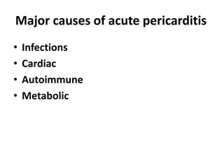
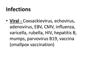
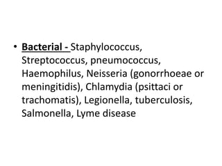
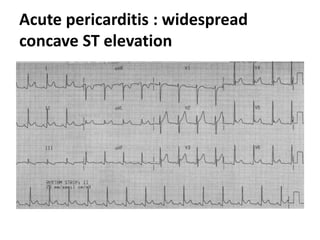
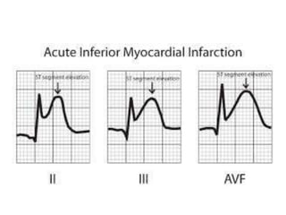
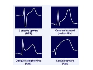
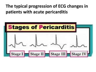
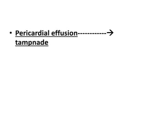
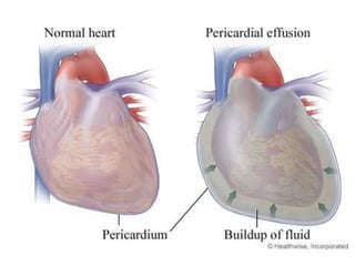
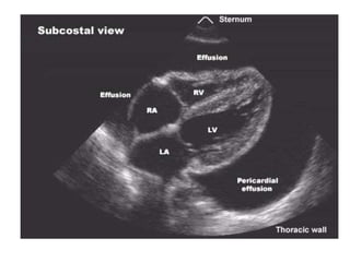
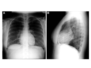
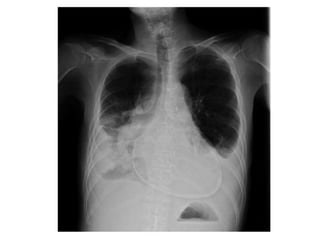
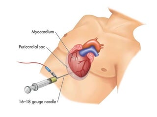
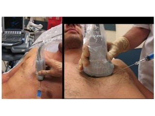
Acute pericarditis
• The pericardium is a fibroelastic sac
made up of visceral and parietal layers
separated by a (potential) space, the
pericardial cavity.
• In healthy individuals, the pericardial
cavity contains 15 to 50 mL of an
ultrafiltrate of plasma
Diseases of the pericardium present
clinically in one of four ways
• Acute pericarditis
• Pericardial effusion without major
hemodynamic compromise
• Cardiac tamponade
• Constrictive pericarditis
• Acute pericarditis is the most
common disorder involving the
pericardium
Major causes of acute pericarditis
• Infections
• Cardiac
• Autoimmune
• Metabolic
• Neoplasm
• Drugs
• Trauma
• Idiopathic
Infections
• Viral - Coxsackievirus, echovirus,
adenovirus, EBV, CMV, influenza,
varicella, rubella, HIV, hepatitis B,
mumps, parvovirus B19, vaccina
(smallpox vaccination)
• Bacterial - Staphylococcus,
Streptococcus, pneumococcus,
Haemophilus, Neisseria (gonorrhoeae or
meningitidis), Chlamydia (psittaci or
trachomatis), Legionella, tuberculosis,
Salmonella, Lyme disease
• Mycoplasma
• Fungal - Histoplasmosis, aspergillosis,
blastomycosis, coccidiodomycosis,
actinomycosis, nocardia, candida
• Parasitic - Echinococcus, amebiasis,
toxoplasmosis
Cardiac
• Early infarction pericarditis
• Late postcardiac injury syndrome
(Dressler's syndrome), also seen in other
settings (eg, post-myocardial infarction
and post-cardiac surgery)
Autoimmune
• Rheumatic diseases - Including lupus,
rheumatoid arthritis, vasculitis,
scleroderma, mixed connective disease
• Other - Granulomatosis with polyangiitis
(Wegener's), polyarteritis nodosa,
sarcoidosis, inflammatory bowel disease
(Crohn's, ulcerative colitis), Whipple's,
giant cell arteritis, Behcet's disease,
rheumatic fever
Metabolic
• Hypothyroidism - Primarily pericardial
effusion
• Uremia
• Ovarian hyperstimulation syndrome
Neoplasm
• Metastatic - Lung or breast cancer,
Hodgkin's disease, leukemia, melanoma
• Primary - Rhabdomyosarcoma, teratoma,
fibroma, lipoma, leiomyoma, angioma
• Paraneoplastic
Drugs
• Procainamide, isoniazid, or hydralazine
as part of drug-induced lupus
• Other - Cromolyn sodium, dantrolene,
methysergide, anticoagulants,
thrombolytics, phenytoin, penicillin,
phenylbutazone, doxorubicin
Trauma
• Blunt
• Penetrating
Idiopathic
• In most case series, the majority of
patients are not found to have an
identifiable cause of pericardial disease.
• Frequently such cases are presumed to
have a viral or autoimmune etiology
Clinical manifestations
• The major clinical manifestations of
acute pericarditis include :
• Chest pain – typically sharp and pleuritic,
improved by sitting up and leaning
forward
• Pericardial friction rub – a superficial
scratchy or squeaking sound best heard
with the diaphragm of the stethoscope
over the left sternal border
• Electrocardiogram (ECG) changes – new
widespread concave ST segment
elevation
• In STEM convex ST segment elevation
• The ST segment is usually isoelectric
(ie, zero potential as identified by
the T-P segment) and has a slight
upward concavity.
Acute pericarditis : widespread
concave ST elevation
The typical progression of ECG changes in
patients with acute pericarditis
• Pericardial effusion------------
tampnade
PROGNOSIS
• Patients with acute idiopathic or viral
pericarditis have a good long-term
prognosis.
• Cardiac tamponade rarely occurs in
patients with acute idiopathic pericarditis
and is more common in patients with a
specific underlying etiology such as
malignancy, tuberculosis, or purulent
pericarditis.
• Constrictive pericarditis may occur in
about 1 percent of patients with acute
idiopathic pericarditis, and is also more
common in patients with a specific
etiology.
Treatment
• The therapy of acute pericarditis should
be targeted as much as possible to the
underlying etiology
• Most patients are treated for a
presumptive viral cause with
nonsteroidal anti-inflammatory drugs
(NSAIDS) and colchicine
• Most patients with acute pericarditis can
be managed effectively with medical
therapy alone.
• However, patients with a large pericardial
effusion, a hemodynamically significant
pericardial effusion, a suspicion of a
bacterial or neoplastic etiology, or
evidence of constrictive pericarditis
should be evaluated for invasive
therapies, such as pericardial
drainage and/or pericardiotomy.
Acute pericarditis
Acute pericarditis
Acute pericarditis

Acute pericarditis

  • 1.
  • 2.
    • The pericardiumis a fibroelastic sac made up of visceral and parietal layers separated by a (potential) space, the pericardial cavity.
  • 3.
    • In healthyindividuals, the pericardial cavity contains 15 to 50 mL of an ultrafiltrate of plasma
  • 4.
    Diseases of thepericardium present clinically in one of four ways • Acute pericarditis • Pericardial effusion without major hemodynamic compromise • Cardiac tamponade • Constrictive pericarditis
  • 5.
    • Acute pericarditisis the most common disorder involving the pericardium
  • 6.
    Major causes ofacute pericarditis • Infections • Cardiac • Autoimmune • Metabolic
  • 7.
    • Neoplasm • Drugs •Trauma • Idiopathic
  • 8.
    Infections • Viral -Coxsackievirus, echovirus, adenovirus, EBV, CMV, influenza, varicella, rubella, HIV, hepatitis B, mumps, parvovirus B19, vaccina (smallpox vaccination)
  • 9.
    • Bacterial -Staphylococcus, Streptococcus, pneumococcus, Haemophilus, Neisseria (gonorrhoeae or meningitidis), Chlamydia (psittaci or trachomatis), Legionella, tuberculosis, Salmonella, Lyme disease
  • 10.
  • 11.
    • Fungal -Histoplasmosis, aspergillosis, blastomycosis, coccidiodomycosis, actinomycosis, nocardia, candida
  • 12.
    • Parasitic -Echinococcus, amebiasis, toxoplasmosis
  • 13.
    Cardiac • Early infarctionpericarditis • Late postcardiac injury syndrome (Dressler's syndrome), also seen in other settings (eg, post-myocardial infarction and post-cardiac surgery)
  • 14.
    Autoimmune • Rheumatic diseases- Including lupus, rheumatoid arthritis, vasculitis, scleroderma, mixed connective disease
  • 15.
    • Other -Granulomatosis with polyangiitis (Wegener's), polyarteritis nodosa, sarcoidosis, inflammatory bowel disease (Crohn's, ulcerative colitis), Whipple's, giant cell arteritis, Behcet's disease, rheumatic fever
  • 16.
    Metabolic • Hypothyroidism -Primarily pericardial effusion • Uremia • Ovarian hyperstimulation syndrome
  • 17.
    Neoplasm • Metastatic -Lung or breast cancer, Hodgkin's disease, leukemia, melanoma • Primary - Rhabdomyosarcoma, teratoma, fibroma, lipoma, leiomyoma, angioma • Paraneoplastic
  • 18.
    Drugs • Procainamide, isoniazid,or hydralazine as part of drug-induced lupus • Other - Cromolyn sodium, dantrolene, methysergide, anticoagulants, thrombolytics, phenytoin, penicillin, phenylbutazone, doxorubicin
  • 19.
  • 20.
    Idiopathic • In mostcase series, the majority of patients are not found to have an identifiable cause of pericardial disease. • Frequently such cases are presumed to have a viral or autoimmune etiology
  • 21.
    Clinical manifestations • Themajor clinical manifestations of acute pericarditis include :
  • 22.
    • Chest pain– typically sharp and pleuritic, improved by sitting up and leaning forward
  • 23.
    • Pericardial frictionrub – a superficial scratchy or squeaking sound best heard with the diaphragm of the stethoscope over the left sternal border
  • 24.
    • Electrocardiogram (ECG)changes – new widespread concave ST segment elevation • In STEM convex ST segment elevation
  • 26.
    • The STsegment is usually isoelectric (ie, zero potential as identified by the T-P segment) and has a slight upward concavity.
  • 28.
    Acute pericarditis :widespread concave ST elevation
  • 31.
    The typical progressionof ECG changes in patients with acute pericarditis
  • 32.
  • 38.
    PROGNOSIS • Patients withacute idiopathic or viral pericarditis have a good long-term prognosis.
  • 39.
    • Cardiac tamponaderarely occurs in patients with acute idiopathic pericarditis and is more common in patients with a specific underlying etiology such as malignancy, tuberculosis, or purulent pericarditis.
  • 40.
    • Constrictive pericarditismay occur in about 1 percent of patients with acute idiopathic pericarditis, and is also more common in patients with a specific etiology.
  • 43.
    Treatment • The therapyof acute pericarditis should be targeted as much as possible to the underlying etiology
  • 44.
    • Most patientsare treated for a presumptive viral cause with nonsteroidal anti-inflammatory drugs (NSAIDS) and colchicine
  • 45.
    • Most patientswith acute pericarditis can be managed effectively with medical therapy alone.
  • 46.
    • However, patientswith a large pericardial effusion, a hemodynamically significant pericardial effusion, a suspicion of a bacterial or neoplastic etiology, or evidence of constrictive pericarditis should be evaluated for invasive therapies, such as pericardial drainage and/or pericardiotomy.