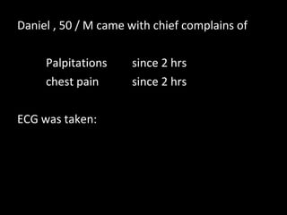
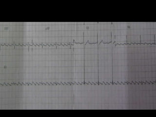
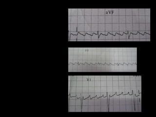
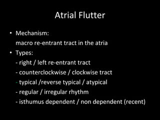
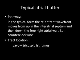
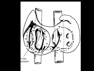
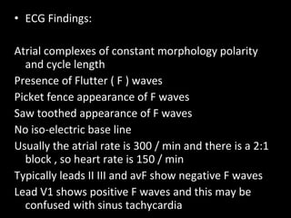
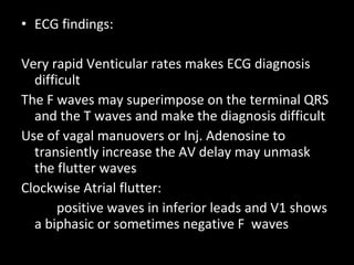
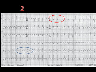
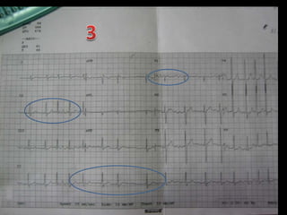
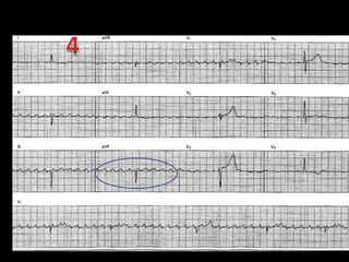
ECG   of the week  Dr. Prof Mageshkumar’s Unit  Devendra Patil
Daniel , 50 / M came with chief complains of  Palpitations  since 2 hrs chest pain since 2 hrs ECG was taken:
 
 
ECG: Atrial rate 300 Heart rate 75/min Regular rhythm 4:1 constant block No iso electric baseline Saw tooth appearance in inferior leads Rsr’ in v1 Impression: Atrial flutter with incomplete RBBB
Atrial Flutter Mechanism: macro re-entrant tract in the atria Types: - right / left re-entrant tract - counterclockwise / clockwise tract -  typical   /reverse typical / atypical  - regular / irregular rhythm - isthumus dependent / non dependent (recent)
Typical atrial flutter Pathway: in the typical form the re-entrant wavefront moves from up in the interatrial septum and then down the free right atrial wall. i.e. counterclockwise Tract location : cavo – tricuspid isthumus
 
ECG Findings: Atrial complexes of constant morphology polarity and cycle length Presence of Flutter ( F ) waves Picket fence appearance of F waves Saw toothed appearance of F waves No iso-electric base line Usually the atrial rate is 300 / min and there is a 2:1 block , so heart rate is 150 / min Typically leads II III and avF show negative F waves Lead V1 shows positive F waves and this may be confused with sinus tachycardia
ECG findings: Very rapid Venticular rates makes ECG diagnosis difficult The F waves may superimpose on the terminal QRS and the T waves and make the diagnosis difficult Use of vagal manuovers or Inj. Adenosine to transiently increase the AV delay may unmask the flutter waves  Clockwise Atrial flutter: positive waves in inferior leads and V1 shows a biphasic or sometimes negative F  waves
Few more  ECG s


Etiology First week after open heart surgery COPD Mitral or tricuspid valve lesions Thyrotoxicosis Surgical correction of congenital heart disease Right atrial enlergement
Treatment Acute: cardioversion Longterm anti-coagulation ( similar to AF ) anti-arrhythmics  Catheter ablation of tract pacemaker insertion
Cardioversion External trans thoracic syncronised DC shock is highly effective Intravenous Ibulitide or procainamide can also be used Care should be taken during use of class 1 esp 1C agents because they may slow the atrial rate and an inadequately suppressed AV node may give way to 1:1 conduction leading to high rates and circulatory collapse.
 
Long term treatment

ECG: Atrial Flutter

  • 1.
    ECG of the week Dr. Prof Mageshkumar’s Unit Devendra Patil
  • 2.
    Daniel , 50/ M came with chief complains of Palpitations since 2 hrs chest pain since 2 hrs ECG was taken:
  • 3.
  • 4.
  • 5.
    ECG: Atrial rate300 Heart rate 75/min Regular rhythm 4:1 constant block No iso electric baseline Saw tooth appearance in inferior leads Rsr’ in v1 Impression: Atrial flutter with incomplete RBBB
  • 6.
    Atrial Flutter Mechanism:macro re-entrant tract in the atria Types: - right / left re-entrant tract - counterclockwise / clockwise tract - typical /reverse typical / atypical - regular / irregular rhythm - isthumus dependent / non dependent (recent)
  • 7.
    Typical atrial flutterPathway: in the typical form the re-entrant wavefront moves from up in the interatrial septum and then down the free right atrial wall. i.e. counterclockwise Tract location : cavo – tricuspid isthumus
  • 8.
  • 9.
    ECG Findings: Atrialcomplexes of constant morphology polarity and cycle length Presence of Flutter ( F ) waves Picket fence appearance of F waves Saw toothed appearance of F waves No iso-electric base line Usually the atrial rate is 300 / min and there is a 2:1 block , so heart rate is 150 / min Typically leads II III and avF show negative F waves Lead V1 shows positive F waves and this may be confused with sinus tachycardia
  • 10.
    ECG findings: Veryrapid Venticular rates makes ECG diagnosis difficult The F waves may superimpose on the terminal QRS and the T waves and make the diagnosis difficult Use of vagal manuovers or Inj. Adenosine to transiently increase the AV delay may unmask the flutter waves Clockwise Atrial flutter: positive waves in inferior leads and V1 shows a biphasic or sometimes negative F waves
  • 11.
    Few more ECG s
  • 12.
  • 13.
  • 14.
  • 15.
  • 16.
    Etiology First weekafter open heart surgery COPD Mitral or tricuspid valve lesions Thyrotoxicosis Surgical correction of congenital heart disease Right atrial enlergement
  • 17.
    Treatment Acute: cardioversionLongterm anti-coagulation ( similar to AF ) anti-arrhythmics Catheter ablation of tract pacemaker insertion
  • 18.
    Cardioversion External transthoracic syncronised DC shock is highly effective Intravenous Ibulitide or procainamide can also be used Care should be taken during use of class 1 esp 1C agents because they may slow the atrial rate and an inadequately suppressed AV node may give way to 1:1 conduction leading to high rates and circulatory collapse.
  • 19.
  • 20.